Figure 1
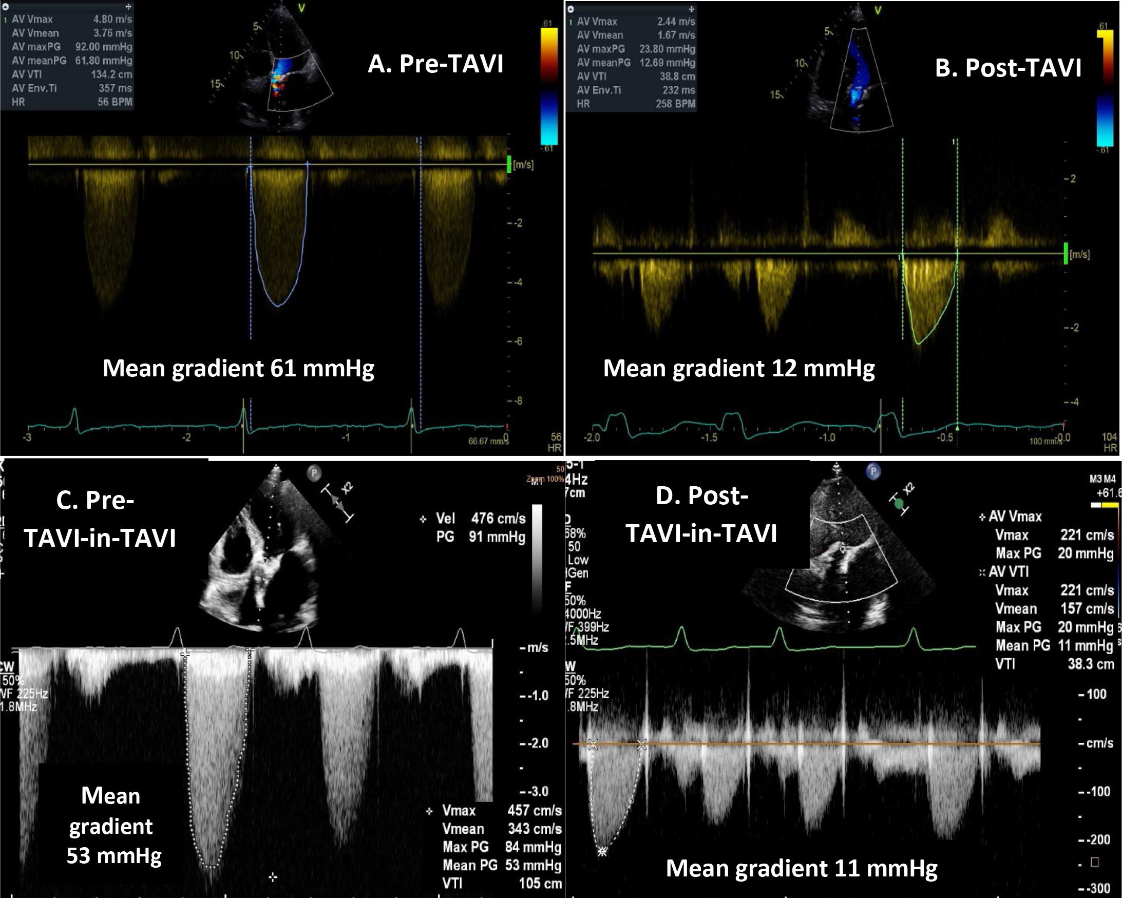
Figure 2
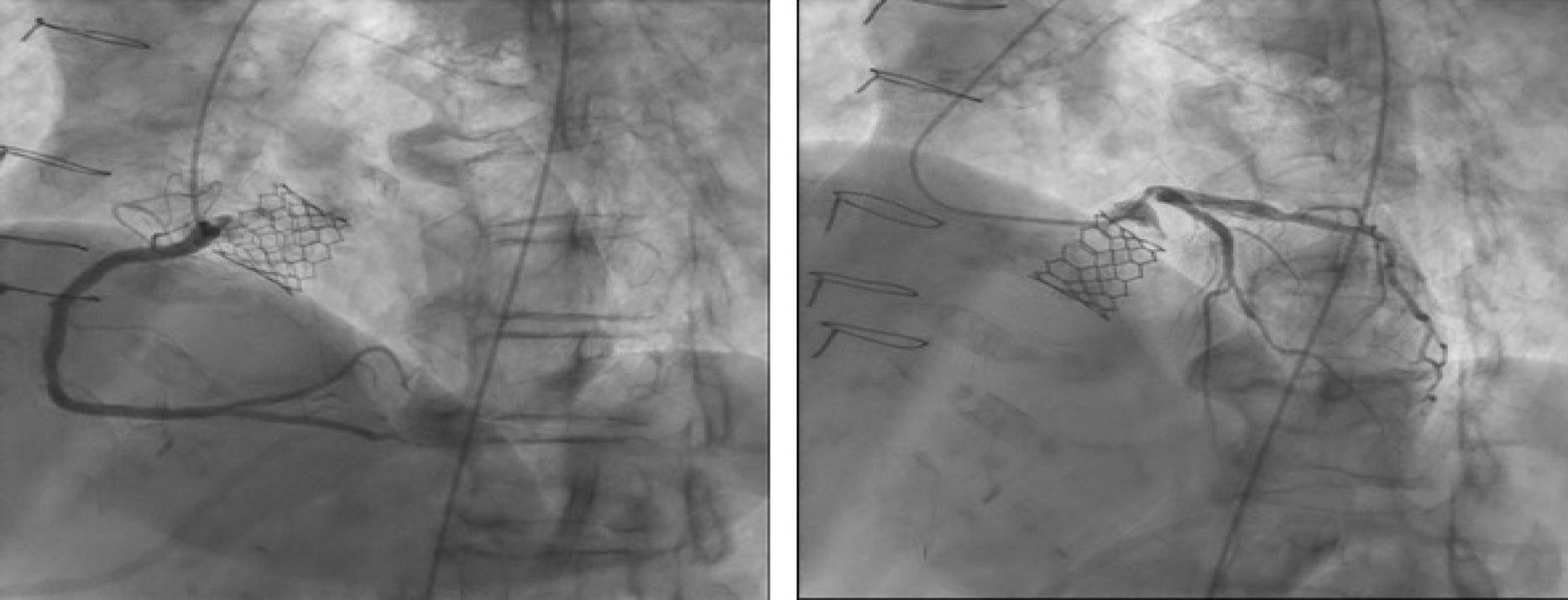
Figure 3
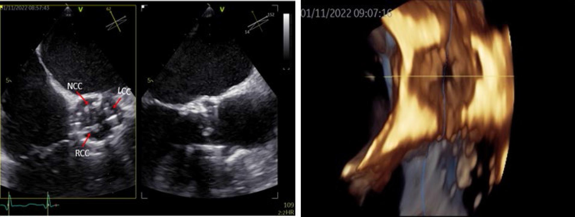
Figure 4
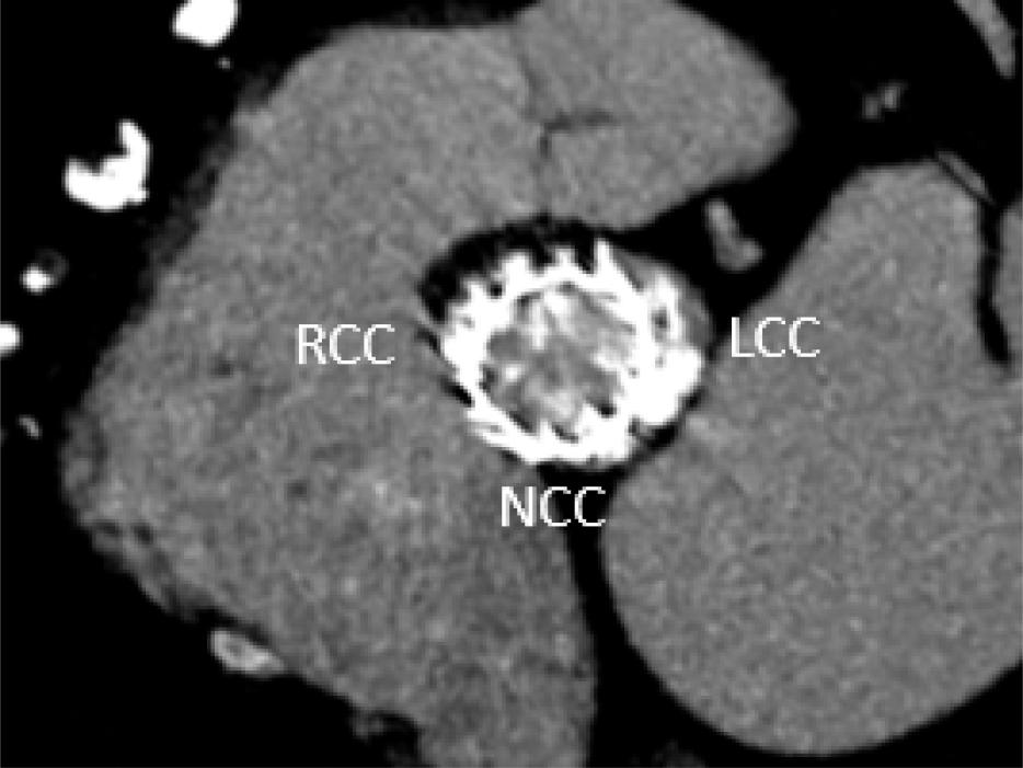
Figure 5
Figure 6
© 2024 Alexandra Apostu, Dan Deleanu, Cătălina Parasca, Răzvan Capșa, Monica Dobrovie, Bogdan Alexandru Popescu, Ovidiu Chioncel, Vlad Anton Iliescu, Ruxandra Jurcuţ, published by Romanian Society of Cardiology
This work is licensed under the Creative Commons Attribution 4.0 License.