
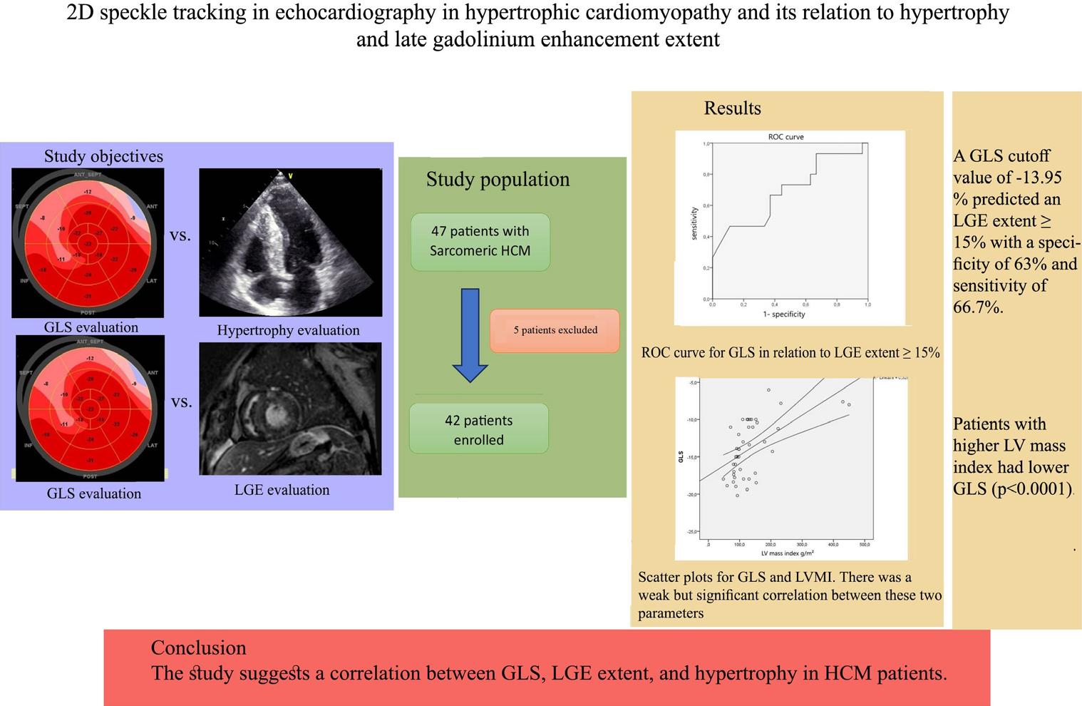
Figure 1:
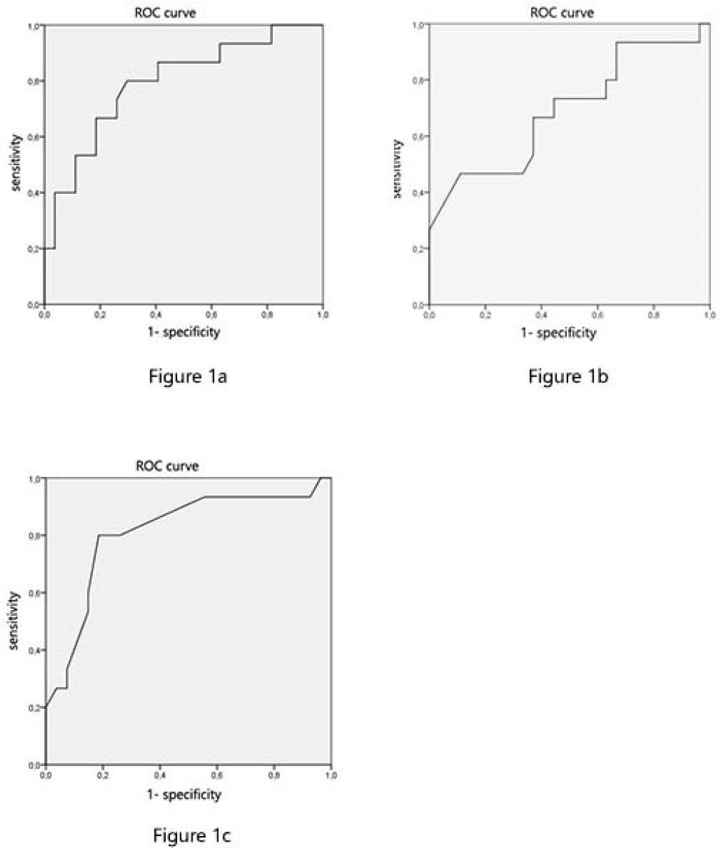
Figure 2
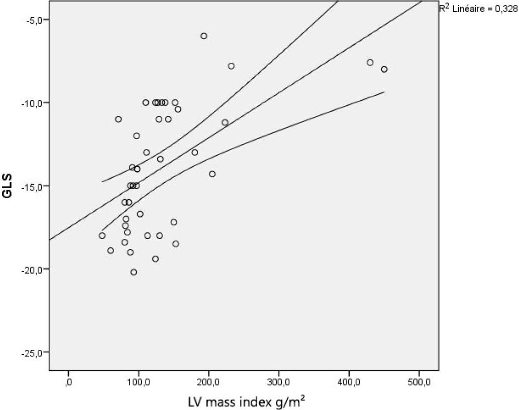
Figure 3
Echocardiographic results in the 42 HCM patients
| All HCM | |
|---|---|
| LVEF, % | 62.5 ± 2 |
| GLS, % | -13.9 ± 1.2 |
| Maximal wall thickness, mm | 22.4 ± 2 |
| LV anteroseptal wall thickness, mm | 17.6 ± 2.4 |
| LV posterolateral wall thickness, mm | 13.1 ± 1.7 |
| Anteroposterior left atrium diameter, mm | 40.7 ± 2.3 |
| LVOT gradient, mmHg | 39.2 ± 15.8 |
MRI parameters according to the extent of LGE
| All HCM (n= 42) | LGE≥15% (n= 15) | LGE<15% (n= 16) | LGE=0 (n= 11) | P value (≥15 vs <15%) | P value (≥15 vs 0) | P value (<15 vs 0) | |
|---|---|---|---|---|---|---|---|
| LVEF, % | 65 ± 2.4 | 66.4 ± 5.2 | 64.6 ±3.4 | 62.5 ± 5.5 | 0.575 | 0.322 | 0.495 |
| LV mass index, g/m2 | 134.5 ± 25.6 | 185.4 ± 56.5 | 109 ± 19.3 | 102.3 ± 18.2 | 0.016 | 0.025 | 0.635 |
| LV EDV, ml/m2 | 86.2 ± 7.1 | 93.9 ± 14.5 | 80.8 ± 8.9 | 85.5 ± 10.2 | 0.131 | 0.423 | 0.415 |
Clinical characteristics in HCM patients
| All HCM (n= 42) | LGE≥15% (n= 15) | LGE<15% (n= 16) | LGE=0 (n= 11) | p value | |
|---|---|---|---|---|---|
| Age, y | 48±5 | 50 ± 9 | 52 ± 8 | 42 ± 9 | 0.265 |
| Male, n (%) | 30 (71%) | 13 (31%) | 9 (21%) | 8 (19%) | 0.268 |
| Hypertension, n (%) | 19 (45%) | 9 (21%) | 5 (12%) | 5 (12%) | 0.275 |
| Coronary artery disease, n (%) | 4 (10%) | 2 (5%) | 0 | 2 (5%) | 0.235 |
| Syncope, n (%) | 8 (19%) | 3 (7%) | 2 (5%) | 3 (7%) | 0.626 |
| Palpitation, n (%) | 18 (43%) | 4 (10%) | 11 (26%) | 3 (7%) | 0.029 |
| History of sustained VT, n (%) | 1 (2%) | 0 | 0 | 1 (2%) | 0.236 |
| Paroxysmal AF, n (%) | 4 (10%) | 2 (5%) | 1 (2%) | 1 (2%) | 0.797 |
| NYHA functional class, n (%) | |||||
| 1-2 | 22 (52%) | 9 (21%) | 9 (21%) | 4 (10%) | 0.266 |
| 3 | 4 (10%) | 1 (2%) | 3 (7%) | 0 | - |
Echocardiographic parameters according to the extent of LGE in HCM patients
| All HCM | LGE≥ 15% | LGE<15% | LGE=0 | P value (≥15 vs <15%) | P value (≥15 vs 0) | P value (<15 vs 0) | |
|---|---|---|---|---|---|---|---|
| LVEF, % | 62.5 ± 2 | 61.5 ± 3.5 | 63.5 ± 3.2 | 62.3 ± 4 | 0.421 | 0.788 | 0.641 |
| GLS, % | -13.9 ± 1.2 | -12.3 ± 2.1 | -13.8 ± 1.6 | -16.7 ± 1.8 | 0.284 | 0.016 | 0.071 |
| Maximal wall thickness, mm | 22.4 ± 2 | 26.7 ± 4.2 | 21.4 ± 1.8 | 17.9 ± 1.51 | 0.027 | 0.003 | 0.015 |
| LV anteroseptal wall thickness, mm | 17.6 ± 2.4 | 23.3 ± 4.8 | 15.1 ± 2.7 | 13.4 ± 2.4 | 0.006 | 0.004 | 0.393 |
| LV posterolateral wall thickness, mm | 13.1 ± 1.7 | 16 ± 3.9 | 11.4 ± 1.9 | 11.7 ± 1.9 | 0.043 | 0.096 | 0.837 |
| Anteroposterior left atrium diameter, mm | 40.7 ± 2.3 | 44.3 ± 5 | 38.5 ± 1.7 | 38.81 ± 4.5 | 0.034 | 0.137 | 0.884 |
| LVOT gradient, mmHg | 39.2 ± 15.8 | 39.5 ± 19.6 | 56.9 ± 35.1 | 13.2 ± 9.4 | 0.409 | 0.044 | 0.058 |