
Figure 1
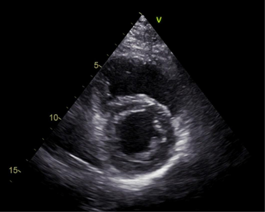
Figure 2
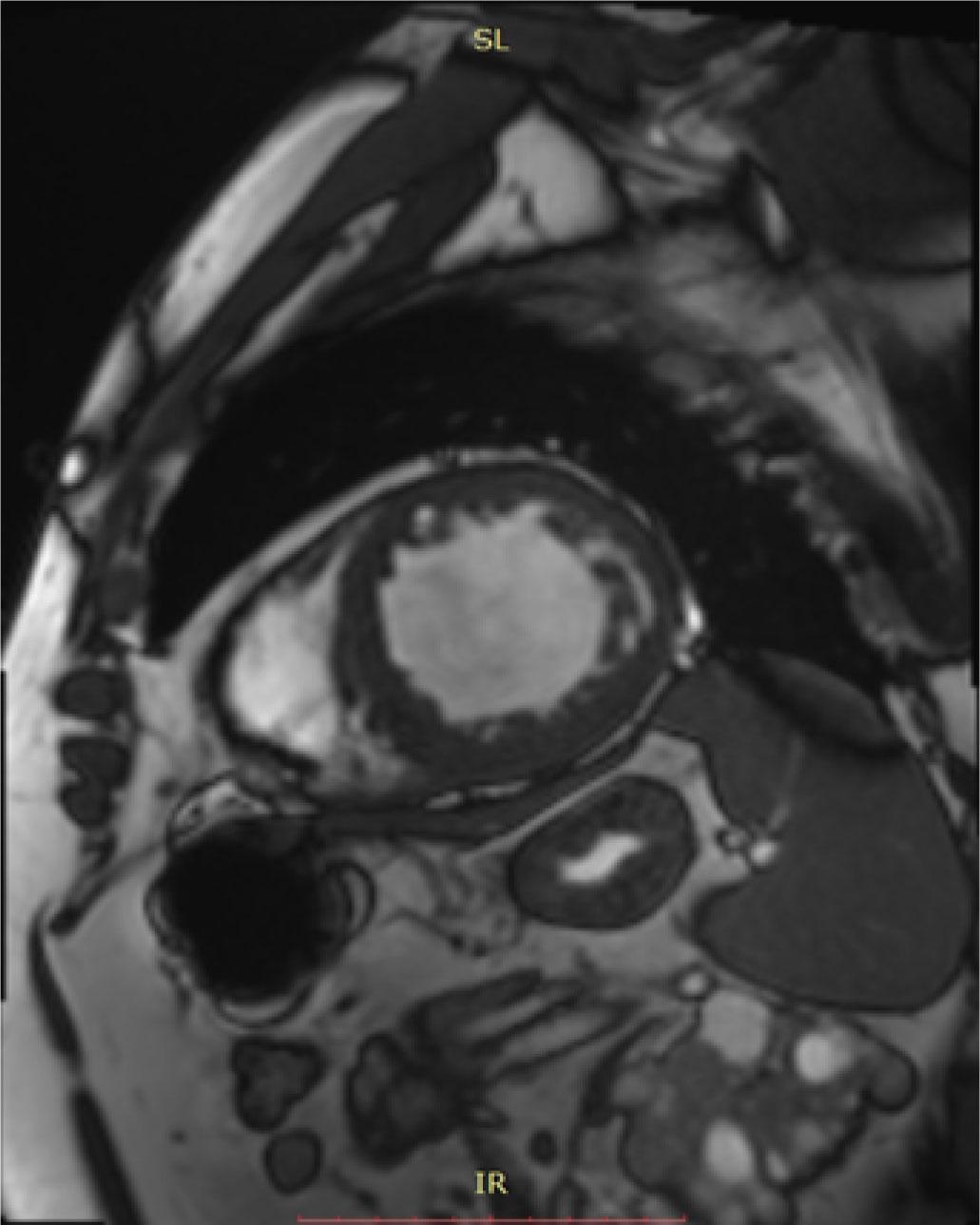
Figure 3
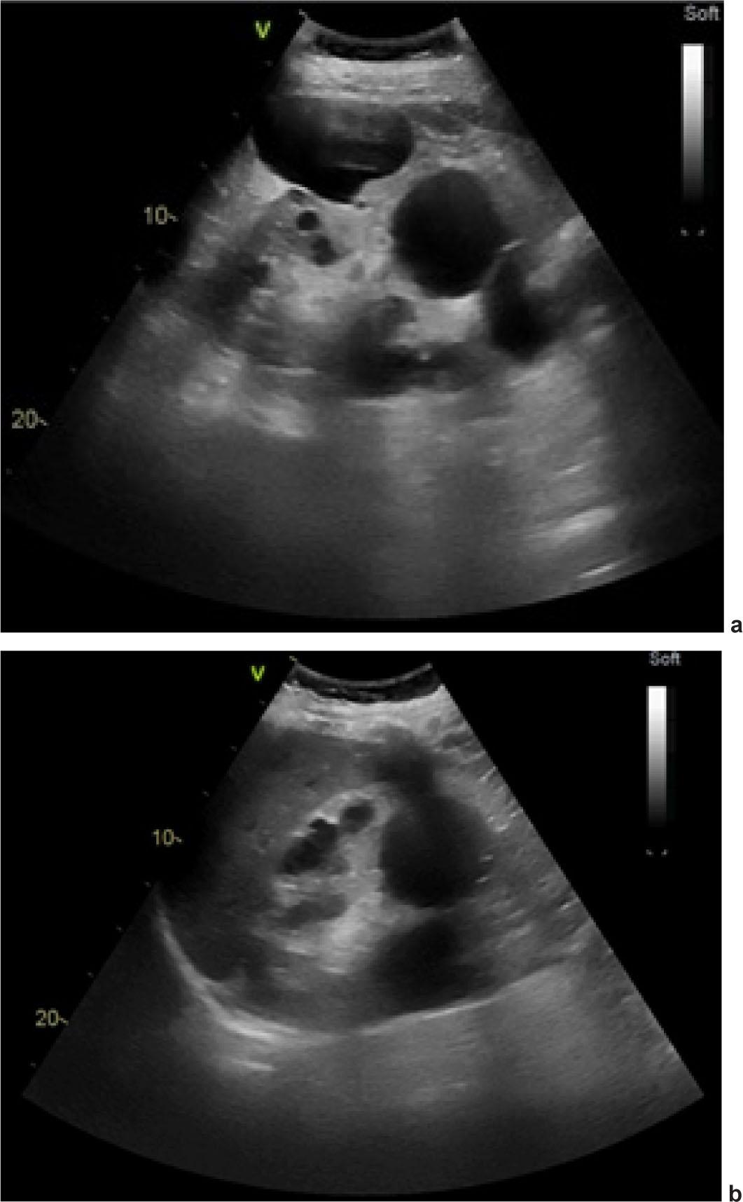
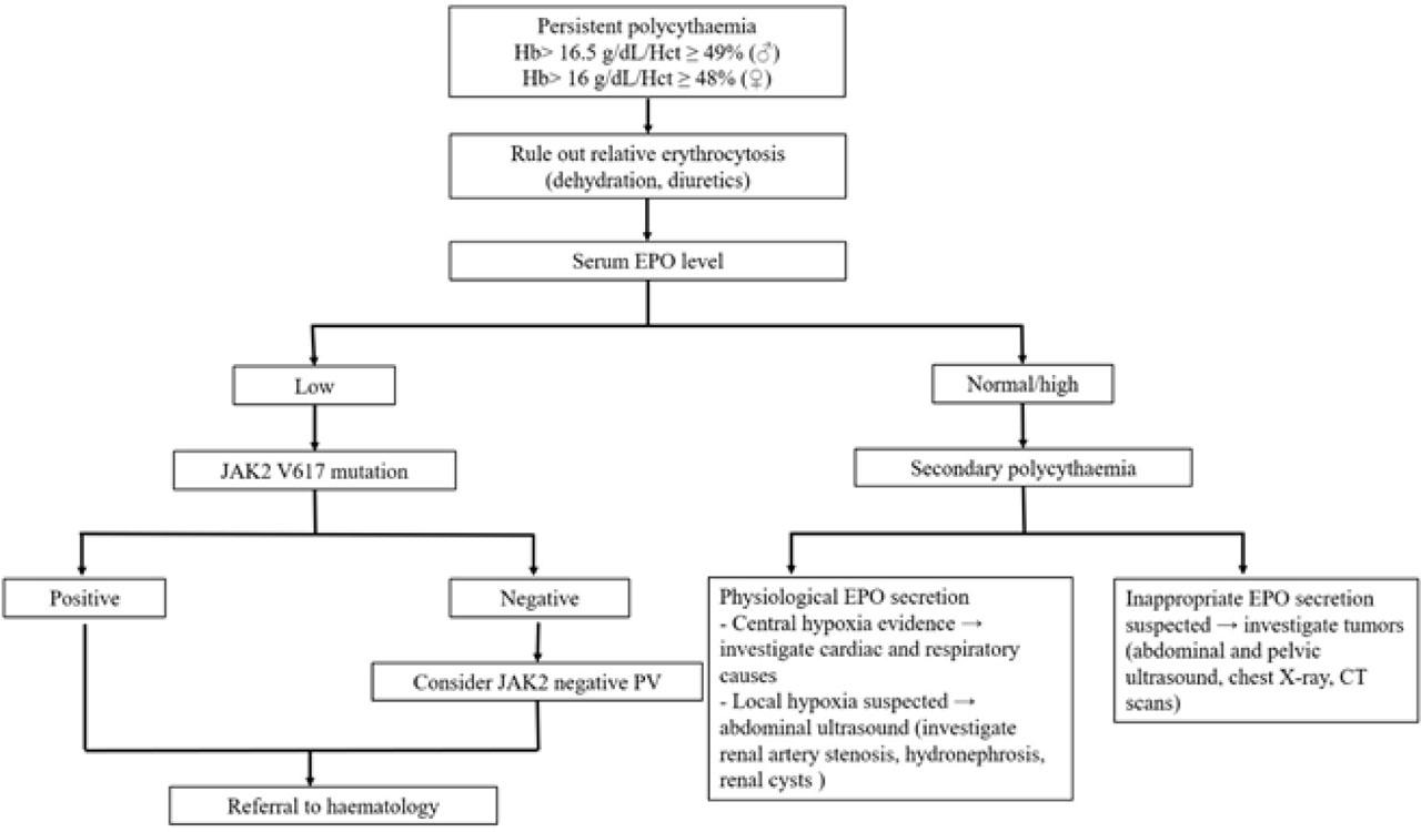
Figure 4
Blood tests results
| Test | Day 1 | Day 3 | Day 7 |
|---|---|---|---|
| Hemoglobin (g/dl) | 18 | 18.7 | 17.8 |
| Erythrocyte count (x106/mm3) | 6.17 | 6.47 | 6.24 |
| Hematocrit (%) | 56 | 58.7 | 55.8 |
| Uric acid (mg/dl) | 9.68 | ||
| Creatinine (mg/dl) | 1.48 | 1.43 | 1.42 |
| eRFG (ml/min/1.73m2) | 52 | 54 | 54 |
| NT-proBNP (pg/ml) | 2346 | ||
| hs-cTnI (pg/ml) | 23.4 | 20.8 | 15.3 |
| O2 pressure in arterial blood (mmHg) | 80 | 87 | 92 |
| Erythropoietin (IU/L) | 40 (N:4.3-29) |
Diagnostic criteria for left ventricular non-compaction cardiomyopathy
| Criteria | Description |
|---|---|
| Echocardiographic criteria | |
| Chin et al. [11] | 1. Prominent trabeculations, deep recesses |
| 2. LV free-wall thickness (ED) augmentation from base to apex | |
| 3. Gradual reduction in the X:Y ratio of myocardial thickness from the mitral valve level to the papillary muscle level (PSAX and apical views) | |
| X – from the epicardial surface to the bottom of the trabeculations | |
| Y – from the epicardial surface to the top of the trabeculations | |
| Stöllberger and Finsterer [12] | 1. Two-layered myocardium with the non-compacted layer thicker than the compacted myocardial layer (ED) |
| 2. >3 trabeculations bulging from the LV apical wall to the papillary muscle | |
| 3. Intertrabecular spaces perfused | |
| Jenni et al. [13] | 1. Bilayered myocardium, multiple prominent trabeculations (ES) |
| 2. Non-compacted to compacted ratio >2:1 | |
| 3. LV cavity communication with the intertrabecular spaces demonstrated by color Doppler | |
| 4. No coexisting cardiac abnormalities | |
| MRI criteria | |
| Petersen et al. [14] | Non-compacted to compacted ratio >2.3 (ED) |
| Jacquier et al. [15] | LV trabecular mass >20% of the global mass |