
Table 1
Types of stem cells and their advantages and disadvantages
| CELL TYPE | ORIGIN | ADVANTAGES | DISADVANTAGES |
|---|---|---|---|
| EMBRYONIC | Zygote. | Higher differentiation potency. Less risk of cell injury during cultivation due to decreased cell-to-cell adhesion. | Subject to ethical issues due to methods of obtaining them. Malignant transformation (teratoma). |
| FETAL | Blastocyst. Fetal structures. Bone marrow. Fetal blood. | High differentiation potency. Less risk due to decreased cell-to-cell adhesion in early fetuses. Lower immunogenic effect. Can be obtained from fetal blood. | Subject to ethical issues due to methods of obtaining them. |
| PERINATAL | Umbilical cord. Placenta. Amniotic fluid. | Multipotent. Can be easily obtained without ethical issues. Lower immunogenic effect. | Differentiation potency is lower than that of fetal cells. |
| ADULT | Bone marrow. Other structures (liver, kidney). | Oligopotent. No ethical issues. More studies have been based on them; they are already in use for certain disorders. | Differentiation potency is the lowest. Higher immunogenic effect (except for mesenchymal stem cells which express lower amounts of antigens). Obtaining these cells can be painful. |
| INDUCED PLURIPOTENT | Adult somatic cells genetically modified to resemble embryonic stem cells. | Obtained from adult and perinatal cells (not an ethical issue). High differentiation potency due to the similarity to embryonic cells. | Immunogenic effect. |
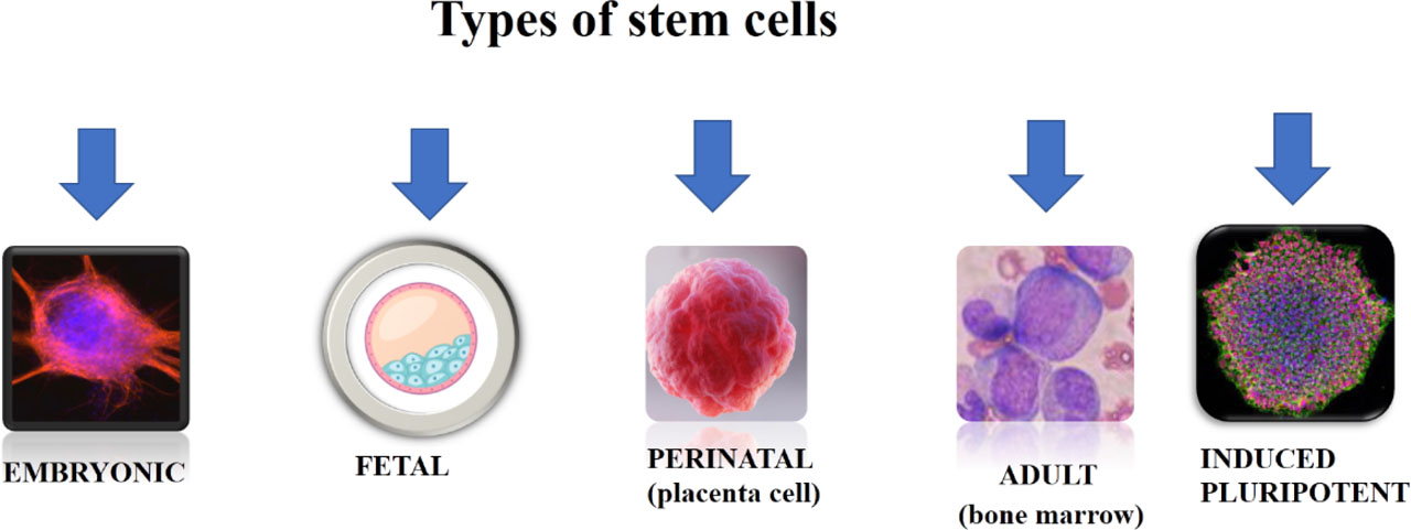
Figure 1
The main types of stem cells
Table 2
Types of stem cells and their effect in reviewed studies
| CELL TYPE [ref] | END POINTS | NO. OF PATIENTS ENROLLED | TIME OF INTERVENTION | NO. OF CELLS | ASSESSMENT METHOD FOR EFFICACY | FINDINGS |
|---|---|---|---|---|---|---|
| Autologous BM-MNC [16] | Safety Efficacy | 15 in study group, 19 in control group. | 24 hours after STEMI. | 8.37 × 10⁶ | LVEDV, LVESV, LVEF by echocardiography (biplane Simpson method – apical two- and four-chamber). | Achieved safety. Nine patients in study group (60%) achieved an improvement in LVEF > 10% at 12-month follow-up. |
| Autologous BM-MSC [14] | Safety Efficacy | 14 in study group, 12 in control group. | 1 month after STEMI. | 7.2 ± 0.9 × 10⁷ | LVEF by SPECT and echocardiography. | Achieved safety. Improvement in LVEF in study group at 4-month follow-up (8.8% vs. 4.8%, p=0.031 by SPECT and 9% vs. 5.3%, p=0.023 by echocardiography). |
| Autologous BM-MNC [18] | Efficacy | 51 in early study group 44 in late study group, 54 in control group. | Early: 5–7 days after STEMI Late: 3–4 weeks after STEMI. | 5 × 10⁷ – 5 × 10⁸ | GLS and GCS by cardiac magnetic resonance. | None of time related treatment proved an improvement in cardiac parameters. |
| Autologous BM-MNC [17] | Efficacy | 66 in study group, 55 in control group. | 6–9 days after STEMI. | 100 × 106 | LVEF, LVEDV, LVESV, infarct size by cardiac magnetic resonance. | Study and control group had similar results at baseline, at 6-month follow-up and between times. |
| Autologous BM-MSC [10] | Safety Efficacy | 21 in study group, 22 in control group. | 14.07 +/−9.53 days after STEMI. | 3.31 ± 1.7 × 10⁶ | LVEF, LVEDV, LVESV by echocardiography (Simpson method) at 12 months and myocardial perfusion and metabolic activity by SPECT at 6-month follow-up. | Achieved safety. No statistically significant difference (p>0,05) between study and control group regarding LVEF, LVEDV and LVESV at 12 months or myocardial perfusion and metabolic defect index at 6-month follow-up. |
| Allogenic CSC [19] | Safety Efficacy | 33 in study group 16 in control group. | Days 5–7 after STEMI. | 35 × 106 | Infarct size, LVEF, LVEDV, LVESV and wall motion score by cardiac magnetic resonance. | Safety was the primary endpoint, and it didn’t reveal any major cardiac adverse events or deaths at 6- and 12-month follow-up. No significant differences in assessed parameters between the groups at baseline and follow-up times. |
| Allogenic CDC [20] | Safety Efficacy | 90 in study group 44 in placebo group. | 4 weeks to 12 months after STEMI. | 2.5 × 10⁶ | Infarct size, LVEDV, LVESV, LVEF by cardiac magnetic resonance. | Safety endpoint was achieved. There was no significant difference in infarct size between groups at 6-month follow-up (p=0.51). Yet there was a reduction in LVEDV (p=0.02) and LVESV (p=0.02) at 6 month follow-up in favor of study group. |
[i] BM-MNC – bone marrow mononuclear stem cells, BM-MSC – bone marrow mesenchymal stem cells, CDC – cardiosphere derived cells, CSC – cardiac stem cells, GCS – global circumferential strain, GLS – global longitudinal strain, LVEDV – left ventricular end diastolic volume, LVEF – left ventricular ejection fraction, LVESV – left ventricular end systolic volume, SPECT – single photon emission computed tomography, STEMI – ST- segment elevation myocardial infarction
Table 3
The biological parameter changes in trials proving the beneficial effect of stem-cell therapy in myocardial infarction
| TRIAL | CELL TYPE | BIOLOGICAL PARAMETER | |
|---|---|---|---|
| Fernandez-Aviles et al., 2018 19 | Allogenic CSC | CRP | Similar baseline values between groups. Higher levels decrease by day 7 in the study group (p=0.04). Similar values at 1-month follow-up in both groups. |
| NT-proBNP | Similar baseline values. No statistically significant value at 12 months (p=0.9). | ||
| CK and TnT | Similar decreasing trend. | ||
| Makkar et al., 2020 20 | Allogenic CDC | NT-proBNP | Greater decreasing level at 6 months in study group (p=0.02). |
| Peregud-Pogorzelska et al., 2020 16 | Autologous BM-MNC | CRP, CK-MB, TnT, BNP | Similar baseline levels between the entire study group and control group but lower CK-MB, TnT, and BNP initial values in responders, with faster decrease in BNP levels in the entire study group and TnT levels in the responders. |
[i] BM-MNC – bone marrow mononuclear stem cells, BNP – brain natriuretic peptide, CDC – cardiosphere derived cells, CK – creatine kinase, CK-MB – isoenzyme MB of creatine kinase, CRP – C reactive protein, CSC – cardiac stem cells, TnT – troponin T