
The incidence of peripheral arterial disease (PAD) has increased substantially over the past decade. Its global prevalence in 2010 was approximately 200 million - a 23% rise from the year 2000 - while reports from 2015 estimated over 237 million cases, representing a further increase of more than 17% from 2010.1 This upward trend is partly attributed to an aging population, but other contributing factors include diabetes mellitus (DM), hyperlipidemia, and chronic kidney disease (CKD).1,2
When PAD is suspected based on clinical findings and foot examination, noninvasive tests such as the ankle-brachial index (ABI) are recommended. An ABI value of < 0.90 is diagnostic for lower limb PAD.3 However, the sensitivity of this test is limited in elderly patients and individuals with diabetes.4 In symptomatic PAD patients, diagnostic imaging plays a crucial role in treatment planning.5
Among noninvasive modalities, cross-sectional imaging techniques such as computed tomography angiography (CTA) and magnetic resonance angiography (MRA) offer more objective and clinically useful data than ultrasound.6
The older population, often with multiple comorbidities, requires a more nuanced approach to PAD imaging. Approximately 40% of PAD patients present with significant renal impairment with varying degrees of kidney dysfunction and are therefore at high risk for iodine contrast-induced nephropathy.4,7 In such patients it is also recommended to avoid the use of Group I gadolinium-based contrast agents, thereby reducing the risk of nephrogenic systemic fibrosis.5 For symptomatic PAD patients, it is essential to avoid further compromising renal function with iodinated contrast agents solely for diagnostic CTA.
Both CTA and MRA are widely used for assessing peripheral arteries in patients with chronic limb-threatening ischemia (CLTI) or claudication and are invaluable for treatment planning. However, each modality has its limitations. Vascular calcification is the primary challenge in CTA, as it produces blooming artifacts that impair image interpretation. MRA, by contrast, is unaffected by calcifications. CTA also carries limitations in patients with impaired renal function and involves exposure to ionizing radiation.
In the early 2000s, contrast-enhanced MRA was recommended for patients with CKD requiring arterial imaging.8 However, after the discovery of nephrogenic systemic fibrosis (NFS) and evidence of gadolinium deposition in soft tissues, brain, and bones, there has been a shift toward non-contrastenhanced (NCE) MRA techniques, particularly those based on time-of-flight (TOF) sequences.8,9 Quiescent-interval single-shot (QISS) non-contrast MR angiography (MRA) has emerged in the last decade as one of TOF sequences.
Digital subtraction angiography (DSA), long considered the gold standard for vascular imaging due to its high spatial resolution and real-time imaging capability, is now primarily reserved for interventional procedures.10 Despite its accuracy, DSA is invasive, involves iodinated contrast, and carries the risk of potentially serious complications.
While QISS MRA has been validated in larger populations, its diagnostic performance in patients with extensive vascular calcification remains underexplored.
In this study, we compared CTA and MRA images of the pelvic, femoral and below-the-knee arteries with corresponding DSA images. Our primary aim was to assess segment-based intermodality agreement between QISS MRA, CTA and DSA for the detection of significant (≥ 50%) stenosis and occlusion. As a secondary, exploratory aim, we evaluated the diagnostic performance of QISS MRA and CTA using DSA as the reference standard, with particular focus on below-the-knee arteries and patients with medial arterial calcification.
This study was approved by the institutional review board (IRB Approval No.UKC-MB-KME-12/25), with waived patient consent due to its retrospective nature.
Twenty-five consecutive patients with symptomatic PAD who underwent clinically indicated QISS MRA were retrospectively included in this study. Thirteen of these patients also had CTA, 19 had DSA and seven patients underwent all three modalities (QISS MRA, CTA and DSA). Six patients had MRA and CTA only, 11 had MRA and DSA. The presence of medial arterial calcification (mediocalcinosis) was assessed qualitatively based on available prior imaging CTA and/or DSA, as documented in the radiology reports; QISS MRA itself was not used to identify calcifications.
Imaging was performed using a 1.5-Tesla MRI system (Magnetom Sola, Siemens Healthcare GmbH, Erlangen, Germany). Data acquisition utilized an optimized 2D ECG-triggered Cartesian QISS-MRA pulse sequence. Patients were positioned feet-first in the supine position, and body, peripheral, and spine matrix phased-array radiofrequency coils were used for signal reception. QISS MRA was performed using an automated “push-button” protocol with an ECG-gated prototype pulse sequence, as previously described.11 The protocol included 8–10 stations with 48 slices each (144 mm z-axis coverage per station), covering the run-off from the aortic bifurcation to the feet. Scanning was performed without breath-holding. If imaging of the abdominal aorta was required, an additional 12 stations from the diaphragm to the iliac crest were acquired with breath-hold commands. Maximum intensity projection (MIP) images were automatically generated and manually corrected if necessary.
CTA was performed on a Siemens Dual Source CT 3rd generation system (SOMATOM Drive, 2019, Siemens). Arterial-phase CTA images were obtained from the distal abdominal aorta to the distal lower extremities. Each patient received 100 mL of iodinated contrast medium (400 mgI/mL, Iomeron, Bracco), followed by 50 mL of saline at a flow rate of 4 mL/s, administered via an automated dual-syringe power injector (Ulrich Medical CT Injection System), in line with current guidelines. Bolus tracking was based on a 200 HU threshold (Care-Bolus, Siemens). The acquisition parameters were: FOV 490 mm; pitch 0.4; collimation 128 × 0.6 mm; tube voltage 120 kV; tube current 84 mAs (CARE Dose 4D and CARE kV on: 70–120 kV, modulated). Data were reconstructed using a soft tissue kernel (Bv38, Siemens) with a slice thickness of 1 mm and an increment of 0.7 mm, using the ADMIRE (Advanced Modeled Iterative Reconstruction) algorithm at strength level 3 (range: 1–5).
DSA was performed using a Philips Allura 2009 system (Philips, Netherlands). In most cases, antegrade femoral artery puncture was used. Contrast medium (350 mgI/mL, Iomeron, Bracco) was administered through a 6-FR femoral sheath. DSA images were acquired for the femoral, popliteal, below-the-knee, and foot arteries. DSA served as the reference imaging modality in this study. Examples of multimodality imaging in the same patient are shown in Figures 1 and 2.
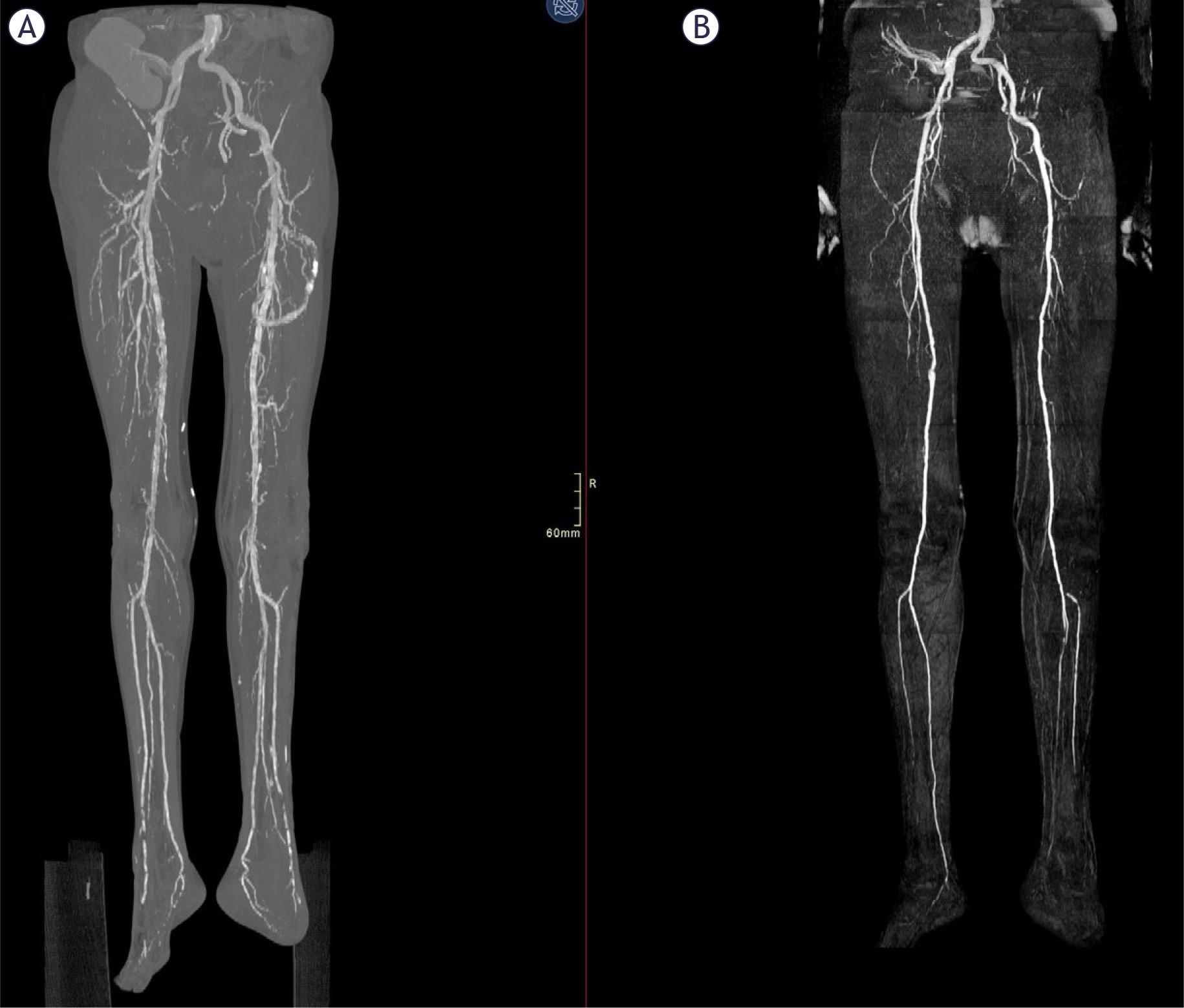
(A) Computed tomography angiography (CTA) of the pelvis and lower extremities demonstrating diffuse arterial wall calcifications, most pronounced in the below-the-knee arteries, limiting accurate assessment of vessel patency due to blooming artefacts. The arrow indicates a heavily calcified proximal anterior tibial artery. (B) Non-contrast quiescent-interval single-shot magnetic resonance angiography (QISS MRA) of the same vessels in the same patient, demonstrating preserved arterial patency without interference from arterial wall calcifications, which are not visualized on QISS MRA. The arrow indicates two significant stenoses of the proximal anterior tibial artery.
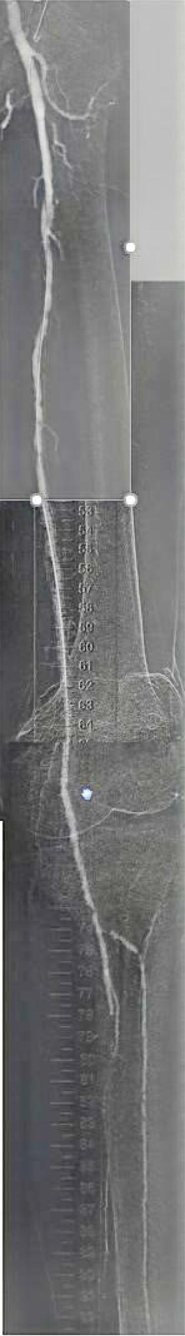
Carbon dioxide angiography (CO2 angiography) in the same patient as Figure 1, showing patency of the femoral arteries as seen on computed tomography angiography (CTA) and non-contrast quiescent-interval single-shot magnetic resonance angiography (QISS MRA). The arrow indicates two significant stenoses of the anterior tibial artery.
For image analysis, the pelvic, femoropopliteal and below-the-knee arterial segments were evaluated on both sides as predefined segments. Each segment was scored on a five-point ordinal scale: 1 = normal patency, 2 = < 50% diameter stenosis, 3 = ≥ 50% stenosis, 4 = occlusion and 5 = non-diagnostic (e.g. due to motion or severe artefacts such as streaks, metallic implants or foreign bodies). Segments scored as 5 were considered non-evaluable and were excluded from further analysis. In segments with multiple stenoses, the most severe stenosis was used for analysis. All readings were performed by a single experienced vascular radiologist.
For the agreement analysis the 5-level scale was dichotomised into non-significant disease (scores 1–2) versus significant stenosis or occlusion (scores 3–4). Only arterial segments that were imaged and evaluable on both modalities under comparison (QISS MRA vs. CTA, QISS MRA vs. DSA, CTA vs. DSA) were included for that particular analysis. Cohen’s kappa (κ) was calculated to describe segment-based inter-modality agreement, and raw percentage agreement was also reported for each modality pair.
Because not all patients underwent all modalities, each pairwise agreement analysis and each DSA-referenced diagnostic performance analysis was performed on the subset of segments available for that specific modality comparison. Consequently, kappa statistics and performance metrics are based on different patient/segment subsets and should not be interpreted as direct head-to-head comparisons between QISS MRA and CTA. To address this, we additionally performed a matched sensitivity analysis restricted to segments imaged by all three modalities.
Agreement was interpreted as follows poor < 0.00; slight = 0.00–0.20; fair = 0.21–0.40; moderate = 0.41–0.60; substantial = 0.61–0.80; almost perfect = 0.81–1.00).
Using DSA as the reference standard, segmentbased sensitivity, specificity, positive predictive value (PPV), negative predictive value (NPV) and overall accuracy for the detection of significant (≥ 50%) stenosis/occlusion were calculated for QISS MRA and CTA, both for all evaluable segments and separately for below-the-knee segments. Because multiple arterial segments per patient were analysed, no adjustment was made for intrapatient clustering; the results should therefore be interpreted as exploratory segment-level estimates. For patients who underwent more than one imaging modality, the time interval between QISS MRA and CTA and between QISS MRA and DSA was calculated in days as the absolute difference between examination dates.
Data analysis was performed using IBM SPSS Statistics for Windows, Version 29.0 (IBM Corp., Armonk, NY, USA).
QISS MRA was performed in 25 patients; 13 of these patients also had CTA and 19 had DSA, with 7 patients undergoing all three modalities. The mean patient age was 67 ± 12 years (range: 32–86). There were 20 men (mean age: 67 ± 11 years; range: 32–83) and 5 women (mean age: 69 ± 14 years; range: 54–86). Twelve patients (48%) had CLTI, and medial arterial calcification (mediocalcinosis) was present in 7 patients (28%).
Among patients with multiple imaging modalities, the mean (± SD) absolute interval between QISS MRA and CTA was 248 ± 353 days (range 28–1155 days; n = 13), and between QISS MRA and DSA 198 ± 300 days (range 0–1081 days; n = 18).
A total of 450 segments were evaluated using QISS MRA, 229 using CTA, and 149 using DSA. Segments excluded due to artifacts accounted for 15% in MRA, 24% in CTA, and 4% in DSA.
QISS MRA was evaluable in 382/450 (84.9%) segments, CTA in 174/450 (38.7%), and DSA in 143/450 (31.8%). CTA evaluability was lowest in below-the-knee segments (52/200, 26.0%). Among DSA-evaluable segments, 47/143 (32.9%) showed significant (≥ 50%) stenosis or occlusion.
On QISS MRA, 54.7% of evaluable segments had no stenosis, 10.2% had non-hemodynamically significant stenosis (< 50%), 13.6% had significant stenosis (≥ 50%) and 21.5% of segments were occluded. On CTA, 55.7% of evaluable segments had no stenosis, 23.6% had non-hemodynamically significant stenosis, 12.6% had significant stenosis and 8.0% were occluded. On DSA, 46.9% of evaluable segments had no stenosis, 20.3% had non-hemodynamically significant stenosis, 15.4% had significant stenosis and 17.5% of segments were occluded.
In patients with significant medial arterial calcification (n = 7), QISS MRA showed stronger agreement with DSA (κ = 0.58) than CTA (κ = 0.32), consistent with the tendency of CTA to underestimate stenosis severity in heavily calcified below-the-knee arteries.
For the detection of significant (≥ 50%) stenosis/occlusion, segment-based agreement between QISS MRA and CTA was moderate (κ = 0.41, raw agreement 66.7%; 156 paired, evaluable segments). Agreement between QISS MRA and DSA was also moderate (κ = 0.49, raw agreement 63.6%; 121 paired segments). Agreement between CTA and DSA was moderate (κ = 0.57 and raw agreement 76.9%; 39 paired segments).
Segment-based diagnostic performance metrics for QISS MRA and CTA relative to DSA, including TP, TN, FP, FN, sensitivity, specificity, PPV, NPV and overall accuracy, are summarized in Table 3.
Results of agreement quiescent-interval slice-selective magnetic resonance angiography (QISS MRA), computer tomography angiography (CTA) and digital subtraction angiography (DSA)
| Comparison | κ Value | 95% CI | p-value | Agreement level |
|---|---|---|---|---|
| QISS MRA vs. CTA | 0.41 | 0.31–0.52 | < 0.001 | Moderate |
| QISS MRA vs. DSA | 0.49 | 0.37–0.60 | < 0.001 | Moderate |
| CTA vs. DSA | 0.57 | 0.34–0.80 | < 0.001 | MODERATE |
Kappa values by arterial segment
| Arterial segment | QISS MRA vs. CTA κ | QISS MRA vs. DSA κ | CTA vs. DSA κ |
|---|---|---|---|
| Pelvic arteries | 0.40 | 0.51 | NA |
| Femoropopliteal arteries | 0.29 | 0.23 | 0.55 |
| Below-the-knee arteries | 0.42 | 0.61 | 0.32 |
CTA =computed tomography angiography, DSA = digital subtraction angiography; κ = kappa; QISS MRA = quiescent-interval single-shot MR angiography
Segment-based diagnostic performance of QISS MRA and CTA for detection of significant (≥ 50%) stenosis/occlusion using DSA as the reference standard
| Modality / territory | Paired segments (n) | TP | TN | FP | FN | Sensitivity (%) | Specificity (%) | PPV (%) | NPV (%) | Accuracy (%) |
|---|---|---|---|---|---|---|---|---|---|---|
| QISS MRA – all segments | 121 | 37 | 64 | 13 | 7 | 84.1 | 83.1 | 74.0 | 90.1 | 83.5 |
| QISS MRA – below-the-knee | 72 | 25 | 36 | 7 | 4 | 86.2 | 83.7 | 78.1 | 90.0 | 84.7 |
| CTA – all segments | 39 | 3 | 31 | 3 | 2 | 60.0 | 91.2 | 50.0 | 93.9 | 87.2 |
| CTA – below-the-knee | 16 | 0 | 14 | 1 | 1 | 0.0 | 93.3 | 0.0 | 93.3 | 87.5 |
CTA = CT angiography; DSA = digital subtraction angiography; FN = false negative; FP = false positive; NPV = negative predictive value; PPV = positive predictive value; TP = true positive; TN = true negative; QISS MRA = quiescent-interval single-shot MR angiography
CTA yielded a substantially lower proportion of evaluable segments than QISS MRA, particularly below-the-knee, which limited CTA–DSA comparisons in this territory.
Matched tri-modality sensitivity analysis: Restricting the analysis to segments imaged by all three modalities (32 segments from 7 patients) yielded κ = 0.45 for QISS MRA vs. DSA and κ = 0.53 for CTA vs. DSA. In this matched subset, both QISS MRA and CTA showed a sensitivity of 60% and a specificity of 88.9% and 92.6%, respectively, for detecting ≥ 50% stenosis/occlusion relative to DSA. These estimates are based on few paired segments and are provided to illustrate the impact of subset selection rather than to support definitive head-to- head comparisons.
In the sub-analysis of below-the-knee arteries, QISS MRA showed stronger agreement with DSA (κ = 0.61) than CTA (κ = 0.32), although CTA-DSA estimates were based on few paired segments. Agreement between QISS MRA and DSA was substantial (κ = 0.61; raw agreement 73.6%; 72 paired segments). CTA-DSA comparisons in this territory were based on only 16 paired segments, reflecting the low CTA evaluability below-the-knee.
These findings underline the tendency of CTA to underestimate the severity of stenosis in heavily calcified below-the-knee arteries, whereas QISS MRA remained highly sensitive in this subgroup.
In our study, QISS MRA demonstrated diagnostic confidence comparable to that of DSA for below-the-knee arteries, while its concordance with CTA was only fair. This discrepancy may be attributable to the small sample size. The moderate agreement we observed across femoro-popliteal and iliac arteries between all modalities aligns with findings by Varga-Szemes et al., who reported comparable diagnostic performance between QISS MRA and DSA in the evaluation of PAD.12
A meta-analysis by Verma also showed that QISS MRA has high accuracy not only in identifying but also in excluding arterial stenosis or occlusion in symptomatic PAD patients. It is therefore considered a preferred imaging modality for patients with renal impairment or other risk factors, such as pregnancy or contrast allergies.13 Similarly, Varga-Szemes et al. reported high diagnostic accuracy of QISS MRA for detecting significant vascular stenosis in PAD, comparable to DSA, and noted that it is less affected by imaging artifacts than CTA. The lower agreement observed between CTA and DSA in our study may be due to blooming artifacts caused by calcifications, which can misrepresent the degree of stenosis. This suggests that patients with heavily calcified arteries stand to benefit most from QISS MRA.
Although QISS MRA effectively visualizes segments with impaired flow, it cannot directly depict vascular calcification.14 Patients with CKD and diabetes are also prone to Mönckeberg’s arteriosclerosis, a condition characterized by medial arterial calcification.15 In this disease, smooth muscle cells in the arterial media transform into osteocytes and subsequently calcify. Calcification from both medial sclerosis and atherosclerotic intimal changes can impair the evaluation of luminal patency in peripheral arteries on CTA.
In another study, Varga-Szemes et al. demonstrated that combining QISS MRA with proton density-weighted, in-phase stack-of-stars magnetic resonance imaging (PDIP-SOS MRI) significantly improves diagnostic accuracy—approaching that of CTA—for PAD detection, although the image quality still lags behind CTA.5 Merging these two techniques through dataset co-registration and fusion may offer CTA-like vascular visualization, which is particularly beneficial for surgical or endovascular treatment planning.
Lebenatus et al. also showed that QISS MRA has potential as a preoperative NCE MRA technique for identifying anatomical variants and pathologies of the lower leg arteries, as well as for assessing fibular perforators in patients with facial malignancies undergoing microsurgical fibular graft transfer.16
Over the past decade, new NCE MRA techniques have emerged. Evolved from TOF methods, QISS uses ECG triggering to minimize in-plane saturation.17 Unfortunately, TOF MRA has relatively long acquisition times and is prone to artifacts and in-plane saturation.18 It applies a presaturation radiofrequency (RF) pulse to suppress stationary tissue signals, followed by a venous suppression pulse. A “quiescent interval” during systole allows unsaturated arterial blood to flow into the imaging slice. Subsequently, a fat saturation pulse is applied, and signal acquisition occurs during diastole using a 2D balanced steady-state free precession (bSSFP) sequence.17 Compared to TOF MRA, QISS MRA offers shorter acquisition times and eliminates the need for scout images.17
In recent years, several QISS MRA variants have emerged. The QISS balanced steady-state free precession (bSSFP) sequence may offer advantages due to its significantly shorter scan times and improved visualization of smaller vessels. Additionally, the QISS fast low-angle shot (FLASH) sequence appears to be a robust alternative for NCE MRA, as it is less sensitive to magnetic field inhomogeneities.19
QISS MRA has also been shown to visualize foot arteries; however, when compared to the NATIVE SPACE sequence—which is specifically designed for imaging the tortuous anatomy of foot arteries—QISS MRA was found to be inferior. The NATIVE SPACE sequence demonstrated better image quality and fewer false positives compared to QISS MRA.20 When comparing QISS MRA with 2D time-of-flight (TOF) MRA, QISS was found to offer superior image quality, fewer artifacts, and shorter scan times.17
Currently, the main limitation of QISS MRA lies in its reduced reliability in patients with arrhythmias, which can cause image artifacts.4 In the future, these issues may be addressed through the development of ungated QISS MRA techniques.4
The primary advantage of non-contrast-enhanced (NCE) MRA lies in the elimination of iodinated or gadolinium-based contrast agents and ionizing radiation—all of which carry rare but potentially serious risks. Additionally, NCE MRA eliminates the costs associated with contrast agents and renal function testing.4,21
QISS MRA also allows for repeated imaging of specific segments, as no intravenous contrast is required. Furthermore, it avoids issues related to insufficient contrast opacification, which in CTA can depend on blood flow velocity and cardiac function.22
The time required to acquire QISS MRA images is comparable to that of CTA. QISS MRA scans take approximately 25 minutes, and maximum intensity projection (MIP) images are automatically generated and sent to the picture archiving and communication system (PACS).8 In contrast, CTA requires IV cannulation and post-processing of images, which typically takes an additional 5–10 minutes.8 Taken together—its reproducibility, accuracy, and ease of use—QISS MRA is well-suited for routine clinical practice.
In recent years, QISS MRA has become the most frequently used NCE MRA technique for evaluating PAD in the lower extremities at our institution, with usage increasing annually.
Our study has several important limitations. First, the sample size was small, and the analyses were performed at the segment level, which limits the precision and generalisability of the findings. Second, because not all patients underwent all three imaging modalities, the agreement analyses and diagnostic performance metrics were derived from different patient/segment subsets, introducing potential selection bias and limiting direct modality-to-modality comparisons; we therefore report these results as exploratory and supplemented them with a matched tri-modality sensitivity analysis. Third, multiple arterial segments per patient were analysed without adjustment for intra-patient clustering, so the confidence intervals around agreement and diagnostic performance metrics are likely to be overly optimistic. Fourth, the time intervals between QISS MRA, CTA and DSA were often substantial, with mean absolute differences of approximately six to eight months and ranges exceeding one year in some cases, which may have allowed for disease progression or interim interventions and thus may have reduced observed concordance. Fifth, all imaging assessments were performed by a single experienced reader and we did not evaluate inter-observer variability. Finally, DSA was available only in a subset of patients and segments, introducing potential selection bias towards more complex or advanced disease.
In conclusion, in this small, exploratory cohort QISS MRA showed promising performance for segment-based assessment of PAD, particularly in below-the-knee arteries and in patients with medial arterial calcification, when compared with CTA and DSA. QISS MRA may be especially useful in patients in whom iodinated or gadoliniumbased contrast agents are undesirable and in those with pronounced below-the-knee calcifications, where CTA is prone to blooming artifacts. Larger, prospective multicentre studies with multi-reader assessment and appropriate statistical handling of clustered segment-level data are warranted to further define the clinical role of QISS MRA in PAD.