
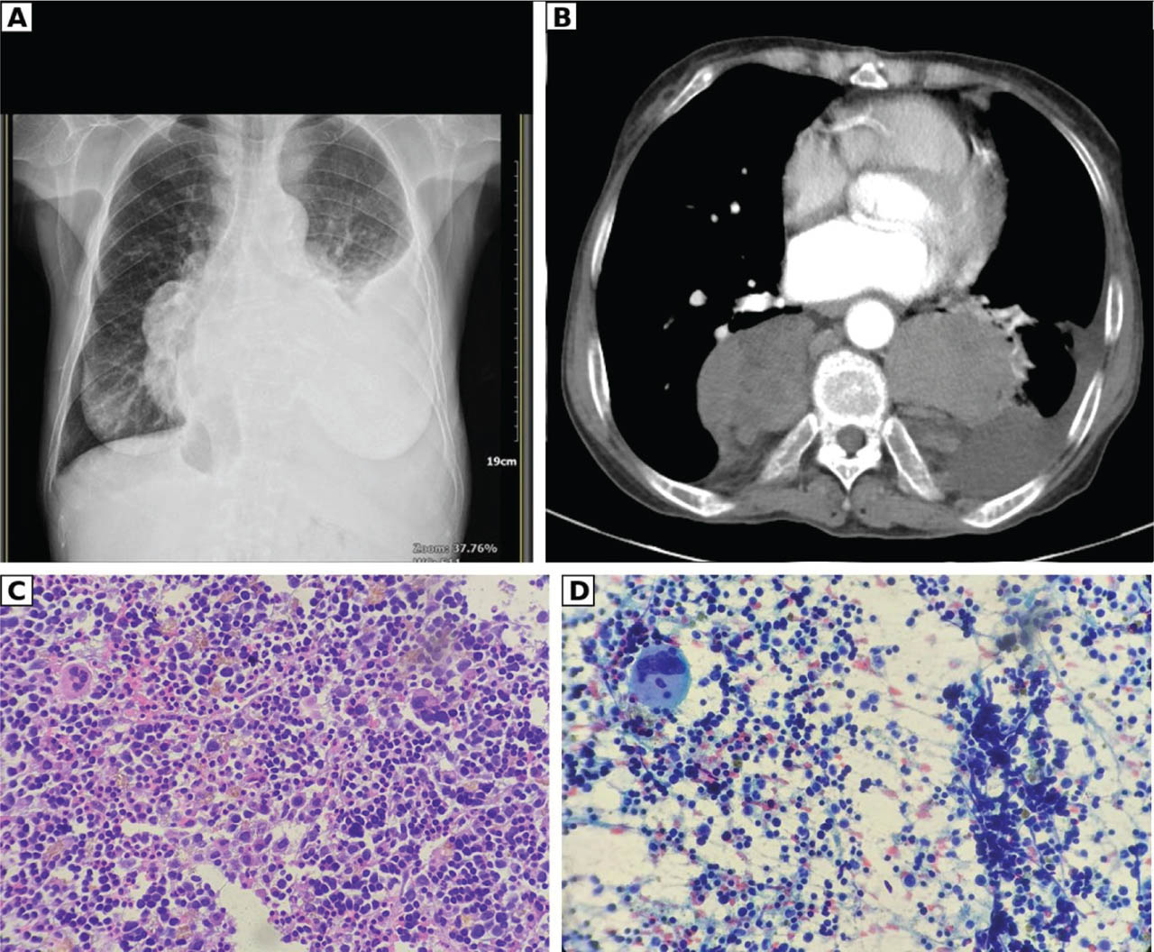
Figure 1.
Patient 1. (A) Posteroanterior chest radiograph showing a right paramediastinal/paravertebral mass with a left pleural effusion. (B) Contrast-enhanced axial CT (mediastinal window) of the whole thorax showing bilateral paravertebral soft-tissue masses flanking the vertebral body behind the heart. (C) H&E-stained section (×400) showing hypercellular trilineage haematopoietic tissue with megakaryocytes. (D) Papanicolaou-stained aspirate (×400) showing a megakaryocyte among haematopoietic precursors. CT, computed tomography; H&E, haematoxylin-eosin.
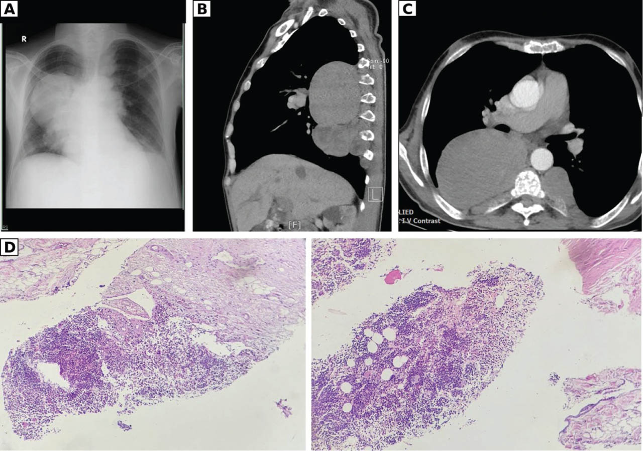
Figure 2.
Patient 2. (A) Posteroanterior chest radiograph showing a right posterior mediastinal mass. (B) Non-contrast CT (sagittal reconstruction) showing lobulated paravertebral masses anterior to the vertebral column. (C) Contrast-enhanced axial CT (mediastinal window) showing a large right posterior mediastinal mass and a left paravertebral mass behind the heart. (D) Core-biopsy histopathology (H&E, ×100) showing hypercellular haematopoietic tissue with erythroid and myeloid precursors and megakaryocytes. CT, computed tomography; H&E, haematoxylin-eosin.
Table 1.
Comparison of the two cases of intrathoracic EMH
| Feature | Case 1 | Case 2 |
|---|---|---|
| Age (years), sex | 59, female | 47, male |
| Thalassaemia type | Beta-thalassaemia; transfused approximately every 3 months for about 30 years | Beta-thalassaemia intermedia with HbE; transfused for about 25 years |
| Relevant comorbidity | None reported | Untreated chronic hepatitis C |
| Presenting symptoms | Progressive dyspnoea, weight loss, reduced appetite | Progressive dyspnoea (5 years), productive cough, chest and back pain, weight loss |
| Examination | Pallor, reduced left breath sounds, left basal dullness, splenomegaly; BMI 16.4 kg/m2 | Hepatomegaly, splenomegaly; BMI 16.6 kg/m2 |
| Haemoglobin | 7.0 g/dL (10.9 g/dL after transfusion) | 8.6 g/dL |
| Platelets | Not reported | 95,000/μL |
| Ferritin; transferrin saturation | Not reported | 3510 ng/mL; 85% |
| Lactate dehydrogenase | Not reported | 308 U/L |
| Haemoglobin analysis | Not reported | HbF 23.7%, HbE 25% |
| Tumour markers | Negative | CEA, AFP, beta-hCG non-contributory |
| Pleural effusion | Left; serohaemorrhagic exudate; cytology and microbiology negative | None reported |
| Imaging | Right paramediastinal/paravertebral mass with left pleural effusion; bilateral paravertebral soft-tissue masses on CT | Bilateral posterior mediastinal and paravertebral masses with left para-aortic extension; dominant mass 8.58 cm × 7.12 cm (axial), up to ~13 cm (craniocaudal) |
| Initial radiological impression | Lung carcinoma with pleural involvement (presumptive cT4N2M1a) versus EMH | Lymphoma |
| Diagnostic procedure | Transbronchial biopsy and CT-guided transthoracic needle aspiration | Bronchoscopy (compressive stenosis of truncus intermedius and right B6), core biopsy and transthoracic needle aspiration |
| Histopathology | Megakaryocytes and haematopoietic cells (EMH) | Hypercellular hematopoietic tissue with erythroid and myeloid precursors and megakaryocytes (EMH) |
| Management | Transfusion and supportive haematologic care | Deferiprone, folic acid, vitamins B12 and C, ramipril, furosemide, continued transfusion |
| Outcome | Symptomatic improvement; referred to tertiary haematology service; lost to follow-up | Symptomatic improvement in dyspnoea and cough; referred for chelation and transfusion; lost to follow-up |