
Introduction
Thalassaemia is one of the most common inherited monogenic disorders worldwide and results from reduced or absent synthesis of one or more globin chains; the estimated annual incidence of symptomatic beta-thalassaemia is approximately 1 in 100,000 globally (1). Beta-thalassaemia is clinically classified as thalassaemia major, thalassaemia intermedia, or thalassaemia minor, graded by the severity of anaemia and by the transfusion requirement (1, 2). Extramedullary haematopoiesis (EMH) is the expansion of haematopoietic stem and progenitor cells outside the bone marrow as a compensatory response to ineffective erythropoiesis, and it occurs predominantly in non-transfusion-dependent phenotypes such as thalassaemia intermedia (2, 3). In thalassaemia, it develops in the liver, spleen and lymph nodes and, less frequently, at paravertebral and intrathoracic sites; the reported frequency in thalassaemia intermedia approaches 20% (3, 4).
Intrathoracic EMH usually presents as one or more posterior mediastinal or paravertebral masses that may be unilateral or bilateral and smooth or lobulated (5,6,7). Parenchymal involvement is rare and is more often associated with myeloproliferative disease (8,9,10,11). On imaging, these lesions may show ill-defined margins and increased fluorodeoxyglucose uptake, features that overlap with neurogenic tumours, lymphoma and carcinoma (4, 7). When the underlying haematologic disorder is unrecognised, an intrathoracic EMH mass may be misclassified as malignancy and lead to unnecessary thoracotomy, resection, or oncologic therapy (5, 6). We present two patients with thalassaemia in whom intrathoracic masses were initially interpreted as suggestive of malignancy and in whom histopathological confirmation of EMH redirected management.
Case presentation
Case 1
A 59-year-old woman with beta-thalassaemia, maintained on packed red-cell transfusions approximately every 3 months for 3 decades, was referred for a right thoracic mass with a left pleural effusion. She reported progressive dyspnoea, weight loss and reduced appetite, without cough, fever, or haemoptysis. She was pale; examination showed reduced left-sided breath sounds, left basal dullness and splenomegaly, with a body-mass index (BMI) of 16.4 kg/m2. Thoracentesis yielded serohaemorrhagic exudative fluid that was cytologically negative for malignant cells and negative on microbiology, including testing for Mycobacterium tuberculosis. Chest radiography showed a right paramediastinal/paravertebral mass with a left pleural effusion (Figure 1A).
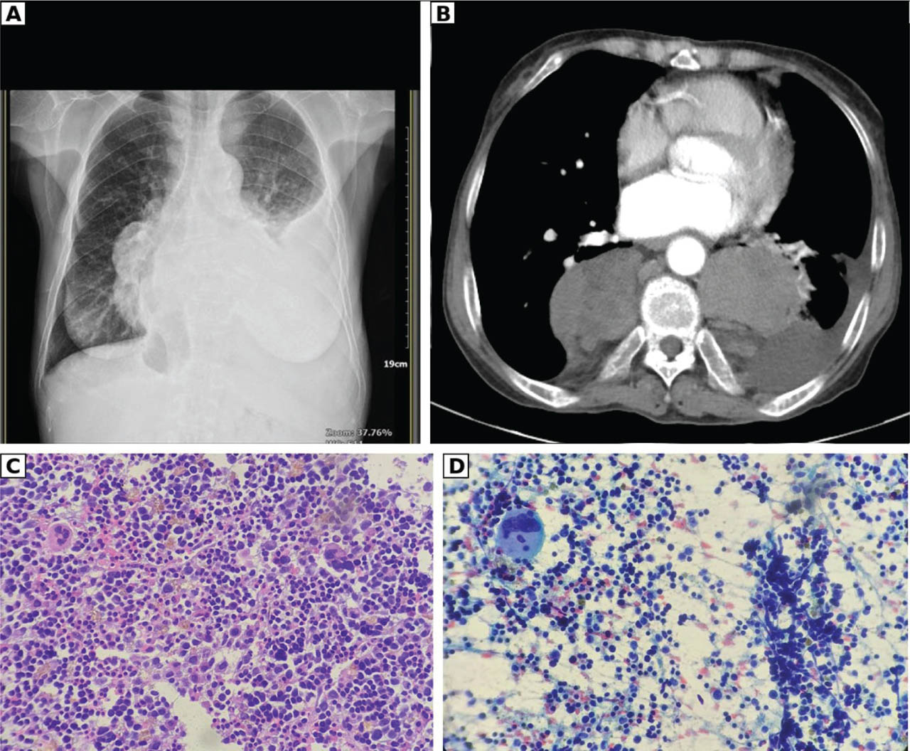
Figure 1.
Patient 1. (A) Posteroanterior chest radiograph showing a right paramediastinal/paravertebral mass with a left pleural effusion. (B) Contrast-enhanced axial CT (mediastinal window) of the whole thorax showing bilateral paravertebral soft-tissue masses flanking the vertebral body behind the heart. (C) H&E-stained section (×400) showing hypercellular trilineage haematopoietic tissue with megakaryocytes. (D) Papanicolaou-stained aspirate (×400) showing a megakaryocyte among haematopoietic precursors. CT, computed tomography; H&E, haematoxylin-eosin.
Three months later she was admitted with worsening dyspnoea, recurrent effusion and severe microcytic hypochromic anaemia (haemoglobin 7.0 g/dL), which corrected to 10.9 g/dL after transfusion. Contrast-enhanced computed tomography (CT) confirmed paravertebral soft-tissue masses with a left pleural effusion (Figure 1B). The radiological differential diagnosis included a right lung carcinoma with pleural involvement versus EMH. On the presumption of malignancy, the mass was provisionally assigned a radiological clinical stage of cT4N2M1a (stage IVA); this was a presumptive imaging-based stage pending tissue diagnosis and not a confirmed oncologic diagnosis. Bronchoscopic transbronchial biopsy and CT-guided transthoracic needle aspiration demonstrated clusters of megakaryocytes and haematopoietic cells, establishing EMH (Figures 1C and 1D). She was managed with transfusion and supportive haematologic care, without resection or radiotherapy, with symptomatic improvement. She was subsequently referred to a tertiary centre with comprehensive haematology services for definitive long-term management and was lost to follow-up at our institution.
Case 2
A 47-year-old man, lifelong non-smoker and teacher, with beta-thalassaemia intermedia (haemoglobin E [HbE]) treated by repeated transfusion for 25 years and untreated chronic hepatitis C, was referred for a suspected mediastinal tumour. He described 5 years of progressive dyspnoea with productive cough, chest and back pain and weight loss, without fever or haemoptysis. Examination showed hepatomegaly and splenomegaly without respiratory distress; the BMI was 16.6 kg/m2. Laboratory studies showed bicytopenia (haemoglobin 8.6 g/dL, platelets 95,000/μL), markedly elevated ferritin (3510 ng/mL) with transferrin saturation of 85%, lactate dehydrogenase of 308 U/L and non-contributory tumour markers (carcinoembryonic antigen, alpha-fetoprotein and beta-human chorionic gonadotropin). Haemoglobin analysis showed foetal haemoglobin (HbF) 23.7% and HbE 25%, confirming beta-thalassaemia intermedia with HbE.
Chest radiography showed a right posterior mediastinal mass (Figure 2A). Non-contrast CT demonstrated lobulated paravertebral masses with additional left para-aortic masses, interpreted as suggestive of lymphoma (Figure 2B). The dominant right posterior mediastinal and paravertebral mass measured 8.58 cm × 7.12 cm in axial cross-section and extended up to approximately 13 cm in craniocaudal dimension; these values represent different measurement axes of the same lesion rather than interval enlargement. Contrast-enhanced CT confirmed the bilateral posterior mediastinal masses, with hepatosplenomegaly and marrow expansion (Figure 2C). Bronchoscopy showed compressive stenosis of the truncus intermedius and the right B6 segment; transbronchial and bronchial-washing samples were negative for malignancy and tuberculosis. Core biopsy and transthoracic needle aspiration showed hypercellular haematopoietic tissue with erythroid and myeloid precursors and megakaryocytes, confirming pulmonary EMH (Figure 2D). He was treated with deferiprone (1000 mg three times daily), folic acid, vitamins B12 and C, ramipril and furosemide, with continued transfusion, and his dyspnoea and cough improved. He was likewise referred to a centre with comprehensive haematology services for ongoing iron chelation and transfusion management and was subsequently lost to follow-up at our institution. The clinical, laboratory, imaging and histopathological features of both patients are compared in Table 1.
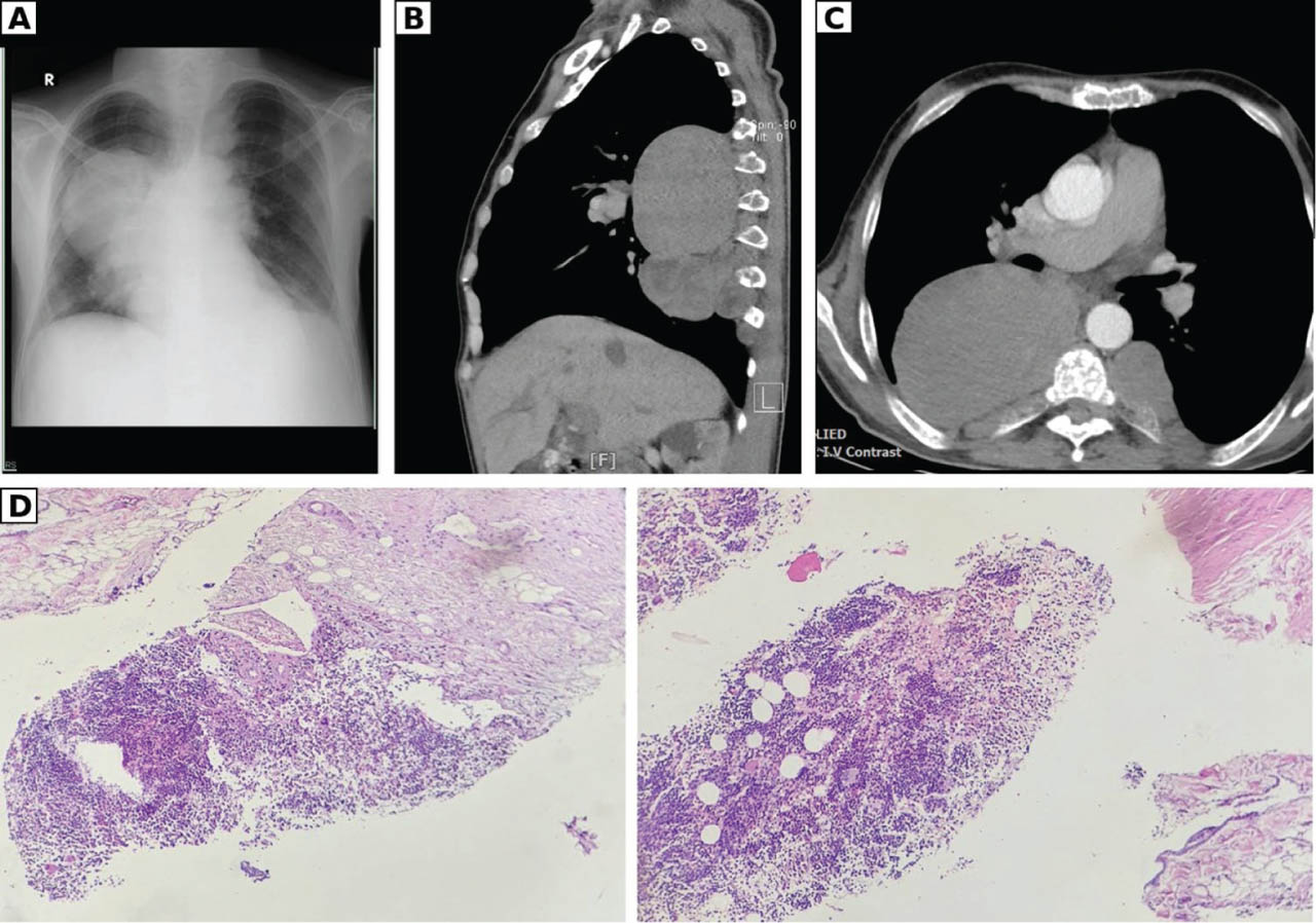
Figure 2.
Patient 2. (A) Posteroanterior chest radiograph showing a right posterior mediastinal mass. (B) Non-contrast CT (sagittal reconstruction) showing lobulated paravertebral masses anterior to the vertebral column. (C) Contrast-enhanced axial CT (mediastinal window) showing a large right posterior mediastinal mass and a left paravertebral mass behind the heart. (D) Core-biopsy histopathology (H&E, ×100) showing hypercellular haematopoietic tissue with erythroid and myeloid precursors and megakaryocytes. CT, computed tomography; H&E, haematoxylin-eosin.
Table 1.
Comparison of the two cases of intrathoracic EMH
| Feature | Case 1 | Case 2 |
|---|---|---|
| Age (years), sex | 59, female | 47, male |
| Thalassaemia type | Beta-thalassaemia; transfused approximately every 3 months for about 30 years | Beta-thalassaemia intermedia with HbE; transfused for about 25 years |
| Relevant comorbidity | None reported | Untreated chronic hepatitis C |
| Presenting symptoms | Progressive dyspnoea, weight loss, reduced appetite | Progressive dyspnoea (5 years), productive cough, chest and back pain, weight loss |
| Examination | Pallor, reduced left breath sounds, left basal dullness, splenomegaly; BMI 16.4 kg/m2 | Hepatomegaly, splenomegaly; BMI 16.6 kg/m2 |
| Haemoglobin | 7.0 g/dL (10.9 g/dL after transfusion) | 8.6 g/dL |
| Platelets | Not reported | 95,000/μL |
| Ferritin; transferrin saturation | Not reported | 3510 ng/mL; 85% |
| Lactate dehydrogenase | Not reported | 308 U/L |
| Haemoglobin analysis | Not reported | HbF 23.7%, HbE 25% |
| Tumour markers | Negative | CEA, AFP, beta-hCG non-contributory |
| Pleural effusion | Left; serohaemorrhagic exudate; cytology and microbiology negative | None reported |
| Imaging | Right paramediastinal/paravertebral mass with left pleural effusion; bilateral paravertebral soft-tissue masses on CT | Bilateral posterior mediastinal and paravertebral masses with left para-aortic extension; dominant mass 8.58 cm × 7.12 cm (axial), up to ~13 cm (craniocaudal) |
| Initial radiological impression | Lung carcinoma with pleural involvement (presumptive cT4N2M1a) versus EMH | Lymphoma |
| Diagnostic procedure | Transbronchial biopsy and CT-guided transthoracic needle aspiration | Bronchoscopy (compressive stenosis of truncus intermedius and right B6), core biopsy and transthoracic needle aspiration |
| Histopathology | Megakaryocytes and haematopoietic cells (EMH) | Hypercellular hematopoietic tissue with erythroid and myeloid precursors and megakaryocytes (EMH) |
| Management | Transfusion and supportive haematologic care | Deferiprone, folic acid, vitamins B12 and C, ramipril, furosemide, continued transfusion |
| Outcome | Symptomatic improvement; referred to tertiary haematology service; lost to follow-up | Symptomatic improvement in dyspnoea and cough; referred for chelation and transfusion; lost to follow-up |
Discussion
The mechanism underlying both cases is chronic ineffective erythropoiesis, in which persistent anaemia and elevated erythropoietin drive proliferation of haematopoietic precursors beyond the marrow; this compensatory tissue localises to the paravertebral and posterior mediastinal regions and produces the masses observed here (3, 4). The haematologic phenotype supported this mechanism, particularly in Patient 2, who had markedly elevated HbF, iron overload, marrow expansion and hepatosplenomegaly (3).
On imaging, intrathoracic EMH tends to form well-defined posterior mediastinal or paravertebral masses that may contain fat and usually spare the adjacent bone, although ill-defined margins and variable fluorodeoxyglucose uptake are also described and reduce the specificity of imaging (4, 7). Magnetic resonance imaging is valuable for characterising paraspinal lesions and for assessing possible spinal cord involvement (3). These appearances must be distinguished from neurogenic tumours, lymphoma, meningocele and metastatic disease (4, 5). In both of our patients, the imaging was read as suggestive of malignancy, consistent with published reports in which intrathoracic EMH was mistaken for neurogenic tumours, lymphoma, or lung cancer (4,5,6,7). Fluorine-18 fluorodeoxyglucose positron emission tomography/computed tomography (FDG-PET/CT) was not performed in either patient owing to resource constraints; although EMH can show variable FDG uptake that contributes to this malignancy mimicry (4), the diagnosis was ultimately established by histology.
Because imaging is non-specific, the diagnosis depends on clinical context and tissue confirmation. A known haemoglobinopathy, non-contributory tumour markers and negative cytology lower the probability of carcinoma and germ-cell tumour and support biopsy over resection. Tissue sampling (by transbronchial biopsy, CT-guided percutaneous core or fine-needle aspiration, or video-assisted thoracoscopic surgery) remains the reference standard and was decisive in both patients (8). Biopsy also carries a bleeding risk, because EMH tissue is vascular and many patients have concurrent thrombocytopenia and splenomegaly; this risk is weighed against the value of a definitive diagnosis that averts unnecessary thoracotomy or oncologic therapy.
Management is directed at the erythropoietic drive rather than at the mass itself. Adequate transfusion suppresses the stimulus that sustains EMH, and both of our patients improved with correction of anaemia and supportive haematologic therapy rather than resection (3, 12). Because haematopoietic tissue is radiosensitive and transfusion-responsive, symptomatic intrathoracic EMH can also be treated with agents that induce HbF, such as hydroxyurea, or with low-dose radiotherapy, with surgery reserved for selected indications such as cord compression (3, 12, 13).
This series has several limitations. FDG-PET/CT was not performed, and pleural-fluid analysis was limited to cytology and microbiology; these were the investigations available at our centre. Biopsy carried a bleeding risk in these thrombocytopenic and splenomegalic patients. Both patients were referred to centres with comprehensive haematology services and were lost to follow-up at our institution, so long-term outcome and imaging response could not be assessed.
Conclusion
Intrathoracic EMH can develop in patients with thalassaemia and closely mimic thoracic malignancy. It should be systematically included in the differential diagnosis of posterior mediastinal and paravertebral masses in patients with haemoglobinopathies, alongside neurogenic tumours, lymphoma, meningocele and metastatic disease. Tissue sampling is paramount, both for preventing unnecessary surgical or oncologic intervention and for facilitating appropriate medical management, as EMH can often be managed medically.
Acknowledgements
The authors thank the staff of the Department of Pulmonology and Respiratory Medicine and the Department of Anatomic Pathology, Persahabatan National Respiratory Center Hospital, for their assistance with the clinical care and specimen handling of these patients.
Notes
[3] Contributed by Author's contributions
Kevin Aristyo contributed to concept, planning and design, data collection and processing, analysis and interpretation, literature review, and writing of the manuscript. Arini Purwono contributed to planning and design, data collection and processing, and literature review. Dea Putri Audina contributed to data collection and processing, analysis and interpretation, and literature review. Ginanjar Arum Desianti contributed to planning and design, and data collection and processing. Tina Reisa contributed to planning and design, data collection and processing, analysis and interpretation, and literature review. Hayatun Nufus contributed to analysis and interpretation, and critical review. Jamal Zaini contributed to concept, supervision, and critical review. Fariz Nurwidya contributed to concept, supervision, and critical review. Eylin Rahardjo contributed to supervision, analysis and interpretation, and critical review. Mia Elhidsi contributed to concept, supervision, and critical review.