
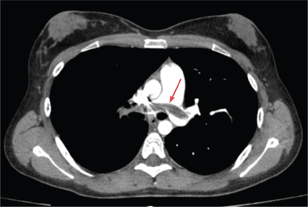
Figure 1.
Contrast-enhanced chest CT scan: massive occlusive PE in the ‘saddle’ of the pulmonary trunk. CT, computed tomography; PE, pulmonary thromboembolism.
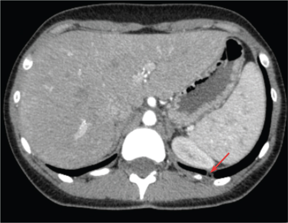
Figure 2.
Contrast enhanced chest CT scan: pulmonary infarctions. CT, computed tomography.
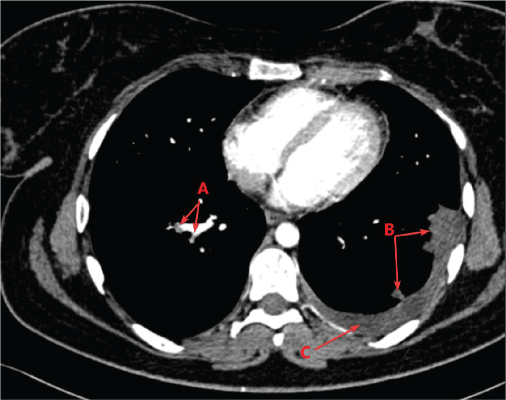
Figure 3.
Contrast-enhanced chest CT scan: (A) PE, (B) pulmonary infarcts, (C) pleural effusion. CT, computed tomography; PE, pulmonary thromboembolism.
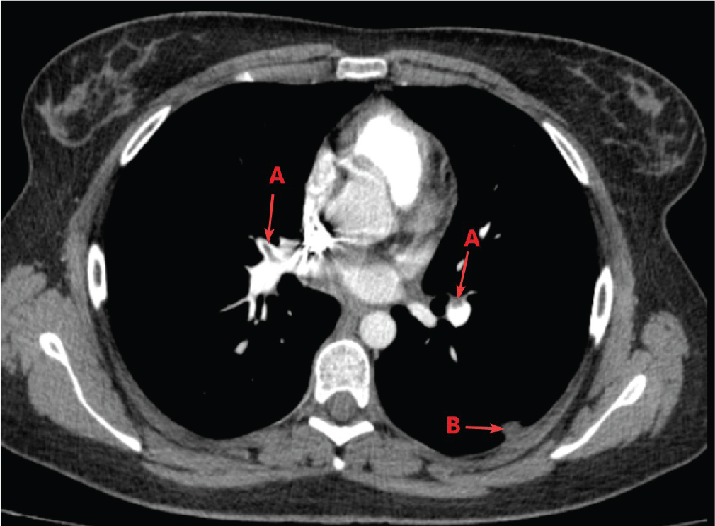
Figure 4.
Contrast-enhanced chest CT scan: (A) PE, (B) pulmonary infarcts. CT, computed tomography; PE, pulmonary thromboembolism.