
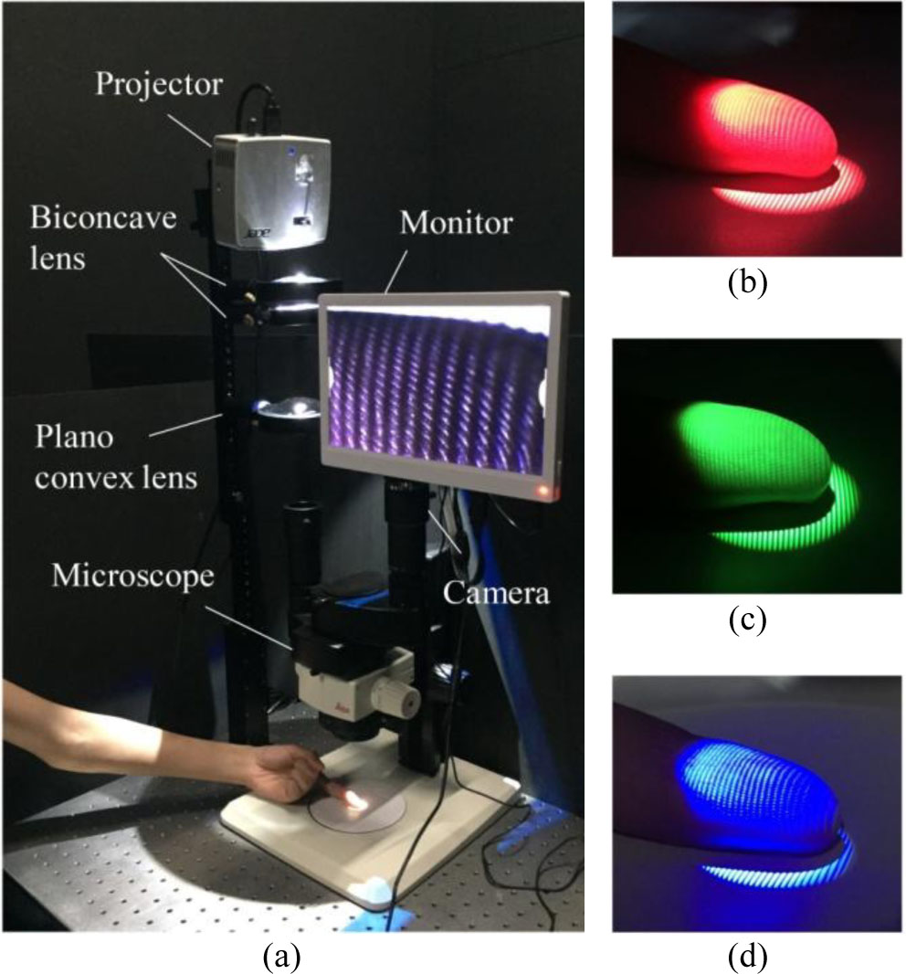
Fig. 1.
(a) Experimental setup of rapid touchless 3D fingerprint imaging, including a projector, optical lenses, a microscope, and a camera; (b) red; (c) green; and (d) blue fringe patterns projected on the finger surface.
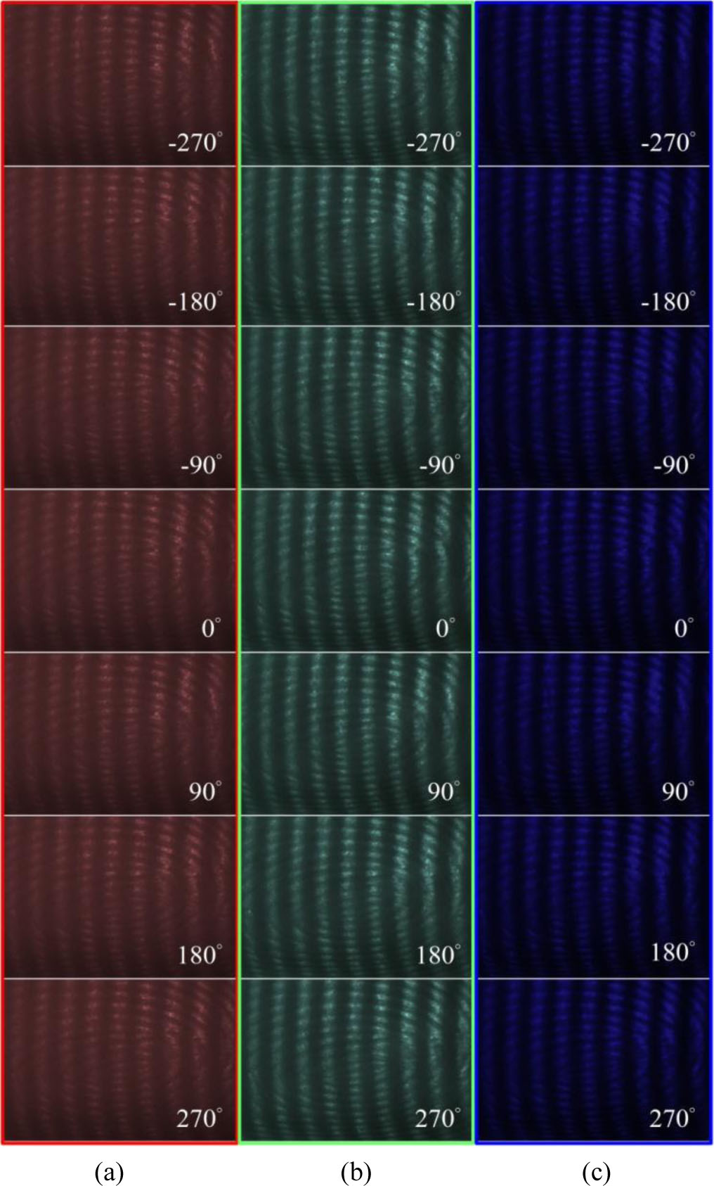
Fig. 2.
Photomicrographs of seven single-color fringe patterns projected on the finger surface: (a) red; (b) green; and (c) blue deformed fringe patterns.
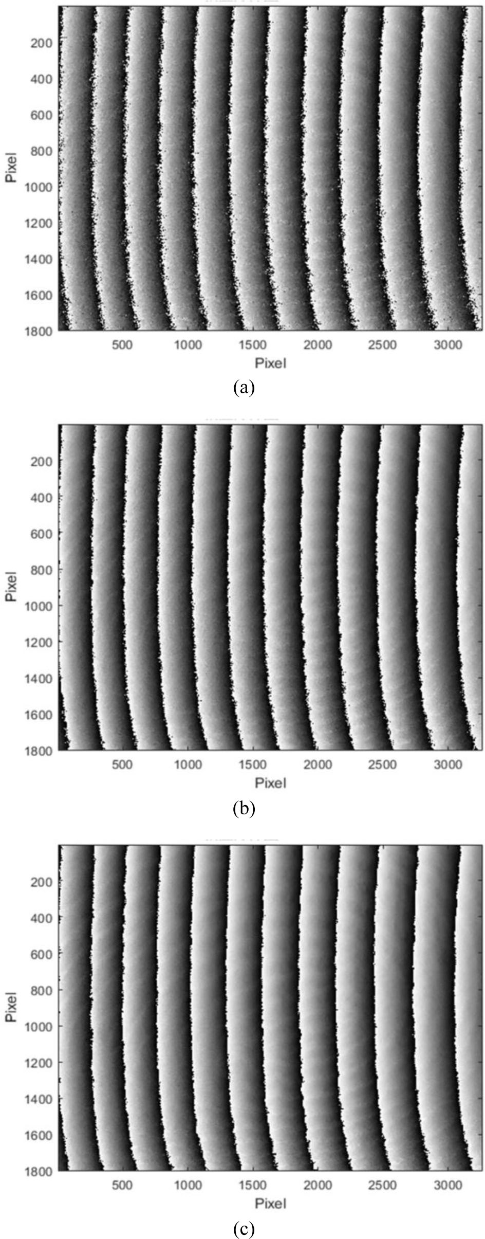
Fig. 3.
Wrapped phase maps based on seven-step phase-shifting calculation with (a) red; (b) green; and (c) blue deformed fringe patterns.
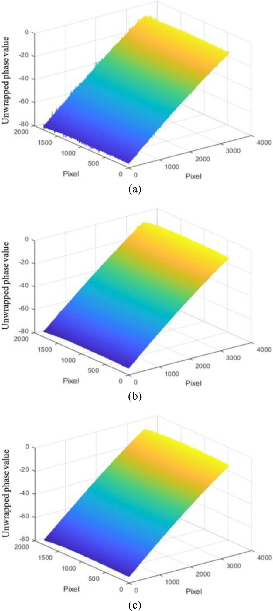
Fig. 4.
Unwrapped phase maps based on seven-step phase-shifting calculation with (a) red; (b) green; and (c) blue deformed fringe patterns.
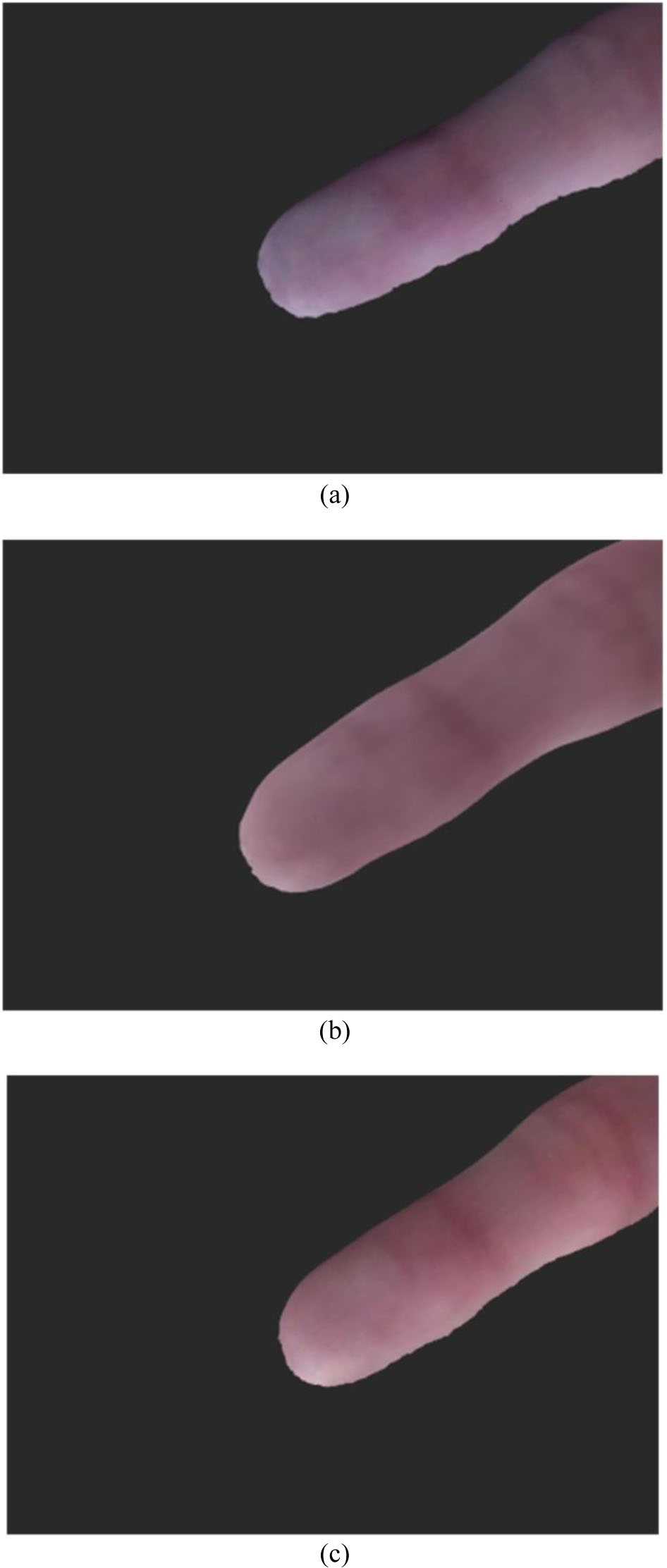
Fig. 5.
3D shape reconstruction of the finger based on seven-step phase-shifting calculation with (a) red, (b) green, and (c) blue deformed fringe patterns.
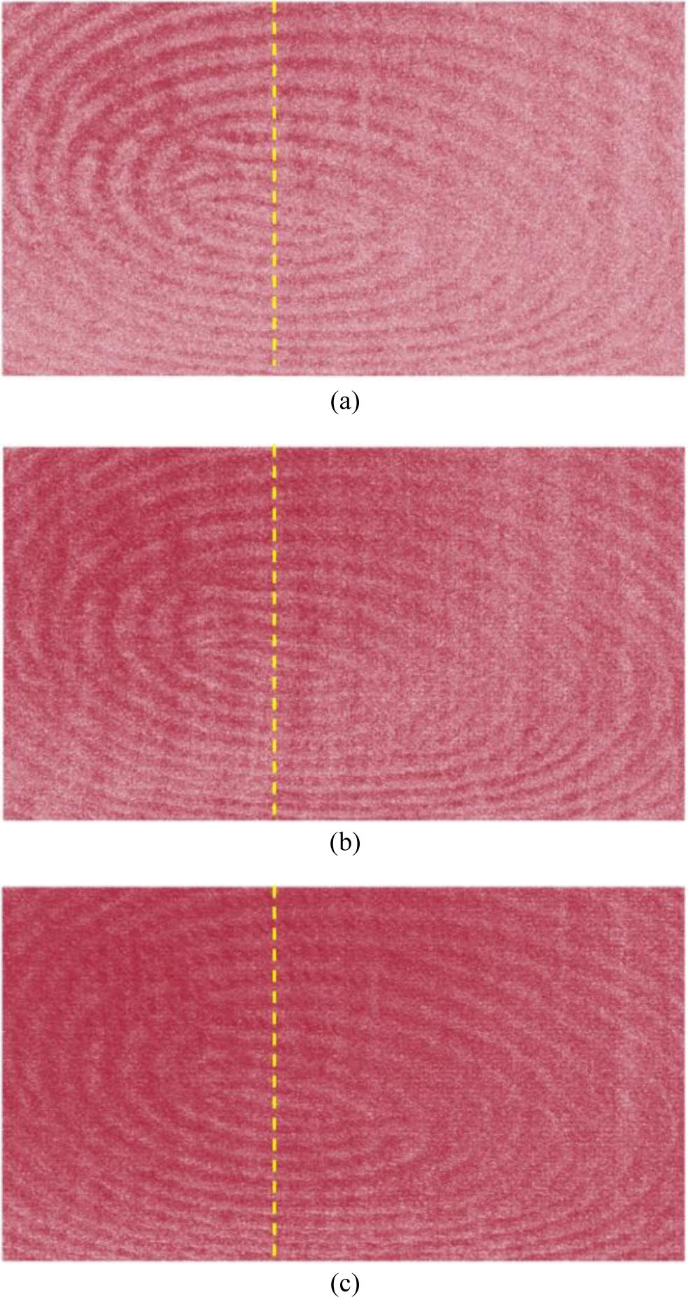
Fig. 6.
3D representation of the captured fingerprint based on seven-step phase-shifting calculation with (a) red; (b) green; and (c) blue deformed fringe patterns.
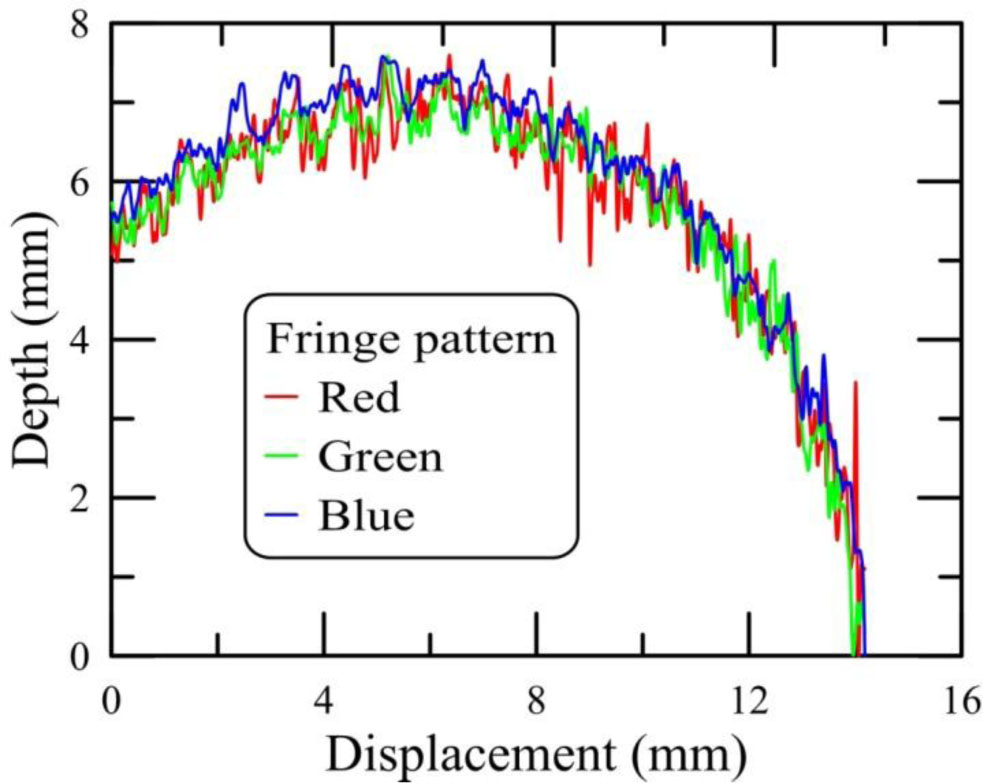
Fig. 7.
Line scan profiles of the 3D fingerprint reconstructed by red, green, and blue deformed fringe patterns. The dashed lines in Fig. 6 indicate the line-scan position.
Table 1.
Comparison of this work with related studies.
| Study | Technique | Resolution [µm] | Level 3 feature capture | Spectral analysis | Acquisition time [s] |
|---|---|---|---|---|---|
| This work | 7-step FPP, RGB illumination | < 40 lateral, < 5 vertical | Yes – pores, ridge edges | Yes – RGB comparison | <1 |
| [7] | Single-shot FPP + biospeckle | ∼100 | Partial | No | ∼1 |
| [23] | Phase measuring profilometry | ∼50 | No | No | ∼0.5 |
| [32] | Full-field sinusoidal FPP | ∼70 | No | No | ∼1.5 |
| [33] | Composite color fringe projection | ∼60 | No | Yes – synthetic color | ∼1 |
| [48] | Level 3 matching (ridge, pores) | N/A | Yes – 2D | No | N/A |