
1. Introduction
In an increasingly globalized and digitized world, the demand for secure and reliable personal identification systems has become more critical than ever [1], [2]. These systems form the backbone of numerous applications, including access control, electronic transactions, law enforcement, and national security [3]. Biometric technologies, which authenticate individuals based on unique physiological or behavioral traits, have emerged as robust and efficient solutions. Among the various modalities, fingerprint recognition remains the most widely adopted due to its universality, permanence, and ease of acquisition [4], [5]. The unique ridge-valley patterns of fingerprints are highly distinctive, stable throughout an individual's lifetime, and exceedingly difficult to replicate. Consequently, fingerprint-based systems are extensively deployed across governmental, commercial, and forensic domains, alongside other biometrics such as iris, facial, and voice recognition. Despite their widespread use, most conventional fingerprint recognition systems rely on two-dimensional (2D) contact-based imaging [6]. These systems are inherently limited by nonlinear distortions caused by variable finger pressure, misalignment, and surface friction, which can alter the spatial configuration of minutiae, critical features in fingerprint matching [7], [8]. Additionally, projecting the fingertip’s three-dimensional (3D) structure onto a 2D plane results in the loss of depth information, further compromising recognition accuracy [9], [10]. Image quality in 2D systems is also highly sensitive to external factors such as illumination conditions, skin moisture, dirt, and diverse dermatological states [10]. Conditions such as excessive dryness, chapping, or perspiration frequently result in poor image capture or false rejections of legitimate users. These limitations not only undermine system accuracy and reliability but also increase susceptibility to increasingly sophisticated spoofing attacks. Therefore, the development of next-generation fingerprint recognition systems that are more accurate, robust, and user-friendly remains an urgent and ongoing challenge in biometric security.
With advances in image acquisition technologies, 3D biometric techniques have garnered increasing attention as promising alternatives to conventional 2D systems. 3D biometrics, including facial recognition [11]–[13], 3D ear [14]–[16], and 3D palmprint recognition [17]–[21], offer several advantages: contactless acquisition, elimination of elastic deformation, enhanced spatial geometric representation, and improved robustness to environmental variations. These advantages address many of the inherent limitations of 2D modalities, particularly in real-world applications. In the domain of 3D fingerprint recognition, early research has established foundational methods for acquiring and processing spatial fingerprint data. For example, Parziale et al. [22] introduced a surround imager system that employs a multi-camera array to capture finger images from multiple viewpoints and reconstructs the 3D fingerprint using the shape-from-silhouette technique. Wang et al. [23] proposed a non-contact phase measuring profilometry (PMP) based approach that captures fine topographical details of fingerprint ridges and valleys. Structured light projection methods, especially fringe projection profilometry (FPP), have emerged as powerful tools for high-precision 3D surface acquisition and have been extensively applied across scientific and engineering domains [24]–[27]. A typical FPP system comprises a projector that casts structured patterns (e.g., sinusoidal or binary fringes) onto the object surface, a camera to capture the deformed patterns, and a computing unit that performs phase estimation and 3D reconstruction. In biometrics, FPP has shown notable effectiveness, particularly in 3D palmprint recognition systems that have employed various FPP techniques, ranging from binary fringe projection to full-field color sinusoidal projection for simultaneous shape and texture acquisition. In the context of 3D fingerprinting, PMP-based methods represent a practical application of FPP, enabling high-resolution, non-contact capture of fine microstructural fingerprint features.
Despite the demonstrated potential of FPP for high-precision 3D data acquisition, its application to rapid, non-contact 3D fingerprint imaging at the microscopic level presents significant challenges. Conventional multi-step phase-shifting techniques, such as those employed in PMP or sinusoidal fringe projection methods [28]–[31], require the sequential capture of multiple images. This inherently increases acquisition time and introduces susceptibility to motion artifacts. Reported acquisition durations of 0.5 seconds or less may still be inadequate for real-time biometric applications [32]. Moreover, achieving complete finger immobilization during imaging, especially when capturing fine features such as pores, is inherently difficult without physical stabilization mechanisms. Although certain studies have explored channel-wise optimization to enhance contrast using individual RGB components [32], [33], a comprehensive investigation into the influence of illumination wavelength on 3D reconstruction quality at the microscopic scale remains largely unexplored. Given the wavelength-dependent nature of light-skin interactions, including absorption and scattering phenomena [34]–[37], selecting optimal spectral bands can substantially improve ridge contrast and depth reconstruction accuracy. While prior work has established the feasibility of digital projection-based fingerprint topography capture, there remains a critical gap in integrating wavelength-optimized illumination strategies with real-time, high-resolution imaging performance. Addressing these challenges is essential for advancing practical 3D fingerprint acquisition systems suitable for deployment in real-world biometric authentication scenarios.
In this study, we developed a rapid, non-contact 3D fingerprint imaging system based on full-field microscopic fringe projection profilometry. The system integrates a digital light processing (DLP) projector, a custom-designed microscope-lens assembly, and a high-resolution CCD camera to project and capture sinusoidal fringe patterns at red, green, and blue wavelengths. A seven-step phase-shifting algorithm was used to generate wrapped phase maps, which were subsequently processed using a quality-guided phase unwrapping technique to reconstruct the 3D topography of the fingertip surface. To assess the impact of illumination wavelength on reconstruction quality, we performed comparative analyses of the wrapped phase, unwrapped phase, 3D surface reconstructions, and line profiles across the three color channels. Experimental results indicated that green fringe projection produced the most distinct deformation patterns and yielded the highest 3D reconstruction accuracy, whereas red fringe projection showed the weakest performance. The system was capable of resolving fine anatomical features, such as ridge contours and pores, and completed the entire acquisition process within one second. These results demonstrate the feasibility and effectiveness of combining microscopic FPP with wavelength-optimized illumination for real-time, high-resolution 3D fingerprint imaging on live fingers. The proposed approach offers a promising direction for advancing non-contact biometric identification technologies.
2. Experimental Arrangement
The proposed rapid, non-contact 3D fingerprint imaging system is designed to capture microscale surface topography of fingerprints using a full-field sinusoidal fringe projection approach. As depicted in Fig. 1(a), the system comprises a DLP projector (Acer K132+) for generating sinusoidal fringe patterns, a custom-configured optical lens assembly, a stereo microscope (Leica M50), and a high-resolution CCD camera (Tucsen TrueChrome II, 1920 × 1080 pixels). The optical path includes a biconvex lens, a biconcave lens, and a plano-convex lens, all precisely aligned based on ray-tracing simulations. This alignment ensures appropriate image demagnification, beam collimation, and fringe resizing, thereby optimizing the pattern projection onto the sample surface. The stereo microscope employed in this study offers five discrete magnification settings, namely 6.3 ×, 10 ×, 16 ×, 25 ×, and 40 ×, with a working distance of 89.6 mm and an effective measurement area ranging from 36.5 mm to 5.75 mm, depending on the selected magnification. Within the proposed imaging system, the stereo microscope performs two essential functions: it projects the structured fringe patterns onto the fingertip surface and collects the deformed fringe patterns reflected from the skin. The captured fringe images are then recorded by the CCD camera and transmitted to an industrial-grade computer for system synchronization, image acquisition, phase calculation, and 3D surface reconstruction. In addition, a display monitor is used for real-time sample alignment and visualization throughout the measurement process. No significant defocus was observed within the effective imaging range used for fingerprint measurement.
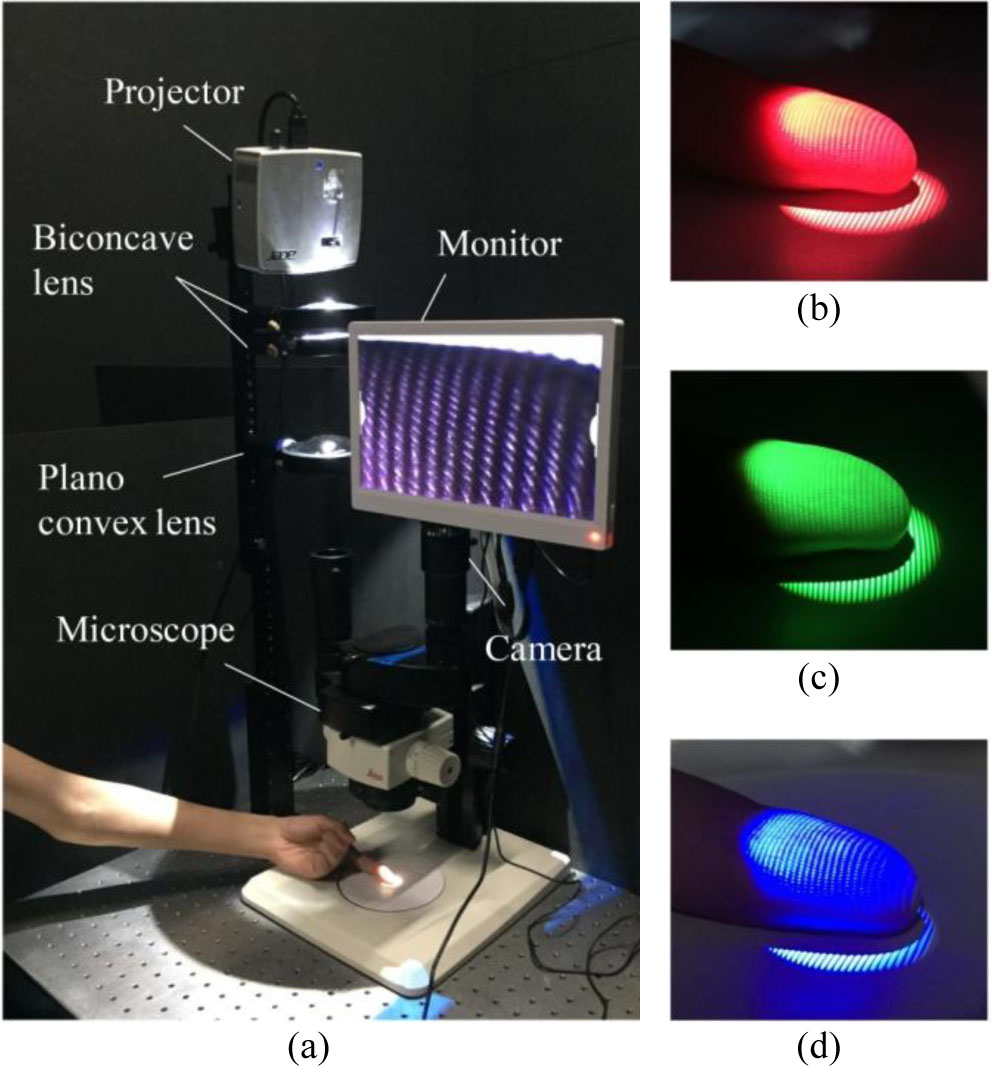
Fig. 1.
(a) Experimental setup of rapid touchless 3D fingerprint imaging, including a projector, optical lenses, a microscope, and a camera; (b) red; (c) green; and (d) blue fringe patterns projected on the finger surface.
To investigate the influence of illumination wavelength on 3D fingerprint reconstruction accuracy, sinusoidal fringe patterns were projected using red, green, and blue light sources, corresponding to distinct regions of the visible spectrum. For each 3D reconstruction, only one illumination wavelength was used, and the reconstructed surface was generated from the corresponding seven deformed fringe patterns of that single color channel. These wavelengths were selected to enable a comparative assessment of how wave-length-dependent light absorption and scattering in human skin affect fringe deformation and reconstruction fidelity. Red light, with its longer wavelength, penetrates deeper into the skin layers, potentially attenuating surface contrast and diminishing the visibility of fine features. In contrast, green light offers a favorable balance between penetration depth and surface reflectance, and is widely recognized for its ability to enhance contrast in skin texture imaging. Blue light, possessing the shortest wavelength, interacts primarily with surface structures, providing strong surface contrast but exhibiting increased sensitivity to noise and superficial imperfections. Fig. 1(b)–Fig. 1(d) present the projected sinusoidal fringe patterns on a live fingertip under red, green, and blue illumination, respectively. These captured images served as the basis for subsequent phase analysis and 3D surface reconstruction, enabling a quantitative evaluation of each wavelength’s impact on fringe deformation, phase stability, and the spatial resolution of fine anatomical features such as ridge contours and sweat pores.
3. Results and Discussion
Multi-step phase-shifting algorithms are widely used in FPP to extract accurate, high-resolution phase information from deformed fringe patterns [38]–[45]. In the present study, a seven-step phase-shifting approach is adopted to improve the spatial resolution, smoothness, and robustness of the reconstructed 3D fingerprint surface [46]. Compared to the conventional three-step method, the seven-step algorithm offers greater noise resilience and improved continuity of phase maps, making it well-suited for capturing fine surface microstructures, such as fingerprint ridges and pores.
The digital sinusoidal fringe patterns are generated and modulated by an industrial computer, allowing precise control over the phase shifts and pattern encoding. To enable multi-wavelength data acquisition within a single exposure, three distinct sets of seven-step fringe patterns are spectrally multiplexed and encoded into the red, green, and blue channels, respectively. This color-coded projection scheme facilitates simultaneous acquisition of full-field fringe data across different wavelengths, significantly reducing motion-induced artifacts and enhancing overall measurement efficiency. The projected fringe intensity at each pixel on the object surface can be modeled by the general phase-shifting expression:
where i and j denote the pixel coordinates along the horizontal and vertical directions, respectively; IN(i,j) represents the recorded intensity at pixel (i,j) in the N-th frame; Ig(i,j) is the background intensity at the pixel; Im(i,j) denotes the modulation amplitude of the fringe pattern; ϕ(i,j) is the phase value associated with the surface height of the object; and βN is the known phase shift introduced during the N-th step of the phase-shifting sequence.For the seven-step method, the phase shifts applied are βN = −3π/2, −π, −π/2,0, π/2, π, 3π/2. This corresponds to phase angles of −270°, −180°, −90°, 0°, 90°, 180°, and 270°. The captured deformed fringe patterns for each color are shown in Fig. 2. The corresponding intensity values can be written as:
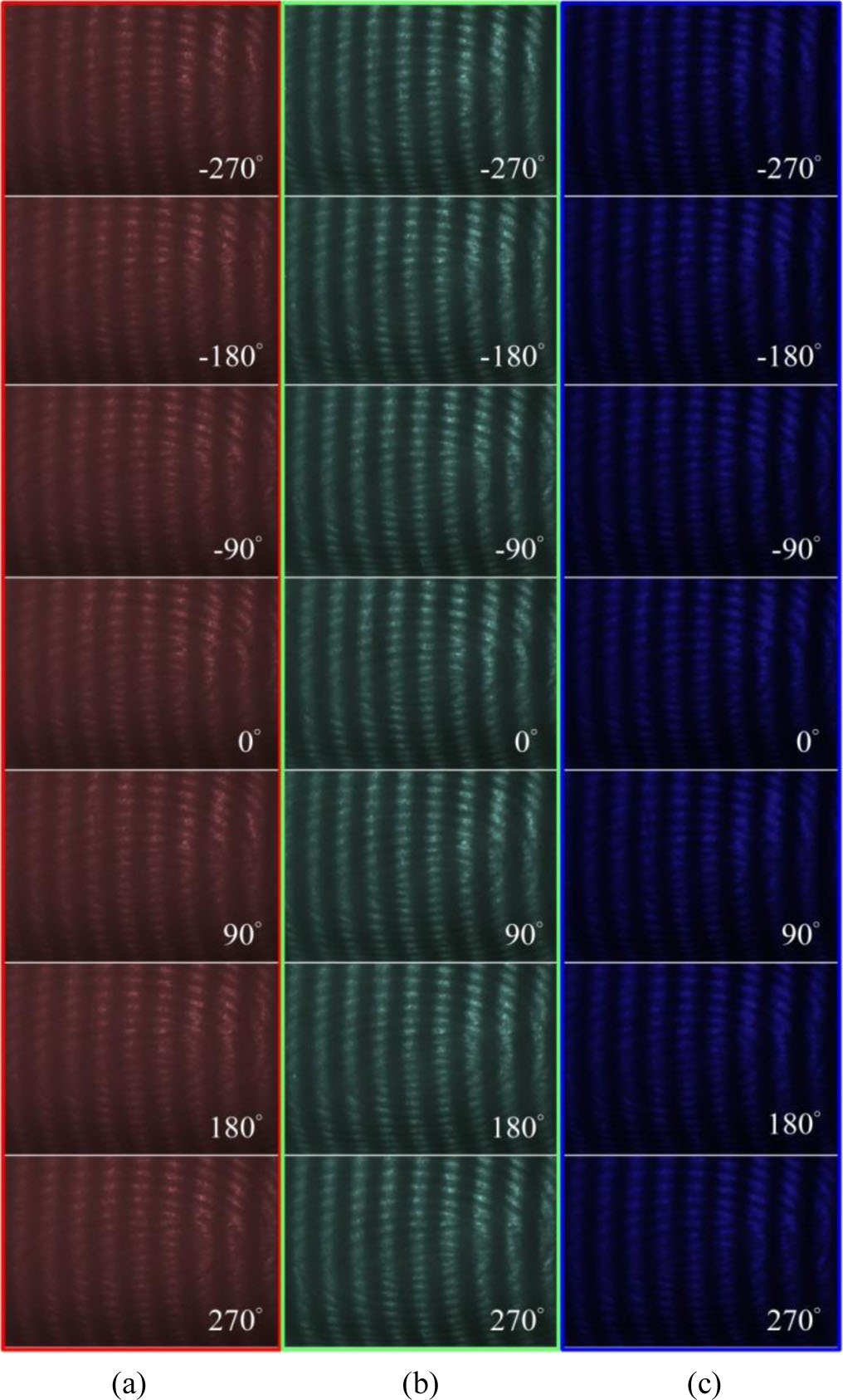
Fig. 2.
Photomicrographs of seven single-color fringe patterns projected on the finger surface: (a) red; (b) green; and (c) blue deformed fringe patterns.
Using the seven captured intensity frames, the wrapped phase ϕw(i,j) at each pixel is computed using a standard trigonometric phase-shifting algorithm that involves a weighted summation followed by an arctangent operation. The resulting phase values are constrained within the interval [−π,π], ensuring local phase continuity but lacking global height information. Each color channel (red, green, and blue) is processed independently, yielding three separate wrapped phase maps. These maps are then subjected to a quality-guided phase unwrapping algorithm to obtain absolute phase distributions, which are linearly proportional to the surface topography of the fingerprint. This processing pipeline enables high-resolution reconstruction of micro-scale surface features and facilitates a comparative evaluation of how different illumination wavelengths affect the accuracy of phase retrieval and the fidelity of reconstructed ridge contours, groove depths, and pore structures. Fig. 2 presents the captured sinusoidal fringe patterns projected onto a live fingertip under red, green, and blue illumination. Among the three, the green illumination produces the highest fringe contrast and sharpness, clearly delineating ridge-valley structures with minimal background interference. This improved visibility enhances the accuracy of phase extraction and contributes to the robustness of the resulting wrapped phase map. In comparison, the fringe pattern under red illumination appears more diffused and blurred, primarily due to increased subsurface scattering and deeper skin penetration at longer wavelengths, which diminishes fringe modulation and edge definition. The blue illumination yields better surface contrast than red, capturing finer details. However, it is more susceptible to noise and surface artifacts such as micro-wrinkles or specular reflections, which may compromise phase stability in some regions.
To reconstruct the 3D surface from captured fringe images, the wrapped phase ϕ(i,j) is first calculated using the following equation:
This formulation assumes local linearity within the coherence envelope and neglects minor intensity fluctuations arising from light source instability and fringe modulation, consistent with the approach proposed by Sandoz [46]. The computed phase is initially wrapped within a 2π interval, resulting in a discontinuous representation. Because adjacent fringe orders differ by integer multiples of 2π, phase unwrapping is necessary to retrieve a continuous and physically meaningful phase distribution. In this study, the quality-guided phase unwrapping algorithm introduced by Herráez et al. [38] is employed. The algorithm evaluates pixel-wise reliability based on the local second-order difference D, with reliability defined as R = 1/D. Smaller D values indicate smoother phase transitions and higher reliability. Reliability values are also assigned to edges between neighboring pixels by summing the reliabilities of adjacent pixels. The unwrapping process is initiated from the most reliable edges, thereby minimizing error propagation from noisy or low-quality regions and improving the overall robustness of the unwrapped phase map.
Fig. 3 illustrates the wrapped phase maps obtained under red, green, and blue structured light illumination. Each map possesses a spatial resolution of approximately 3200 × 1800 pixels, corresponding to more than 5.7 million measurement points. Among the three channels, green illumination yields the best results. The fringe pattern exhibits uniform visibility and continuity across the entire field of view, with minimal distortion at the image boundaries. Quantitatively, the green channel achieves an average fringe modulation contrast of 87.3 %, a standard deviation of 0.07 radians in the wrapped phase, and a signal-to-noise ratio (SNR) exceeding 25 dB. In comparison, the red channel demonstrates significantly reduced fringe visibility, with an average contrast of 61.5 %, a standard deviation of 0.14 radians, and an SNR below 18 dB. The blue channel performs intermediately, with a contrast of 73.8 % and a standard deviation of 0.10 radians. However, it exhibits localized instability in high-texture regions, likely due to increased sensitivity to surface imperfections and noise.
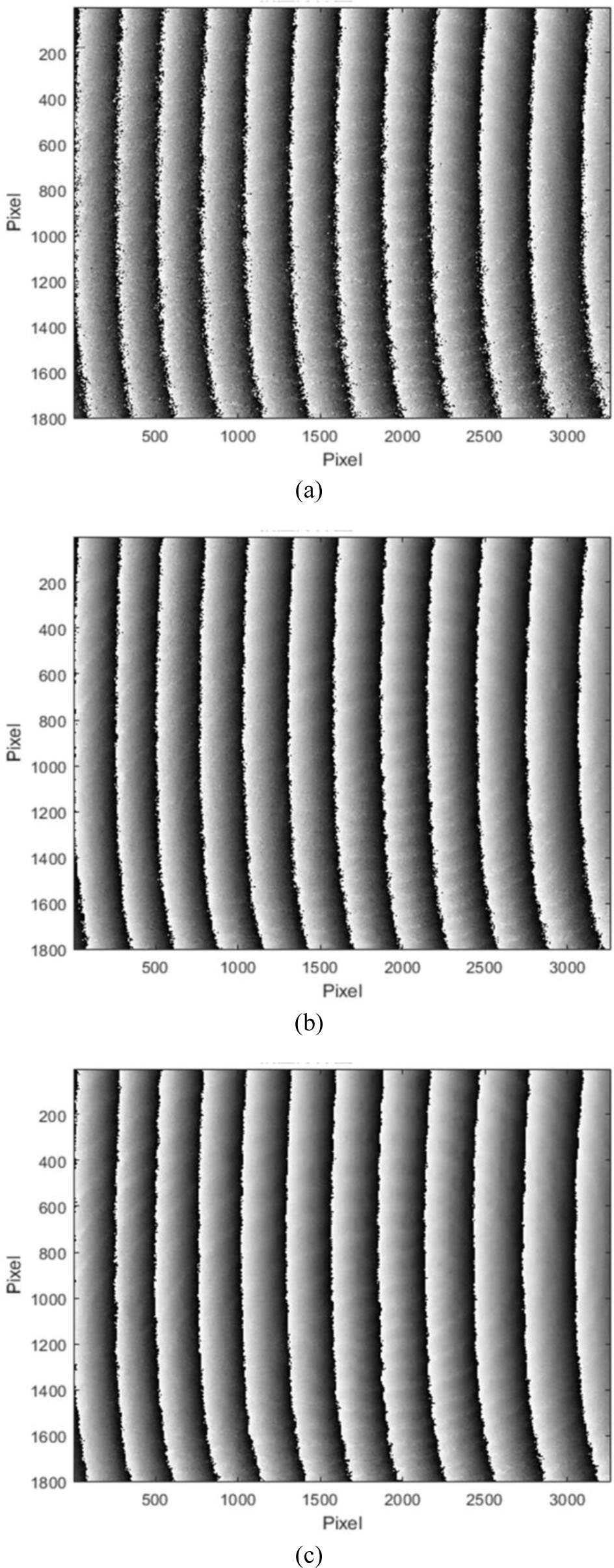
Fig. 3.
Wrapped phase maps based on seven-step phase-shifting calculation with (a) red; (b) green; and (c) blue deformed fringe patterns.
Fig. 4 presents the unwrapped phase maps corresponding to the red, green, and blue illumination channels. Among them, the green-channel result (Fig. 4(b)) yields the most continuous and stable absolute phase distribution, accurately preserving both the global topographic slope and fine ridge-valley structures. The root-mean-square (RMS) error for the green channel is 1.8 pixels, indicating high consistency and phase fidelity. In contrast, the red-channel map (Fig. 4(a)) exhibits noticeable slope distortion, particularly in low-contrast regions, and yields a significantly higher RMS error of 5.1 pixels. The blue-channel result (Fig. 4(c)) retains finer surface details than red but introduces localized noise and phase inconsistencies, resulting in an RMS error of 3.2 pixels. These comparative findings across Fig. 3 and Fig. 4 underscore the critical influence of illumination wavelength on phase quality and unwrapping performance, demonstrating that green light offers the optimal balance between surface contrast and noise robustness for accurate 3D fingerprint reconstruction in micro-scale biometric applications.
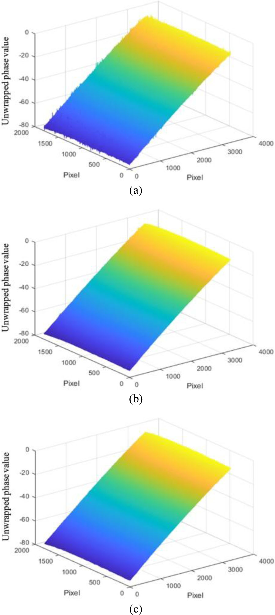
Fig. 4.
Unwrapped phase maps based on seven-step phase-shifting calculation with (a) red; (b) green; and (c) blue deformed fringe patterns.
Once unwrapped, the absolute phase ϕ(i,j) is converted to physical height h(i,j) using triangulation:
Here, P is the projected fringe pitch and θ is the projection angle relative to the measurement plane.
To eliminate surface tilt and isolate the intrinsic topographic features of the fingerprint, a reference plane is estimated and subtracted from the unwrapped phase map, following the method proposed by Liu [47]. Let the unwrapped phase map be denoted as M. To normalize the baseline, the minimum value in M is first subtracted from all elements, ensuring the lowest phase value is zero. A zero-initialized matrix M0 of the same dimensions is then created to represent the reference plane. The modal value of the bottom row of M is selected as Imin, and that of the top row as Imax. The slope N of the reference plane is computed using the following expression:
Here, ΔX denotes the horizontal pixel distance between Imin and Imax. Each row of the reference matrix M0 is then populated with linearly interpolated values spanning these two points to approximate the tilt-induced phase gradient. The final tilt-compensated absolute phase map is obtained by subtracting M0 from M. Experimental validation was per-formed using the right index finger of the author. For each of the three illumination wavelengths (red, green, and blue), seven phase-shifted fringe images were acquired using the DLP projector and CCD camera under identical fringe pitch conditions.
To evaluate the effectiveness of the proposed system in reconstructing micro-scale fingerprint features, a comparative analysis was performed under three spectral illumination conditions: red, green, and blue. The assessment focused on geometric fidelity, visibility of third-level features, and topographic consistency. Fig. 5 presents the reconstructed 3D fingerprint surfaces based on unwrapped phase data for each illumination wavelength. The reconstruction under red illumination (Fig. 5(a)) exhibits significant degradation in ridge-valley contrast and poor delineation of fine structures. This deterioration is attributed to enhanced subsurface scattering and reduced surface reflectance, which diminish fringe modulation and compromise phase accuracy. In contrast, the reconstruction obtained with green illumination (Fig. 5(b)) shows the most accurate and coherent surface morphology. Ridge lines are smooth and continuous, valleys are well-resolved, and third-level features such as sweat pores are clearly identifiable. This superior performance is attributed to the favorable optical interaction of green light with skin tissue, which provides an optimal balance between absorption and scattering, enhancing fringe visibility and phase stability. The blue-illuminated reconstruction (Fig. 5(c)) reveals enhanced edge sharpness and higher spatial resolution along ridge boundaries. However, it introduces pronounced surface granularity. While blue light's sensitivity to fine surface texture improves local contrast, it reduces continuity in areas with low texture density.
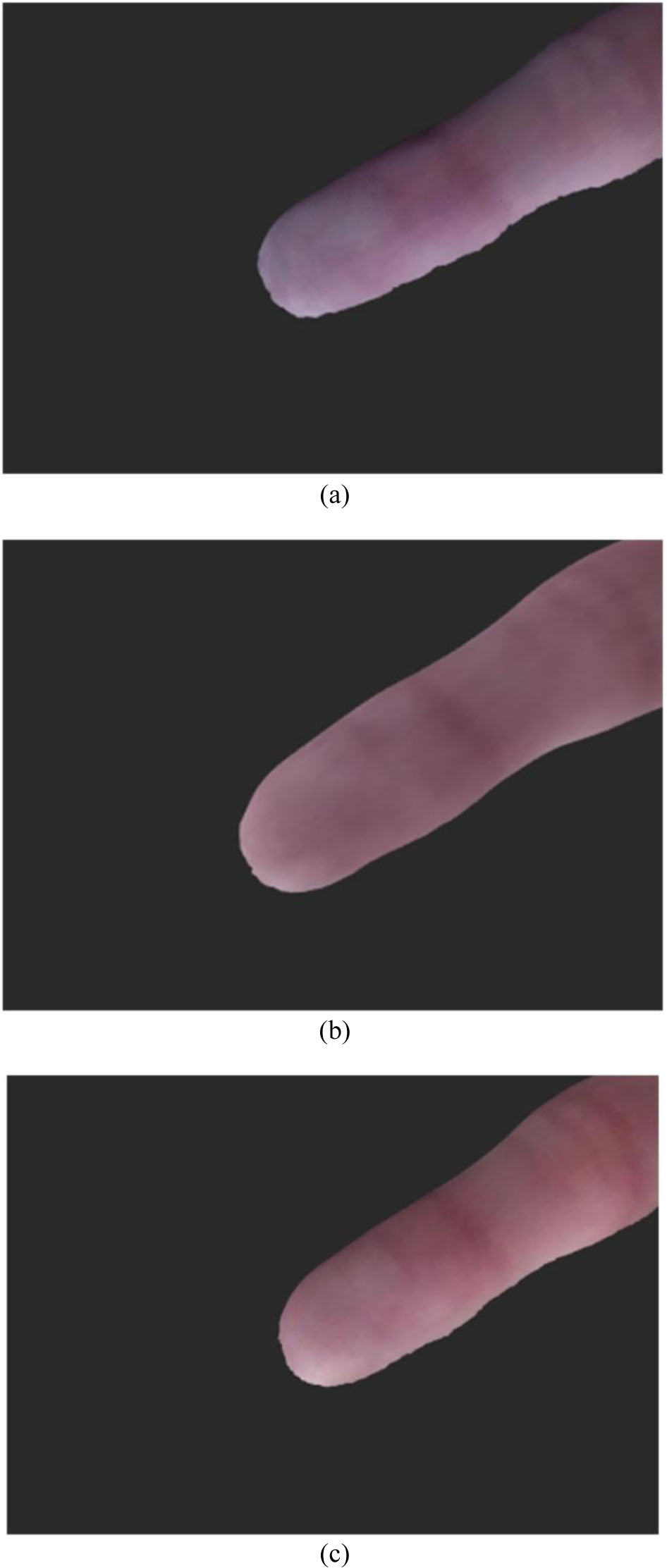
Fig. 5.
3D shape reconstruction of the finger based on seven-step phase-shifting calculation with (a) red, (b) green, and (c) blue deformed fringe patterns.
Fig. 6 presents textured renderings of the reconstructed fingerprint surfaces under the three illumination conditions. The rendering in Fig. 6(a), corresponding to red illumination, appears blurred and lacks fine detail, with minimal tonal variation along the ridge flow, indicating limited geometric and photometric fidelity. In contrast, Fig. 6(b), obtained using green illumination, exhibits a visually coherent, photo-realistic rendering. Structural integrity is well-preserved, and the surface reflectance appears consistent, with balanced shading and discernible skin texture, suggesting high reconstruction accuracy. The rendering under blue illumination in Fig. 6(c) shows sharper ridge contours than in the red channel. However, the presence of artifacts, such as exaggerated gradient transitions and uneven reflectivity, compromises the natural appearance of the surface. These distortions may adversely affect the robustness of feature extraction and matching in biometric recognition pipelines.
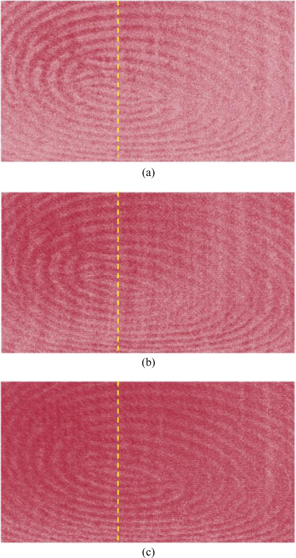
Fig. 6.
3D representation of the captured fingerprint based on seven-step phase-shifting calculation with (a) red; (b) green; and (c) blue deformed fringe patterns.
Fig. 7 presents a comparative analysis of the cross-sectional depth profiles reconstructed under the three spectral illumination conditions. The dashed lines in Fig. 6 indicate the corresponding line-scan positions. The red-channel profile exhibits pronounced noise, irregular ridge shapes, and inconsistent periodicity, indicating reduced fringe modulation sensitivity and compromised phase stability. In contrast, the green-channel profile shows smooth, well-defined ridge-valley transitions, with minimal baseline drift and a relatively uniform amplitude. These characteristics highlight the superior depth resolution and reconstruction consistency achieved under green illumination. The blue-channel profile reveals sharper ridge peaks and enhanced more pronounced representation of high-frequency surface features. However, it also exhibits noticeable phase fluctuations in relatively flat regions, suggesting that although blue illumination improves local detail, it may reduce overall surface continuity and depth accuracy. This behavior can be attributed to the higher sensitivity of the blue channel to superficial texture, local reflectance variations, and high-frequency surface irregularities. As a result, ridge-like features may appear sharper or more pronounced in the blue channel., In contrast, the corresponding structures in the red and green channels may appear smoother or less prominent due to differences in wavelength-dependent optical interactions with the skin.
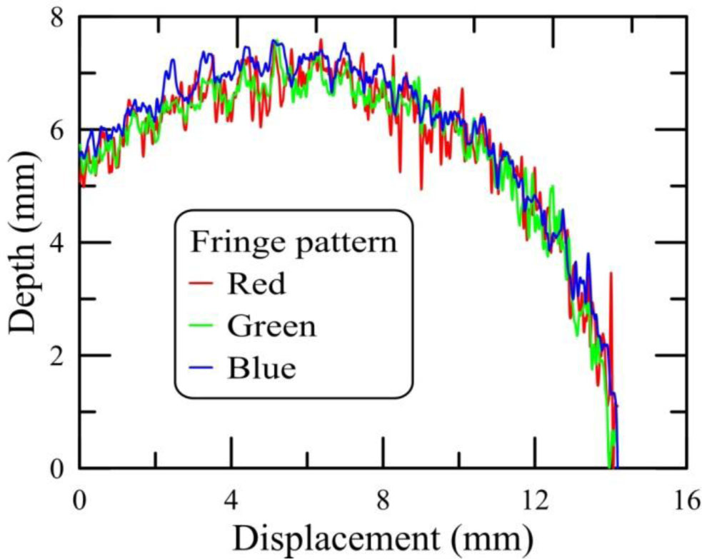
Fig. 7.
Line scan profiles of the 3D fingerprint reconstructed by red, green, and blue deformed fringe patterns. The dashed lines in Fig. 6 indicate the line-scan position.
To contextualize the performance of the proposed approach, Table 1 provides a comparative overview of representative 3D fingerprint and biometric imaging techniques reported in the literature. Both quantitative and qualitative evaluations indicate that the proposed system not only delivers superior geometric reconstruction under green illumination but also outperforms existing methods across key metrics, including spatial resolution, acquisition speed, and the ability to resolve higher-order fingerprint features. The integration of full-field fringe projection, a multi-step phase-shifting algorithm, and optimized spectral selection facilitates accurate and reliable reconstruction of both macro- and micro-scale fingerprint structures. This level of precision underscores the system’s potential for deployment in high-security biometric scenarios, including anti-spoofing detection, forensic identification, and robust contactless authentication.
Table 1.
Comparison of this work with related studies.
| Study | Technique | Resolution [µm] | Level 3 feature capture | Spectral analysis | Acquisition time [s] |
|---|---|---|---|---|---|
| This work | 7-step FPP, RGB illumination | < 40 lateral, < 5 vertical | Yes – pores, ridge edges | Yes – RGB comparison | <1 |
| [7] | Single-shot FPP + biospeckle | ∼100 | Partial | No | ∼1 |
| [23] | Phase measuring profilometry | ∼50 | No | No | ∼0.5 |
| [32] | Full-field sinusoidal FPP | ∼70 | No | No | ∼1.5 |
| [33] | Composite color fringe projection | ∼60 | No | Yes – synthetic color | ∼1 |
| [48] | Level 3 matching (ridge, pores) | N/A | Yes – 2D | No | N/A |
The experimental results obtained from the proposed system demonstrate significant advancements over existing 3D fingerprint imaging techniques, particularly in terms of spatial resolution, feature fidelity, and adaptability to microstructured surfaces. In contrast to prior approaches that use either monochromatic fringe projection [23], [32] or composite color encoding [33], the presented method integrates full-field multi-step phase-shifting with spectral wavelength tuning. This combination enables high-precision reconstruction of both macro-scale ridge patterns and third-level features, such as sweat pores and ridge contours. Achieving lateral and vertical resolutions of <40 μm and <5 μm, respectively, the system surpasses the performance of most previously reported non-contact fingerprint imaging platforms. Notably, the proposed spectral comparison framework represents a key innovation, allowing systematic evaluation of the influence of illumination wavelength on reconstruction quality. Both quantitative metrics and qualitative assessments confirm that green illumination offers superior fringe modulation contrast, phase stability, and depth reconstruction accuracy. These findings are consistent with the known optical interaction characteristics of human skin, which exhibits an optimal balance of reflectance and scattering in the green spectral band [7]. In comparison, red illumination suffers from excessive subsurface penetration and reduced surface contrast, while blue illumination, though capable of capturing fine surface details, is more susceptible to noise and artifacts in low-texture regions. These insights provide a robust basis for the development of spectrally optimized 3D biometric imaging systems, with potential applications in high-accuracy fingerprint recognition, spoof detection, and contactless identity verification.
In practical applications, the proposed system is particularly well-suited for high-security biometric scenarios where contact-based acquisition is either impractical or undesirable due to concerns related to hygiene, spoofing vulnerability, or fingerprint surface deformation. Potential deployment environments include airport border control, access control for secure facilities, and mobile forensic investigations. Moreover, the system’s capacity to capture third-level fingerprint features in three dimensions enables advanced forensic analysis techniques, such as pore-level matching and authentication independent of contact pressure or finger orientation [48]. Despite its demonstrated advantages, the system exhibits certain limitations. First, the current acquisition protocol requires multiple phase-shifted exposures, which, although executable within one second, may still be susceptible to motion artifacts in completely unconstrained or mobile settings. Second, while the system yields high-fidelity depth maps, the reconstruction pipeline presently depends on external calibration and offline computation. Achieving real-time performance would necessitate further optimization in computational efficiency and hardware synchronization. Lastly, although this study evaluated three discrete wavelengths, future research may explore adaptive spectral selection strategies tailored to individual skin characteristics or varying ambient lighting conditions to further enhance robustness and accuracy.
4. Conclusion
This study presents a rapid, non-contact 3D fingerprint imaging system that combines a seven-step phase-shifting fringe projection approach with a microscope-integrated optical configuration and structured RGB illumination. The system achieves lateral resolution better than 40 μm and vertical resolution below 5 μm, enabling accurate reconstruction of both macro-level ridge-valley topography and third-level fingerprint features, such as sweat pores and ridge-edge contours. A comprehensive spectral analysis demonstrated that green illumination provides the most reliable performance, yielding a wrapped phase standard deviation of 0.07 rad and a SNR exceeding 25 dB. Unwrapped phase maps under green illumination exhibited an RMS error of 1.8 pixels, outperforming the blue and red channels, which showed RMS errors of 3.2 and 5.1 pixels, respectively. Cross-sectional depth profiles confirmed the system’s ability to capture ridge heights ranging from 45–65 μm and to resolve pore structures with diameters of 80–150 μm and depth variations exceeding 10 μm. Compared with existing systems, the proposed method demonstrates superior preservation of fine-scale biometric features and enhanced phase fidelity. Beyond achieving higher spatial resolution and improved detail retention, this work introduces a systematic investigation into the influence of illumination wavelength on 3D reconstruction accuracy, an aspect that has been largely overlooked in previous studies. With an acquisition time of less than one second and reliable recovery of third-level fingerprint features, the system shows strong potential for deployment in high-security access control, forensic identification, and advanced anti-spoofing applications. Future research will focus on integrating real-time processing capabilities, reducing system size for increased portability, and improving motion robustness to facilitate broader adoption in dynamic, unconstrained operational environments.
Acknowledgment
The authors would like to thank the National Science and Technology Council of Taiwan (111-2221-E-A49-102-MY2, 113-2223-E-007-005, 114-2221-E-A49-064-MY3) and Veterans General Hospitals and University System of Taiwan Joint Research Program (VGHUST113-G7-5-1, VGHUST114-G7-4-1, VGHUST115-G7-4-1) to sponsor this study.