
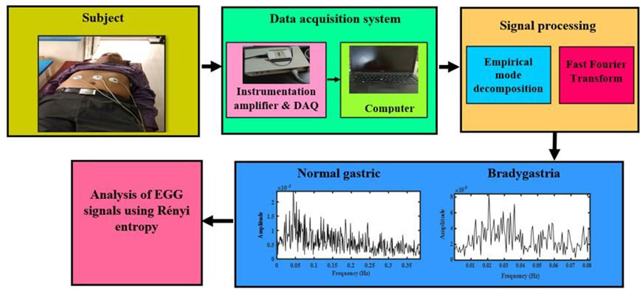
Fig. 1.
The process of recording the EGG signals from normal and bradygastric participants.
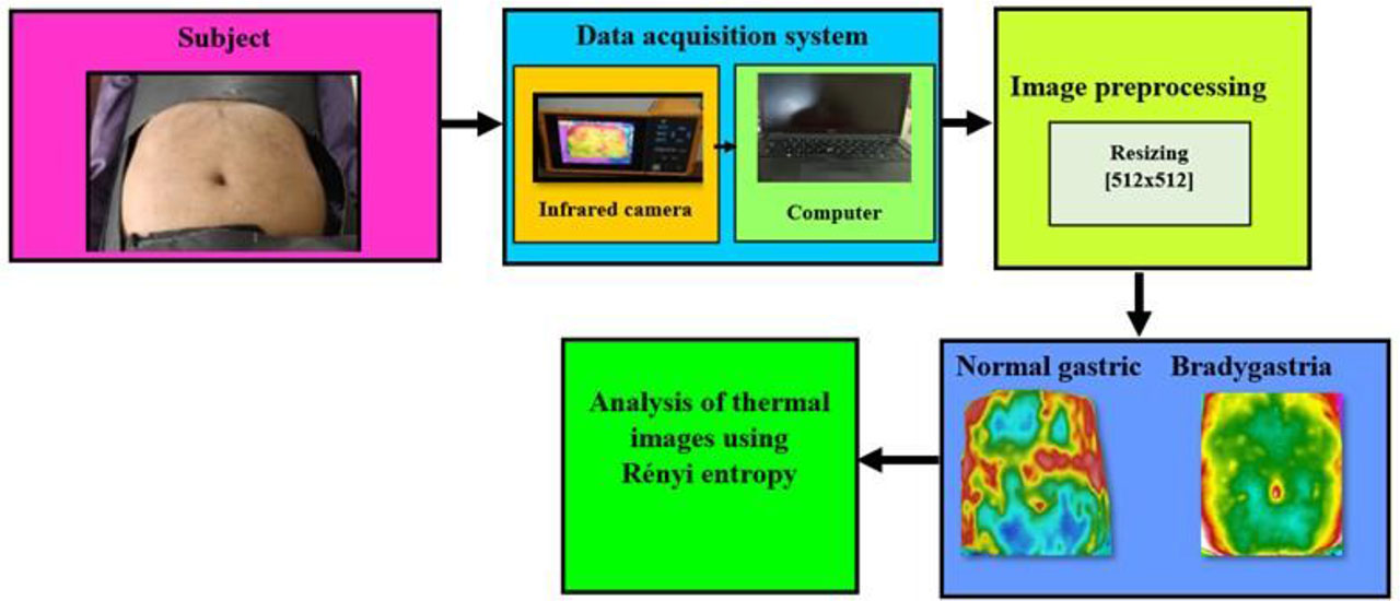
Fig. 2.
The process of IRT image acquisition.
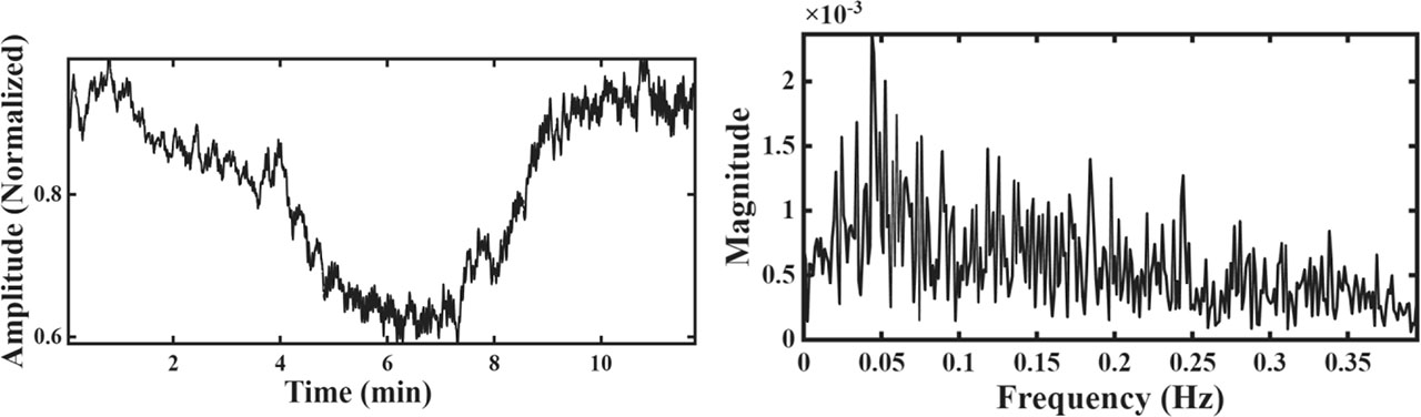
Fig. 3.
(a) Typical normalized electrogastrogram of a normal participant and (b) its FFT.
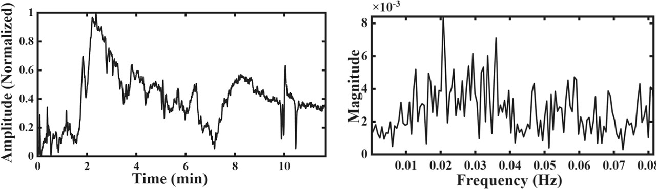
Fig. 4.
(a) Typical normalized electrogastrogram of a bradygastric participant and (b) its FFT.
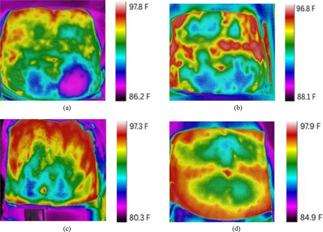
Fig. 5.
Typical abdominal thermal images of normal participants.
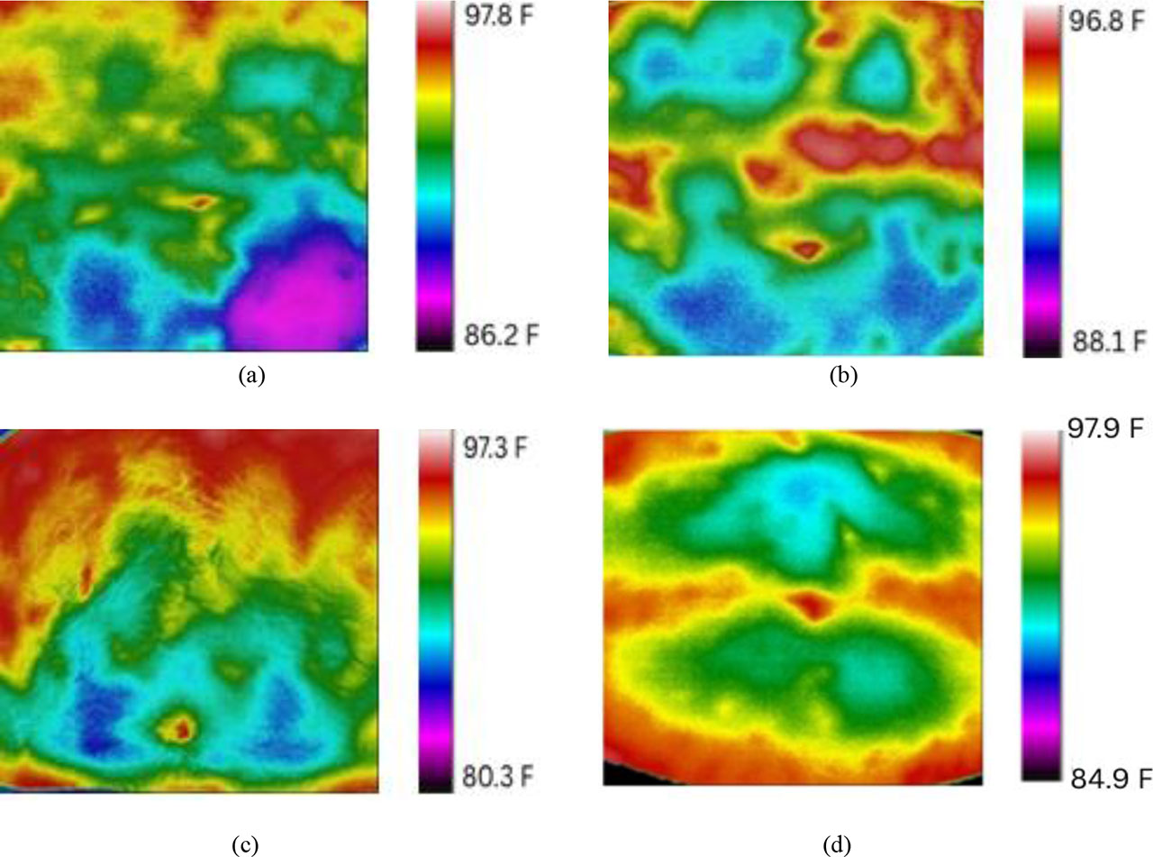
Fig. 6.
Pre-processed thermal images of normal participants.
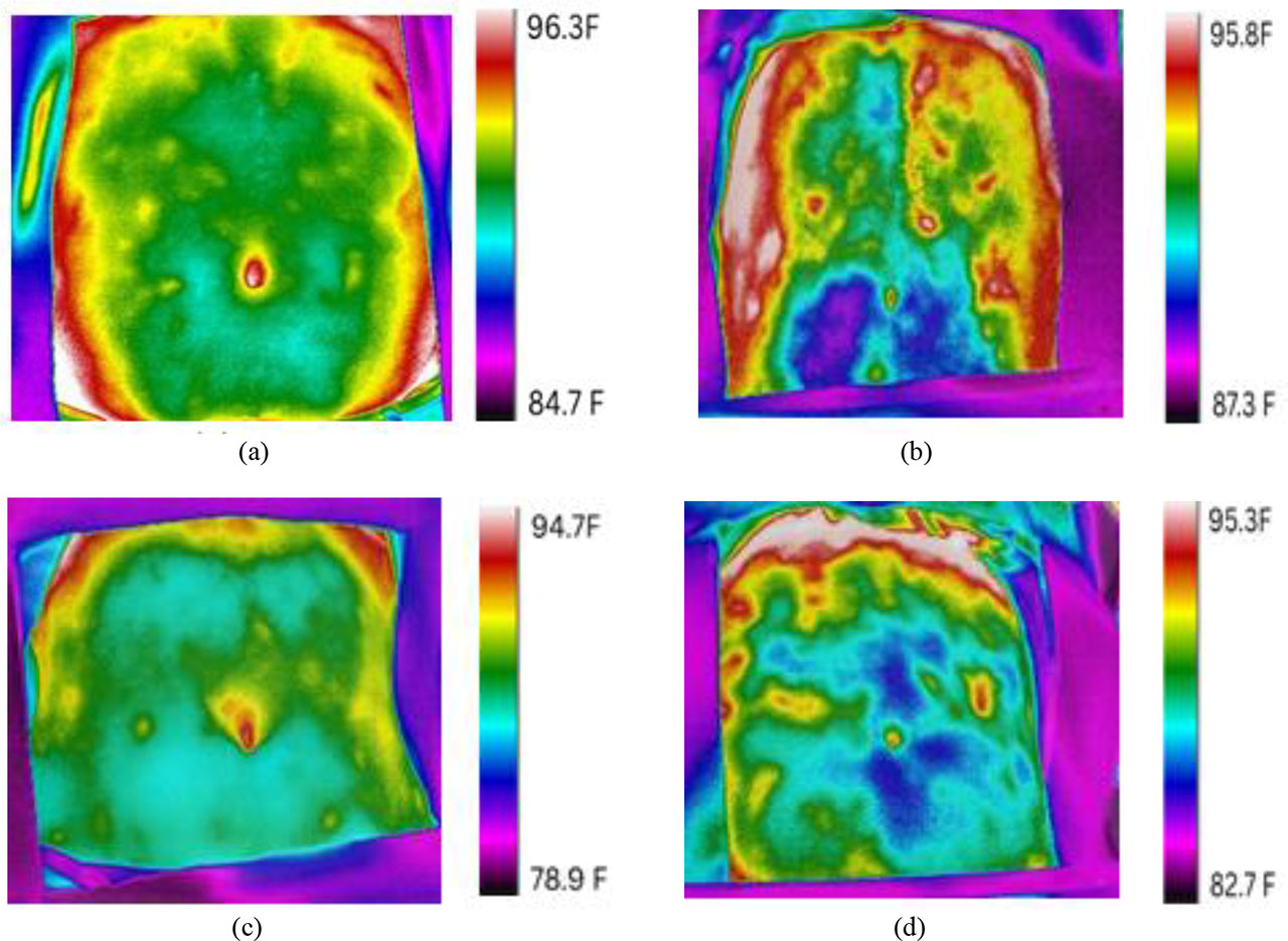
Fig. 7.
Typical abdominal thermal images of bradygastric participants.
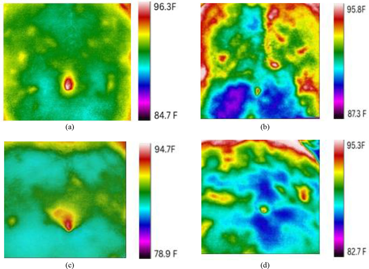
Fig. 8.
Pre-processed thermal images of bradygastric participants.
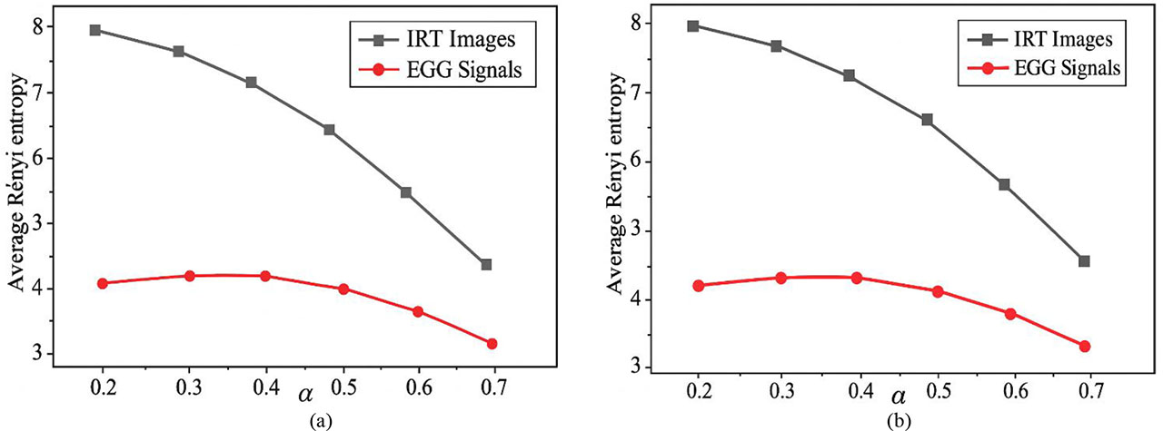
Fig. 9.
Variation of an average Rényi entropy of IRT images and an average Rényi entropy of electrogastrograms shown as a function of α for (a) normal and (b) bradygastric participants.
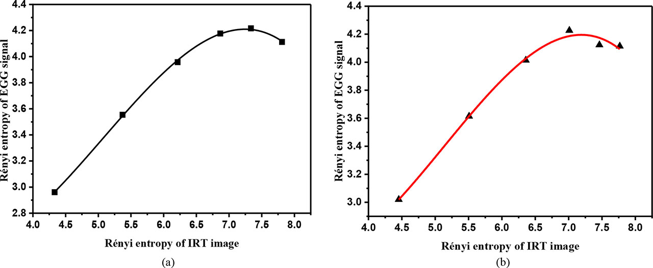
Fig. 10.
Variation of the Rényi entropy of IRT images as a function of the Rényi entropy of electrogastrograms for typical (a) normal participants and (b) bradygastric participants.
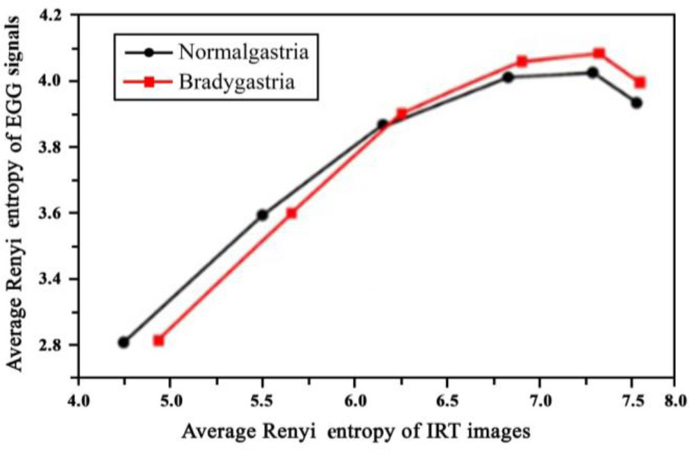
Fig. 11.
Average variation of the Rényi entropy of IRT images as a function of the average Rényi entropy of electrogastrograms for normal subjects and bradygastric subjects.