
1. Introduction
Copper ferrite nanoparticles (CuFe2O4 NPs) have received significant attention due to their unique properties. As a member of the spinel ferrite family, CuFe2O4 typically shows an inverse spinel structure, where Cu+2 ions primarily occupy octahedral sites, while Fe+3 ions are distributed between tetrahedral and octahedral sites. This unique structural configuration gives it distinctive physicochemical and biological properties, such as chemical and thermal stability, making it suitable for harsh environments [1,2], excellent bandgap energy of 2.22 eV, enabling efficient catalysis for the removal of organic contaminants [3], as well as strong magnetic behavior, electronic conductivity, and biocompatibility [4]. Therefore, CuFe2O4 NPs could be a promising candidate for applications in catalysis, environmental remediation, and biomedical fields.
CuFe2O4 NPs have been synthesized using various chemical and physical methods, such as co-precipitation [1], high-energy ball milling method [2], sol–gel [3], microwave-assisted combustion method [4], hydrothermal [5], and solvothermal methods [6]. Although these conventional approaches are effective in controlling particle size and crystallinity degree, they often involve hazardous chemicals, high energy consumption, and complex processing conditions. In this context, these restrictions have led to increased interest in the green approach, which has emerged as a viable, eco-friendly, and sustainable alternative, consistent with the principles of green chemistry, aiming to reduce toxic reagents, lower energy consumption, and minimize negative environmental impact [7]. This approach relies primarily on natural biological resources in the synthesis of NPs, where biomolecules are extracted from plants, bacteria, or fungi, enabling the production of bio-NPs from aqueous solutions containing suitable salts [8–12]. Among these natural biological resources, the biosynthesis of NPs by plants has received considerable attention due to its simplicity, cost-effectiveness, and scalability. This method relies on the diverse phytochemical constituents found in different parts of the plant, including leaves, stems, fruits, flowers, and roots, to reduce and stabilize metal ions [13,14].
Several recent studies have reported the successful green synthesis of CuFe2O4 NPs using different plant extracts. For instance, Aloe vera [3], Monsonia burkeana [12], Nasturtium officinale [13], and Morus alba leaf [14] extracts have been employed to produce these NPs with enhanced functional properties. In addition, alternative biological materials, such as egg whites, have also been used as eco-friendly fuels in the green synthesis of CuFe2O4 NPs [15]. Despite these advances, the choice of plant sources remains a critical factor affecting the morphology, size distribution, and physicochemical properties of NPs due to differences in phytochemical composition. Capsicum annuum (sweet green pepper) is a nutrient-rich plant, known for its high content of phenolic acids, flavonoids, alkaloids, vitamins, and other antioxidant compounds [16,17]. These components showed great effectiveness as natural reducing, stabilizing, and capping agents in the eco-friendly production of NPs [18–20]. Through electron transfer and complexation mechanisms, the biomolecules present in its aqueous extract contribute to the reduction of metal precursors, such as Fe+3 and Cu+2, to their corresponding ferritic forms. These biomolecules simultaneously cap the growing NPs to enhance their colloidal stability and avoid agglomeration, highlighting their strong reducing capability and biological activity [21,22].
Despite the extensive use of plant extracts in the green synthesis of ferrite NPs, limited studies have explored the use of Capsicum annuum extract in the synthesis of CuFe2O4 NPs. Moreover, the influence of its rich phytochemical composition on the structural, optical, magnetic, and biomedical properties of CuFe2O4 NPs has not been extensively studied. This gap highlights the need for further systematic studies on the role of Capsicum annuum extract in modifying the properties of these NPs. Based on the literature, this study is one of the first attempts to use Capsicum annuum extract as a reducing and stabilizing agent for the green synthesis of CuFe2O4 NPs, which has been reported on a limited scale. Therefore, the aim of the present study is to synthesize CuFe2O4 NPs using Capsicum annuum extract via a green synthesis approach, along with a systematic study of the effect of these NPs on physicochemical and biomedical properties.
2. Experimental method
2.1. Capsicum annuum extract
In this study, 200 g of sweet green peppers (GP) were collected from local markets in Baghdad, chopped into small slices, and mashed for 10 min to obtain a juice-like consistency. The mixture was then filtered using Whatman filter paper (Grade 1) with a particle retention of 11 µm to obtain the extract. Subsequently, 50 mL of distilled water was added to the resulting extract, and the mixture was heated to 60°C for 30 min with continuous stirring. After cooling, the resulting extract was stored at 4°C for later use.
2.2. Green synthesis of CuFe2O₄ NPs
In separate beakers, 50 mL of distilled water was used to dissolve the raw materials CuSO₄ and Fe(NO₃)₃·9H₂O, supplied by British Drug Houses Chemicals Ltd with a purity of approximately 98%, for 15 min. To ensure thorough mixing, the solutions were collected and mixed for 30 min using a magnetic stirrer. Subsequently, 50 mL of GP-extract was added to the mixture, resulting in a pH of 6. The mixtures were then heated on a hot plate at 60°C with continuous stirring for 90 min, until the solvent had completely evaporated, leaving a dry solid powder. To remove any remaining ions and organic compounds associated with the powder resulting from the mixtures, it was centrifuged and then washed several times with distilled water. Finally, the collected solid powder was sintered at 800°C for 2 h, with a heating rate of 13°C/min and a soaking duration of 1 h, using a furnace from Nabertherm GmbH (Germany).
2.3. Characterization
The atomic structure of CuFe2O4 NPs was analyzed by X-ray diffraction (XRD) using a Panalytical X’Pert PRO (UK), with Cu-Kα radiation at a wavelength of λ = 1.5406 Å. Fourier transform infrared spectroscopy (FTIR) was carried out in the range of 400–4,000 cm‒1 using a PerkinElmer 100 Optica spectrometer equipped with an attenuated total reflection (ATR) accessory to identify the characteristic functional groups and bioactive organic compounds in the plant extract and to confirm their role in the formation and stabilization of the synthesized CuFe2O4 nanoparticles. The surface morphology was studied by field emission scanning electron microscopy (FESEM) using a ZEISS Sigma VP (Germany). Optical analysis (UV-Vis) was performed in the range of 200–1,000 nm using a Shimadzu UV-1900i (Japan). To prepare the samples for the test, 5 mg CuFe2O4 NPs was dispersed in 10 mL of distilled water. The mixture was subjected to continuous bath sonication for 15 min to ensure a highly homogeneous suspension and reduce particle agglomeration. The pure solvent served as the blank baseline reference, and the stable suspension was immediately transferred to a quartz cuvette with a 1 cm optical path length. Finally, the magnetic properties were evaluated using a vibrating sample magnetometer (VSM) from Meghnatis Daghigh Kavir Company (model LBKFB, Iran), within a magnetic field ranging from –15.000 to +15.000 Oe.
2.4. Antibacterial activity
2.4.1. Preparation of Mueller-Hinton (MH) agar
MH medium was prepared by adding 38 g powder to 1 L of distilled water and heating it with constant shaking on a burner until completely dissolved. The medium was then autoclaved at 121°C for 15 min. The medium was allowed to cool to 50°C, then poured into the Petri dishes and left for about 15 min until solidified. After solidification, the dishes were inverted and refrigerated at 4°C.
2.4.2. Antibacterial activity
The efficacy of prepared CuFe2O4 NPs as an antibacterial agent against gram-negative Escherichia coli (E. coli) and gram-positive Staphylococcus aureus (S. aureus) strains was studied using a well diffusion assay in agar medium. Approximately 20 mL of MH medium was placed in sterile Petri dishes. The bacterial species were collected from their stock cultures using a sterile wire loop [20] and then inoculated onto the agar surface. Subsequently, wells with a diameter of 6 mm and a depth of 4 mm were formed in the dishes using a sterile tip, and these wells were filled with different proportions of CuFe2O4 NPs. The cultured dishes containing bacterial strains and CuFe2O4 NPs were incubated overnight at 37°C, and the average diameter of the inhibition zone was measured [21,22].
2.5. Biofilm activity
The bacterial strains were cultured in 96-well dishes at a concentration of 1 × 10⁶ cells/mL and treated with CuFe2O4 NPs for 24 h. The samples were then removed, and the wells were gently rinsed with phosphate-buffered saline (PBS) to remove any non-adherent cells. The adherent cells were fixed and stained with crystal violet stain at a concentration of 0.1%. The wells were then rinsed twice with distilled water to remove excess stain. To estimate the amount of biofilm formed, 0.2 mL of 95% ethanol was added to each well to dissolve the stain adhering to the cells, and the dishes were incubated with shaking for 2 h. Finally, the light absorption was measured at 595 nm [23].
2.6. Cytotoxicity assays
To determine the cytotoxic effects of CuFe2O4 NPs, the 3-(4,5-dimethylthiazol-2-yl)-2,5-diphenyltetrazolium bromide assay (MTT assay) was performed using 96-well dishes [24,25]. The cells were seeded at a density of 1 × 10⁴ cells/well and incubated for 24 h to allow for adhesion and the formation of a continuous monolayer. Subsequently, the cells were treated with different concentrations of CuFe2O4 NPs for 24 h. To assess cell viability, the medium was removed, and 100 μL of 3 mg/mL MTT solution was added. The cells were then incubated at 37°C for 3 h. After removing the MTT solution, the resulting formazan crystals were dissolved by adding 100 μL of dimethyl sulfoxide (DMSO). The dishes were then incubated at 37°C for 15 min with shaking to ensure complete dissolution [26]. Absorption was measured using a microplate reader at a wavelength of 492 nm. The test was performed three times (n = 3), and the results were expressed as mean value ± standard deviation (SD). Finally, the cytotoxicity ratio was calculated using the following equation:
where A is the optical density of the control and B is the optical density of the samples [27].2.6.1. Maintenance of cell cultures
MCF-10 cells were cultured in RPMI-1640 medium supplemented with 10% fetal bovine serum, 100 units/mL of penicillin and 100 μg/mL of streptomycin. The cells were incubated at 37°C in an incubator containing 5% CO₂. When the cells reached a confluence of approximately 80%, they were separated using trypsin-EDTA solution, and then re-cultured twice a week to maintain their growth [27].
2.6.2. Statistical analysis
The obtained data were statistically analyzed by an unpaired t-test using GraphPad Prism 6 software. The results were presented as a mean value ± SD of three replicates (n = 3).
where A and B are the optical density of the control and the test, respectively.2.7. Hemolysis test
Hemolysis test was performed on prepared CuFe2O4 NPs based on the previously described method [28]. To prepare red blood cells (RBCs), blood samples were collected, and the plasma was separated from the RBCs using a centrifuge. The RBCs were then washed with PBS at a pH of 7.4 to remove any impurities. Subsequently, a 2% RBC solution was then prepared by adding 0.2 mL of RBCs and gently stirring to ensure homogeneity. After preparing the required dilutions, 0.2 mL of the sample to be tested was taken, and 0.8 mL of RBCs were added. The tubes were incubated at 37°C for a specified time, usually 90 min. After incubation, the RBCs were separated from the solution by centrifugation, and the supernatant from each tube was transferred to a microplate. The absorbance was then measured using an ELISA device.
3. Results and discussion
3.1. Topological and morphological analysis
Figure 1 shows the FESEM images of CuFe2O4 NPs at different magnifications, which were synthesized using GP-extract assistance by the co-precipitation technique. At low magnification (Figure 1a), distinct agglomerations of irregular and semi-spherical NPs can be observed. This agglomerated behavior is expected to occur in magnetic ferrites such as CuFe2O4 NPs, especially those produced biosynthetically, due to the mutual magnetic forces between the NPs, leading to their attraction and aggregation. The bioactive compounds found in GP-extract, like phenols and carotenoids, stimulate multinuclear growth, which at this stage leads to the emergence of many small, closely packed, and heterogeneous NPs. At higher magnification (Figure 1b), the morphology of NPs becomes more pronounced, signifying that they do not completely separate but tend to aggregate due to the presence of organic residues and surface bonds from the bio-extract. The image also shows that the NPs take on semi-spherical to ellipsoidal shapes with a clear degree of size heterogeneity, a common behavior in plant-based materials resulting from the influence of bio-capping agents on the particle growth rate. In Figure 1c, direct measurements of NPs’ diameters show a range of 110–170 nm, reflecting the dominance of bio-capping agents in limiting particle growth, as well as the tendency of the CuFe2O4 spinel phase itself to form low-surface-energy spherical structures. This coherent nanoscale size confirms the success of the green synthesis process using GP-extract.
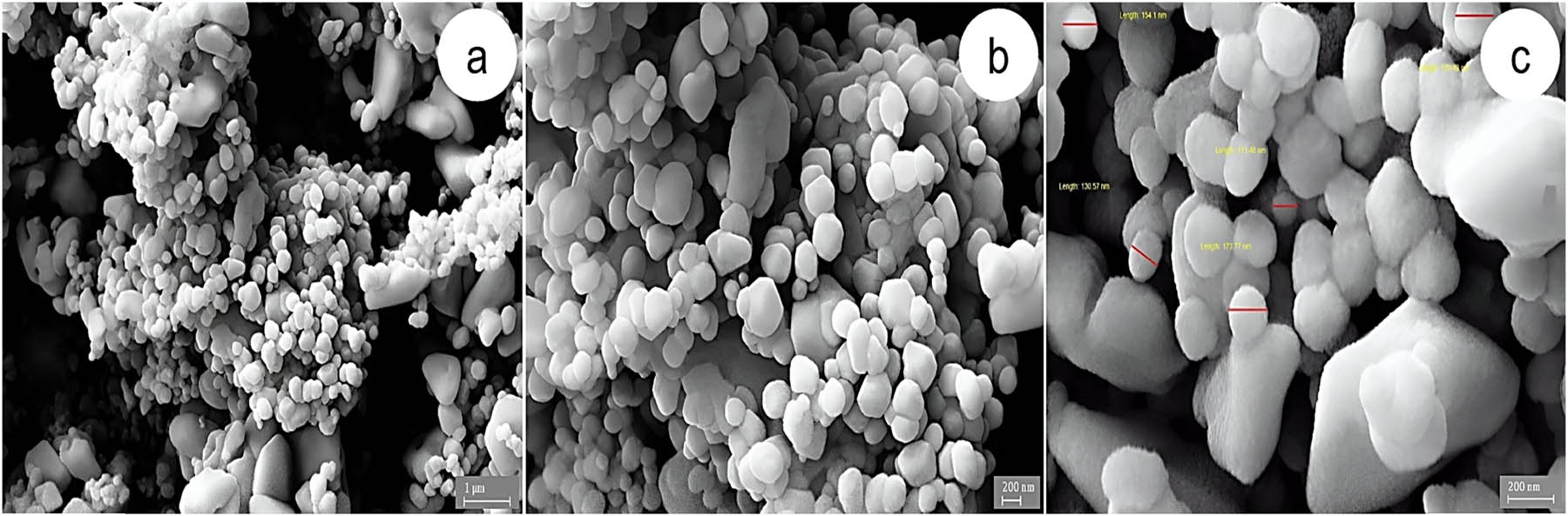
Figure 1
FESEM images of CuFe2O4 NPs at different magnifications: (a) low magnification showing agglomerated nanoparticles 10k×, (b) higher magnification showing semi-spherical/ellipsoidal morphology 20k×, and (c) particle size measurement image showing nanoparticles in the range of 110–170 nm, 50k×.
3.2. Structural and physicochemical properties
The crystallinity of spinel ferrite CuFe2O4 was studied by XRD. Figure 2a shows the resulting diffraction patterns. The results were compared with standard data from the Joint Committee on Powder Diffraction Standards (JCPDS) PDF card No. 01-077-0010. Additionally, the crystallite size (D) was determined using the Scherrer–Debye equation:
where D is the crystal size (nm); k = 0.9, which is the crystal form factor; λ = 0.15406 nm, which represents X-ray’s wavelength; θ is the diffraction angle (°); and β is the full-width at half-maximum (FWHM) value (rad). Moreover, Bragg’s equation was used to calculate the lattice constant (a) from the peak plane (311):The microstrain (ε) developed through the synthesis steps was also estimated using the following relationship [29]:where ε is the microstrain and β is the FWHM.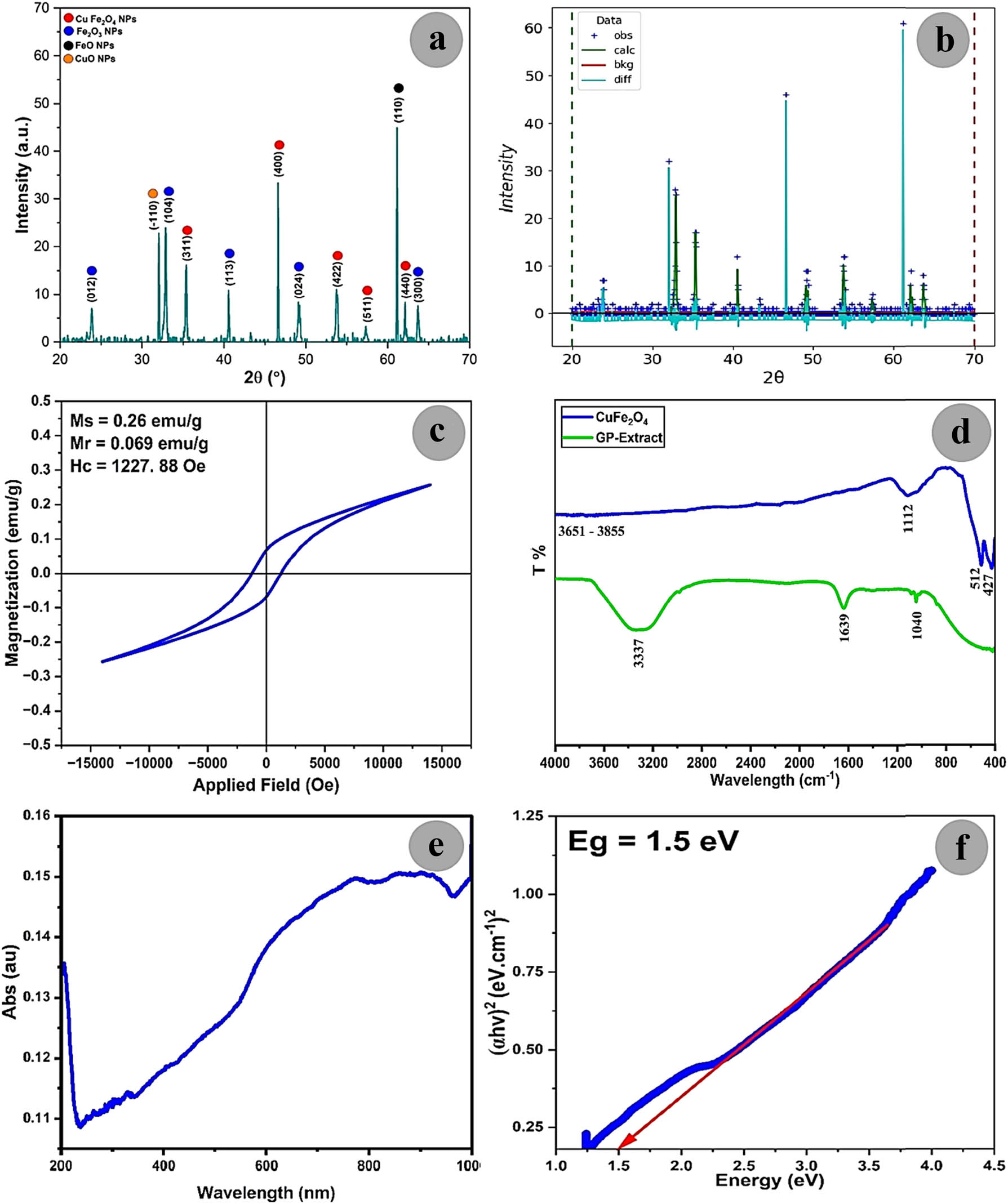
Figure 2
Structural, magnetic, and optical properties of Capsicum annuum extract-assisted synthesized CuFe2O4 nanoparticles sintered at 800℃ for 2 h: (a) XRD pattern, (b) Rietveld refinement of XRD data, (c) magnetic hysteresis loop derived from VSM analysis, (d) FTIR-ATR spectroscopy, (e) UV-Vis absorption spectrum of NPs dispersed in distilled water, and (f) Tauc plot for bandgap energy determination.
Figure 2a shows the XRD pattern of GP-extract assisted synthesized CuFe2O4 NPs, sintered at 800°C for 2 h, using the co-precipitation method. The sharp peaks in the diffraction pattern indicate the high degree of crystallinity for the synthesized CuFe2O4 NPs. CuFe2O4 NPs showed a cubic spinel structure (Fd-3m space group) with a unit cell volume of a = b = c = 8.39 Å. The observed crystalline planes with Miller indices (hkl) (311), (400), (422), (511), and (440) are located at 2θ values of 35.34°, 47.58°, 53.79°, 57.35°, and 62.17°, respectively, confirming the production of spinel-type CuFe2O4 NPs. The observed additional peaks are located at angles 32.05°, 32.87°, 40.47°, 49.17°, 62.58°, and 63.71°, corresponding to crystalline planes (‒110), (104), (113), (024), (110), and (300), as a result of the formation of rhombohedral Fe₂O₃ NPs, cubic FeO NPs, and monoclinic CuO NPs according to JCPDS No. 00-003-0800, JCPDS No. 00-006-0711, and JCPDS No. 45-0937, respectively [30]. Numerous studies have documented that the green synthesis of ferrite NPs often yields secondary phases, such as Fe₂O₃ and FeO [31,32]. This is mainly due to the sensitive nature of phytochemicals to redox reactions during crystal formation, and their role as mild reducing/oxidizing agents. The estimated average crystalline size (D = 26.45 nm) in this study was higher than the crystalline size (D = 18.33 nm) of Ag-doped CuFe2O4 NPs, synthesized with Monsonia burkeana extract assistance, as reported by Makofane et al. [12]. In another study by Meenakshi et al. [11], the crystalline size (D = 19.30 nm) of Aloe Vera-assisted synthesized CuFe2O4 NPs was almost similar to that reported by Makofane et al. [12]. In contrast, the calculated values were lower than values reported using conventional synthesis methods, such as sol-gel (D = 58.22 nm) [33] and Ce+3-doped CuFe2O4 NPs via co-precipitation (D = 47.38 nm) [1]. These results indicate that the value of “D” is significantly affected by the synthesis method and extract type. Moreover, the crystal lattice constant was found to be 8.39 Å, which is higher than the standard value (8.37 Å) and the values reported in other studies, 7.17 Å [12] and 8.23–8.31 Å [34]. The microstrain (ε) was estimated to be 2 × 10‒3, where the sample contains a microstrain value within the normal range for ferrite NPs, i.e., around 10‒3.
Figure 2b shows the Rietveld refinement analysis of the XRD pattern of the CuFe2O4 sample sintered at 800°C for 2 h using GSAS-II software. The structural analysis of the CuFe2O4 sample, derived from Rietveld refinement, confirms the formation of a spinel ferrite phase with a cubic crystal structure (space group Fd-3m). The refined lattice parameter (a = 8.39 Å) is in good agreement with reported values for copper ferrite, indicating the successful incorporation of Cu+2 ions into the spinel lattice. The low refinement indicators (χ 2 < 2, R wp < 10%, and R exp < 8%) suggest a satisfactory fit between the observed and calculated diffraction patterns, confirming the reliability of the structural model. The cation distribution reveals that Fe+3 ions occupy the tetrahedral (A) sites, while Cu+2 and Fe+3 ions are distributed over the octahedral (B) sites, corresponding to an inverse spinel configuration with an inversion parameter of one (ϕ = 1), as shown in Table 1. This distribution is mainly subject to the strong Jahn–Teller distortion associated with Cu+2 ions, which energetically prefers their occupation of octahedral sites. The obtained cell volume (590.59 Å3) is also consistent with the refined crystal lattice parameter, indicating structural stability and the absence of significant lattice distortion. The inverse spinel structure plays a pivotal role in determining the physical properties of CuFe2O4 NPs, particularly its magnetic behavior, where superexchange interactions between A and B sites (Fe+3–O‒2–Fe+3 and Fe+3–O‒2–Cu+2) control the overall magnetic arrangement. These results confirm that the synthesized CuFe2O4 NPs show a pronounced spinelic structure with a cationic distribution consistent with reported literature.
Table 1
Cations distribution derived from Rietveld refinement of CuFe2O4 sintered at 800°C for 2 h.
| Spinel ferrite | a (Å) | χ 2 | R wp (%) | R exp (%) | Cell volume (V), (Å3) | A-site (tetrahedral) | B-site (octahedral) | Final chemical formula | Inversion parameter (ϕ) |
|---|---|---|---|---|---|---|---|---|---|
| CuFe2O4 | 8.39 | 1.8 | 9.3 | 6.88 | 590.59 | () | [] | ()A []B | 1 |
Table 2 shows the phasic composition and crystallographic parameters of the synthesized CuFe2O4 sample sintered at 800°C for 2 h, obtained from Rietveld refinement of XRD data. The results confirm that CuFe2O4 is the dominant phase, estimated at ≈75%, indicating the successful formation of the spinel ferrite structure. The refined lattice parameter (a = 8.39 Å) is consistent with previously reported values, suggesting good crystallinity and proper incorporation of Cu+2 ions into the spinel lattice. In addition to the primary phase, secondary phases such as Fe₂O₃ (≈12%), FeO (≈8%), and CuO (≈5%) were identified. The secondary Fe₂O₃ phase is described in the rhombohedral crystal system using the hexagonal representation (a = b ≠ c; γ = 120°), which is commonly adopted in XRD analysis. The presence of this secondary phase indicates the partial oxidation of iron species during the synthesis process. In contrast, the presence of the secondary FeO phase implies localized reduction conditions or incomplete oxidation, while the presence of the secondary CuO phase may indicate an incomplete reaction or copper oxide residues that did not fully participate in spinel formation. The coexistence of these secondary phases is frequently reported in spinel ferrites synthesized via chemical or low-temperature routes, where kinetic constraints and non-equilibrium conditions can completely inhibit phase formation. Despite the presence of these secondary phases, the high weight fraction of the primary CuFe2O4 phase confirms the predominance of the desired spinel phase. From a structural perspective, the variation in crystal systems among the detected phases (cubic for CuFe2O4 and FeO, rhombohedral for Fe₂O₃ in a hexagonal setting, and monoclinic for CuO) reflects the complexity of phase evolution during the synthesis process. These secondary phases may affect the overall physical properties, particularly magnetic and electrical behavior, due to their differences in crystal symmetry and electronic structure.
Table 2
Phasic composition and crystallographic parameters of the synthesized CuFe2O4 sample.
| Spinel ferrite | Phasic chemical composition | Estimated weight fraction (%) | Crystal system | Space group | Lattice parameters |
|---|---|---|---|---|---|
| CuFe2O4 | CuFe2O4 | ≈75 | Cubic | Fd-3m | a = b = c = 8.39 Å; α = β = γ = 90° |
| Fe2O3 | ≈12 | Rhombohedral (hexagonal setting) | R-3c | a = b = 5.03 Å, c = 13.75 Å; α = β = 90°, γ = 120° | |
| FeO | ≈8 | Cubic | Fm-3m | a = b = c = 4.33 Å; α = β = γ = 90° | |
| CuO | ≈5 | Monoclinic | C2/c | a = 4.68 Å, b = 3.42 Å, c = 5.13 Å; α = γ = 90°, β ≈ 99.5° |
Figure 2c shows magnetic hysteresis loops at room temperature, derived from VSM analysis of CuFe2O4 NPs, which show that these NPs have a hard ferromagnetic behavior with a low saturation magnetization (M s) of 0.26 emu/g, residual magnetization (M r) of 0.06959 emu/g, and elevated coercivity (Hc) of about 1227.88 Oe. The synthesized NPs are characterized by a significantly low Ms value, which can be explained based on cation redistribution and the presence of secondary phases such as Fe₂O₃, FeO, and CuO NPs. Moreover, according to the magnetic domain theory, an elevated Hc value corresponds to single-domain NPs. In such ferrite systems, the magnetostatic energy is reduced by the formation of multiple domains. Within small-sized NPs (D = 26.25 as confirmed by XRD results), a single domain has no domain walls, which requires a high magnetic energy to spin the magnetic moment throughout the material, resulting in a high Hc value. In addition, components of GP-extract, such as phenolic acids, flavonoids, alkaloids, and vitamins, act as reducing, capping, and stabilizing agents. These components primarily worked to reduce Cu+2 and Fe+3 to metal oxides, controlling the growth of NPs to form small and uniform NPs, forming a thin organic layer on the surface of NPs. This creates a core–shell model, where the atoms of the NPs’ core are well-ordered. In contrast, the atoms on the surface are coated with a thin organic layer to spin with disorder. The observed core-shell exchange coupling in these materials results in a high Hc, indicating hard magnetic properties. These high Hc values make CuFe2O4 NPs particularly suitable for various medical applications. Figure 2d shows the FTIR-ATR spectra of GP-extract and CuFe2O4 NPs at room temperature within the wavelength range of 400–4000 cm‒1. Three main peaks were observed for the GP-extract. The first peak, at 3,337 cm‒1, corresponds to the hydroxyl (OH) group on the NPs’ surface. The peak at 1,639 cm‒1 is due to the stretching vibration of the carbonyl (C═O) bond, which is associated with the amide-I group found in Capsicum annuum [35]. The third peak, at 1,040 cm‒1, is attributed to the antisymmetric stretching vibration of the C–O and C–O–C bonds, as well as the bending vibration of the C–O–H bond, which may be associated with polysaccharides, chlorophyll, or phenolic compounds (–OH) [35,36]. The third peak shifted toward a higher wavelength in spinel ferrite NPs, where it is located at 1,112 cm‒1 in CuFe2O4 NPs, indicating an interaction between the functional groups and the nanomaterial. The observed sharp peaks at 427 and 512 cm‒1 are consistent with metal–oxygen (M–O) bond vibrations at the tetrahedral (A-sites) and the octahedral (B-sites), confirming the formation of the spinelic structure of CuFe₂O₄ NPs [37,38].
The UV absorption spectrum data of CuFe₂O₄ NPs were used to compute the bandgap energy and absorption spectrum results. The bandgap energy of these NPs was calculated according to the optical absorption spectrum using Tauc’s equation [39]:
where α is the absorption coefficient and hν is the photon energy (n = 2 for direct bandgap and ½ for indirect bandgap).Figure 2e and f shows the UV–Vis absorption spectra of the synthesized CuFe₂O₄ NPs in the range of 200–1,000 nm and their corresponding bandgap energies. A pronounced difference in the absorption behavior was observed, which is attributed to the interaction of electronic band structures, cation/anion distribution, and defect density. CuFe₂O₄ NPs showed an increase in absorption with increasing wavelength, particularly in the visible region (400–700 nm), resulting in the appearance of sub-absorption tails from the bandgap energy. Such red/near-infrared (NIR) absorption has been linked to mixed valence states (Cu+2/Cu+1 and Fe+2/Fe+3), secondary phases such as FeO, Fe₂O₃, and CuO, and cation redistribution. Moreover, the spin-allowed d–d electronic transitions of Cu+2 ions residing in the octahedral (Oh) sites of the spinel lattice structure are mainly responsible for this wide absorption in the NIR range (750–1,000 nm). Due to the oxygen chromophores acting as weak-field ligands, these transitions are shifted into the low-energy NIR band by the comparatively low crystal field splitting energy, while accompanying Jahn–Teller distortions may cause the broadening of the band. Consequently, the bandgap energy (Eg) was estimated to be 1.5 eV, which is within the typical experimental value reported for CuFe₂O₄ NPs, which ranges between 1.3 and 2.2 eV [40,41,42]. This low Eg may be due to cation redistribution and defect density resulting from the GP-extract, which modified the electronic interactions between Fe–O–Fe and Cu–O–Fe, resulting in the generation of a sub-bandgap energy, and thus a decrease in the energy required to excite the electron, resulting in a lower Eg [43].
3.3. Antibacterial activity
Antibacterial testing was conducted using the well-diffusion method. The Muller’s medium was prepared by following the manufacturer’s guidelines, and the preparation was allowed to solidify. Microbial suspensions were prepared and standardized against McFarland. Bacteria were evenly spread across the agar using a sterile cotton swab, and wells were created in the agar using a sterile well cutter. Each well was filled with 100 μL of CuFe₂O₄ NPs. The result showed higher antibacterial activity of CuFe₂O₄ NPs against Gram-positive bacteria than against Gram-negative bacteria. This is due to differences in the components of the cell wall. Gram-positive bacteria have a rigid and thick peptidoglycan layer that is absent in Gram-negative bacteria, while the gram-negative have a 10 nm thick lipopolysaccharide layer covering the peptidoglycan outer layer, thus hindering the NPs’ access to the cell wall [44].
Figure 3 shows the inhibition zone diameter (IZD) after 24 h of incubation for all samples. The zone of inhibition is measured from the edge of the clear area, across the disc, to the other edge of the clear area. Antibacterial activity is shown by all the prepared samples. The IZD results, listed in Table 3, indicate that the bactericidal potency of NPs improved with increasing CuFe₂O₄ NP content, leading to high IZD formation, such as the decomposition of CuFe₂O₄ NPs and the formation of reactive oxygen species (ROS), electrostatic interaction of nanomaterials with cell walls, and photocatalytic light activation of NPs [45]. However, the mechanism by which NPs are able to penetrate into bacteria is not yet well understood. However, it is possible that by exposing the E. coli bacterium to Cu-substituted cobalt ferrite nanoparticles, the cell membrane is targeted. These NPs stick to the bacterial cell wall and penetrate through the cell membrane. The thick and coarse bacterial cell wall is destroyed by Cu ions, resulting in the degradation and disappearance of cytoplasm, which therefore leads to cell death. Furthermore, high concentrations of Cu nanoparticles demonstrate complete cytotoxicity against E. coli [46]. CuFe₂O₄ NPs, which have a smaller crystalline size and larger surface-to-volume ratio, enhance their bactericidal efficiency, resulting in higher E. coli bacteria killing rates and a larger IZD area.
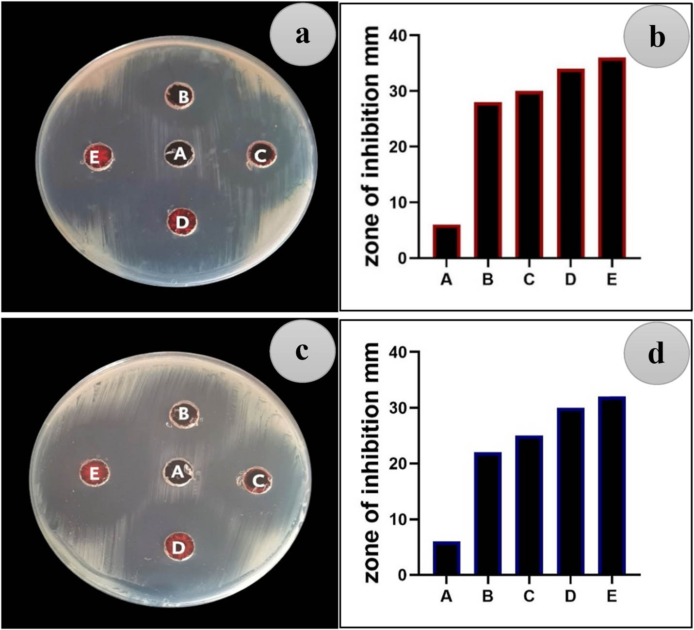
Figure 3
Antibacterial activity of CuFe₂O₄ NPs against (a and b) S. aureus and (c and d) E. coli at different concentrations: (A) control case (deionized water (DIW)), (B) 62.5 µg/mL, (C) 125 µg/mL, (D) 250 µg/mL, and (E) 500 µg/mL.
3.4. Antibiofilm activity
The present study shows that CuFe₂O₄ NPs have considerable antibiofilm activity against both E. coli and S. aureus with a clear concentration-dependent inhibitory effect. The reduction in biofilm biomass confirms the capability of CuFe₂O₄ NPs to break bacterial adhesion and the formation of biofilms. This behavior can be explained by the intrinsic properties of CuFe₂O₄, in particular its redox-active surface and ability to generate ROS, responsible for oxidative stress and damage to the biological cell membranes (Figure 4) [47]. Moreover, metal oxide nanomaterials such as CuFe₂O₄ NPs are known to have large surface areas and the potential to generate reactive radicals, which prevent the growth of bacteria and disrupt the structure of biofilms [48]. The slightly higher sensitivity of S. aureus compared to E. coli can be attributed to differences in the cell wall structures, with Gram-positive bacteria in general more prone to oxidative damage from NPs. This observation is consistent with other investigation work related to ferrite-based nanomaterials with enhanced antibacterial and antibiofilm performances, as shown in Figure 5 [49]. Recent studies highlight the multifunctional antibiofilm mechanisms of advanced ferrite-based nanomaterials, such as EPS disruption, membrane damage, and biofilm maturation inhibition, supporting the results obtained in this work [50].
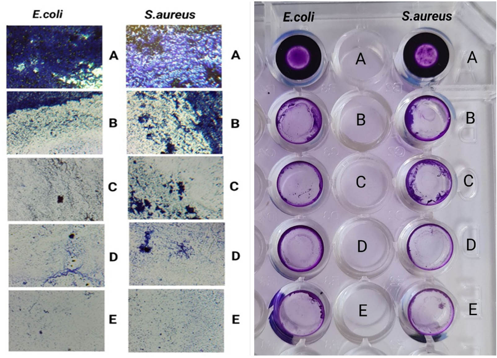
Figure 4
Biofilm formation in E. coli and S. aureus at different concentrations of CuFe₂O₄ NPs: (A) control case, (B) 62.5 µg/mL, (C) 125 µg/mL, (D) 250 µg/mL and (E) 500 µg/mL.
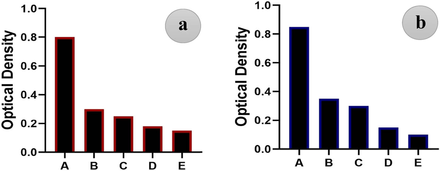
Figure 5
Biofilm formation in (a) E. coli and (b) S. aureus treated with CuFe₂O₄ NPs at different concentrations: (A) control case, (B) 62.5 µg/mL, (C) 125 µg/mL, (D) 250 µg/mL, and (E) 500 µg/mL.
3.5. Cytotoxicity of CuFe₂O₄ NPs
The cytotoxicity of CuFe₂O₄ NPs toward MCF-10 normal breast epithelial cells was evaluated using the MTT assay, which measures cellular metabolic activity as an indicator of cell viability. The assay is based on the ability of mitochondrial dehydrogenase enzymes in viable cells to reduce the yellow tetrazolium salt into insoluble purple formazan crystals. Therefore, a reduction in optical density indicates decreased cell viability and increased cytotoxicity. The obtained results demonstrate that exposure of MCF-10 cells to CuFe₂O₄ NPs resulted in a measurable decrease in cell viability compared with the untreated control cells. However, the reduction in viability was moderate, suggesting that the synthesized nanoparticles exert limited cytotoxic effects on normal breast cells. This observation is important for biomedical applications because materials intended for therapeutic use should ideally exhibit minimal toxicity toward healthy cells while maintaining biological activity [51].
Microscopic observations further supported these findings. The untreated control cells exhibited a typical epithelial morphology characterized by a well-spread cellular structure and strong adherence to the culture surface. In contrast, the cells treated with CuFe₂O₄ NPs showed mild morphological alterations, including slight cell shrinkage and a reduction in cell density. Such morphological changes are commonly associated with nanoparticle-induced cellular stress or early stages of cytotoxic response, as shown in Figure 6. The observed cytotoxicity may be attributed to the intrinsic physicochemical properties of CuFe₂O₄ NPs, particularly their ability to generate ROS within the cellular environment. Transition metal ions such as Cu+2 and Fe+3 can participate in Fenton-like reactions, which enhance intracellular oxidative stress and lead to mitochondrial dysfunction, membrane damage, and inhibition of cell proliferation. Excessive ROS production is a well-known mechanism by which metal oxide nanoparticles exert biological effects on cells [52]. Moreover, the relatively low toxicity observed in Figure 7 in normal MCF-10 cells may indicate a degree of biocompatibility of CuFe₂O₄ NPs, which is a desirable feature for potential biomedical applications such as drug delivery systems, magnetic hyperthermia, targeted cancer therapy, and imaging agents. Previous studies have reported that spinel ferrite NPs possess unique magnetic and catalytic properties that enable controlled biological interactions while maintaining acceptable cytotoxic profiles. Overall, the results suggest that CuFe₂O₄ NPs exhibit moderate cytotoxicity toward normal breast epithelial cells, indicating a relatively safe biological profile at the tested concentrations. These findings support the potential use of CuFe₂O₄ nanomaterials in biomedical applications, provided that their concentration and exposure conditions are carefully optimized [53,54].
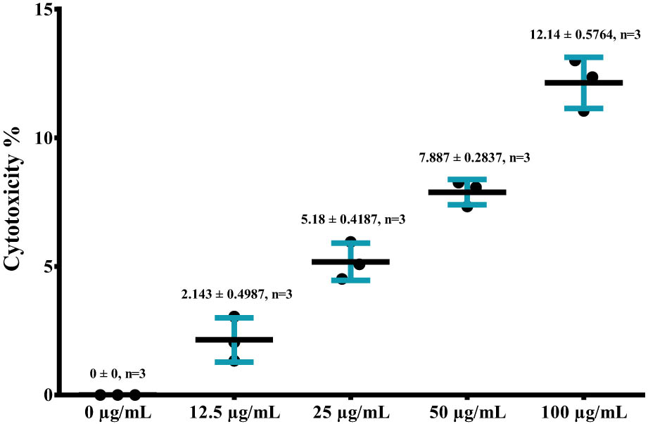
Figure 6
Cytotoxicity effect of CuFe₂O₄ substance in MCF-10 cells.
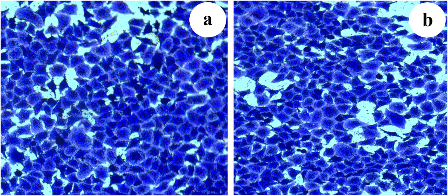
Figure 7
MTT assay: (a) untreated MCF-10 cells and (b) MCF-10 cells after being treated with CuFe₂O₄ NPs.
3.6. Hemolysis test
Hemocompatibility is a critical prerequisite for the clinical translation of biomaterials, particularly for applications involving direct or indirect contact with blood. In the present study, the hemolytic activity of CuFe₂O₄ NPs was evaluated using a standardized RBC hemolysis assay with minor modifications to a previously reported protocol. The obtained results demonstrated that CuFe₂O₄ exhibited minimal hemolytic activity at both tested concentrations (25 and 12.5 µg/mL), with hemolysis percentages of 4.83% and 3.39%, respectively (Table 4). These values fall below the 5% threshold defined by ISO 10993-4 standards, classifying the material as non-hemolytic. The low absorbance values recorded for sample CuFe₂O₄ were comparable to those of the negative control, while remaining significantly lower than the positive control, which confirmed the assay’s sensitivity and reliability [52]. The results were calculated using the following equation:
Table 4
Hemolysis data of CuFe₂O₄ NPs.
| Samples | Result of device | Result after |
|---|---|---|
| 25 µg/mL | 0.045 | 4.83 |
| 12.5 µg/mL | 0.042 | 3.39 |
| Negative control | 0.035 | ـــ |
| Positive control | 0.241 | ـــ |
Figure 8 shows a concentration-dependent trend, indicating that a decrease in the concentration of CuFe₂O₄ NPs is accompanied by a further decrease in hemolytic activity, demonstrating a favorable dose–response relationship. This behavior may be attributed to limited interaction between CuFe₂O₄ NPs and the phospholipid bilayer of erythrocyte membranes, thereby preserving membrane integrity and preventing hemoglobin leakage [53]. From a mechanistic perspective, the negligible hemolytic effect suggests that CuFe₂O₄ NPs do not induce oxidative stress, membrane destabilization, or protein denaturation within RBCs. These properties are particularly desired for nanomaterials or bioactive compounds intended for biomedical applications, including drug delivery systems, implant coatings, or diagnostic platforms [55].
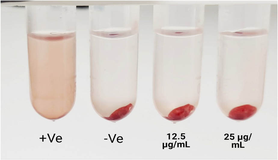
Figure 8
Hemolysis analysis of CuFe₂O₄ NPs at 12.5 and 25 µg/mL.
Similar findings have been reported in recent literature, where biocompatible materials exhibiting less than 5% hemolysis were deemed safe for further in vivo evaluation. The consistency of the present results with previously published hemocompatibility studies further supports the potential application of CuFe₂O₄ NPs in the biomedical field. Nevertheless, while the hemolysis assay provides valuable initial insight into blood compatibility, a comprehensive biocompatibility assessment should include additional investigations such as platelet aggregation, coagulation parameters, cytotoxicity assays, and in vivo hemocompatibility studies. This integrated assessment will be necessary to confirm the long-term safety profile of CuFe₂O₄ NPs [54]. Eventually, the hemolysis data showed that CuFe₂O₄ NPs have excellent hemocompatibility at the tested concentrations, highlighting their potential use in biomedical applications as promising and safe candidate materials when blood-contacting.
4. Conclusion
CuFe₂O₄ NPs were successfully synthesized using an eco-friendly synthesis method based on Capsicum annuum (sweet GP) extract. This study highlights the pivotal role of phytochemicals in controlling the structure and properties of the NPs. The synthesized materials showed tunable optical and magnetic behavior, along with excellent biological performance. Biological studies have shown an improvement in antibacterial activity, particularly against S. aureus bacteria, which is attributed to the ability of NPs to penetrate these bacteria. The cytotoxicity results indicated mild effects on normal cells, confirming the material’s biocompatibility. Hemolysis values of <5% also showed that these materials can be used safely in biomedical applications. Therefore, these results provide a promising foundation for the design of sustainable ferritic nanomaterials with potential applications in advanced biomedical fields.
Acknowledgments
The authors would like to thank Prof. Dr. Nehia Hussein for her support and valuable guidance regarding the biological aspects of this work.
Funding information
The authors received no financial support for the research, authorship, or publication of this article.
Author contributions
Sara H. Shahatha: conceptualization, methodology, biological assays, investigation, writing – original draft, and supervision; Rihab Jabbar: methodology, characterization, data analysis, and writing – review and editing; Mohammed Hayder Ismail Alluaibi: validation, data interpretation, and writing – review and editing. All authors have read and approved the final manuscript.
Conflict of interest statement
The authors declare that they have no known competing financial interests or personal relationships that could have appeared to influence the work reported in this manuscript.
Data availability statement
Data will be made available on request.