Each year, millions of individuals around the globe experience bone defects due to a variety of causes, including joint pain (arthralgia), osteoporosis, tumors, infections, congenital malformations, sports injuries, vehicular accidents, and natural disasters [1,2,3]. Consequently, the field of bone tissue regeneration has become a crucial and continuously evolving area within regenerative medicine and orthopedics. This field focuses on the intricate processes involved in repairing or replacing damaged, injured, or lost bone, offering new hope to those affected by fractures, degenerative bone diseases, traumatic injuries, and congenital skeletal abnormalities.
Although bone possesses a unique capacity for self-regeneration, unlike many soft tissues, this regenerative ability is only partial [4]. When confronted with high-impact injuries or significant bone deformities, the bone inherent self-healing capabilities may prove inadequate, necessitating additional medical interventions [5]. In cases of severe bone tissue damage, the natural healing process is often insufficient to fully restore bone integrity within a patient’s lifetime [6]. Such extensive damage can result from genetic disorders, bone cancers, severe bacterial bone infections, traumatic injuries from road accidents, military combat, and athletic activities.
The limitations of current bone defect treatments, such as limited bone graft availability, the need for multiple surgeries, risks of disease transmission, and immunogenic rejection, have prompted a significant reliance on implants within modern healthcare for tissue restoration. These challenges have also driven the development of bio-nanocomposite materials as part of bone tissue engineering approaches. Bone tissue engineering aims to provide alternative treatment strategies that promote bone repair and regeneration through biomaterials. Ideally, these biomaterials should be biocompatible, closely resemble the natural bone extracellular matrix (ECM), and facilitate adequate vascularization to support the demands of the regenerating tissue. The central concept of tissue engineering is to design biomaterials that interact with cells to harness the body’s inherent capacity for organization and self-repair rather than attempting to recreate complex living tissues outside the body [7].
Synthetic and natural biomaterials, as hydroxyapatite (HAp), β-tricalcium phosphate, bioactive glasses, and composites, have been extensively studied as bone substitutes, leading to several bone graft alternatives [3,8]. While these materials effectively replicate the inorganic components of bone, they often lack the organic counterpart necessary for creating the optimal microenvironment for osteogenesis [9]. Thus, bio-polymers such as alginate, gellan gum, Arabic gum, and carboxymethyl cellulose (CMC) have been employed and studied in the form of bio-composite materials [10,11,12,13,14]. CMC is a natural, biodegradable polymer with moderate tensile strength and good film-forming ability; however, its limited mechanical stiffness and load-bearing capacity restrict its direct use in bone tissue engineering applications [13,14].
Specifically, in the case of CMC copulation with TiO2, the resulting material seems highly important for the study of bone tissue regeneration [15,16]. The inherent characteristics of cellulose make it suitable for such uses. CMC is one of the most effective materials in tissue engineering due to its vast interconnected large pores and high specific surface area. It is essential for providing adequate support and surface for cell attachment and differentiation [17]. Moreover, the high density of surface hydroxyl groups endows cellulose with excellent chemical reactivity, allowing straightforward surface modification and tailoring of specific functional characteristics [18,19]. This physical property of the cellulose makes the compound suitable to be applied as a matrix-reinforcing phase with TiO2 to enhance the characteristics of the composite.
TiO2 has been reported in various studies on applying regenerative material for bone [16,20,21]. However, several important risks must be carefully considered, particularly for implant-related use. One key concern is particle release, where poorly anchored TiO2 nanoparticles or nanotube fragments may detach under mechanical stress or long-term physiological loading, potentially triggering local inflammatory responses or cytotoxic effects [22]. Long-term stability is another critical factor, as prolonged exposure to physiological fluids can lead to surface dissolution, phase transformation, or loss of structural integrity, which may compromise implant performance and safety [23,24]. Therefore, precise control over TiO2 surface properties, and morphology to minimize risks, enhances biological compatibility, and ensures predictable long-term performance in implant applications. TiO2 can be uniformly dispersed within a porous and chemically active cellulose matrix upon incorporation [15]. Besides, CMC is a renewable resource, hydrolyzable, and non-toxic biopolymer, which guarantees its application as implant in the biomedical field [21,25]. Therefore, cellulose incorporation in combination with TiO2 leads to a composite material that has upgraded mechanical strength, osteogenic prospects, and general applicability for bone tissue engineering [16,21,26]. Therefore, the unique properties of cellulose, namely, a porous structure and chemical activity, which are due to its ideal biocompatibility features coupled with TiO2, can offer an approach toward applying this concept in bone bio-materials development (naturally based biomaterial) used in bone tissue engineering. These combine with the fractional properties exhibited by cellulose, making it an ideal constituent for creating biopolymer composites, including TiO2, to improve the quality and performance of scaffolds in bone tissue engineering.
The novelty of this work lies in the strategic incorporation of TiO2 nanotubes (TiO2NT) into the CMC biopolymer to enhance its functional performance. TiO2NT were selected due to their highly ordered tubular morphology, large surface-to-volume ratio, and abundant active sites, which collectively facilitate superior drug loading, controlled release behavior, and improved interfacial interaction with biological systems [27]. Unlike conventional TiO2 nanoparticles, nanotubes provide a unidirectional channel structure that promotes cell adhesion, enhances biomolecule immobilization, and supports improved biological responses [28]. Additionally, the unique electronic properties of TiO2NT contribute to enhanced photocatalytic and antibacterial activities, which are advantageous for biomedical applications such as wound healing, tissue regeneration, and therapeutic delivery [29,30]. By integrating TiO2NT into the film and scaffold, this study introduces a new functional platform that has not been demonstrated in previous literature, thus establishing a meaningful advancement in material design for biomedical applications.
All materials were used as received without further purification. Titanium dioxide (MW 79.87 g/mol, product number 13463-67-7, lot number K45324808505) and sodium hydroxide (MW 40.00 g/mol, product number 1310-73-2, lot number B0783098219) were obtained from Merck. Hydrochloric acid (MW 36.46 g/mol, 37% concentrated, lot number P230312-250612) was purchased from HmbG. CMC (CAS number 9000-07-01, lot number MCFD00081480) and glycerol (product number G5516, lot number STBC1888V) were supplied by Sigma-Aldrich. Calcium chloride (CAS number 10043-52-4, lot number K47117278604) was used as received.
In this study, commercial TiO2 powder (MERCK) was used as precursors for TiO2NT synthesis. A 2.0 g of commercial TiO2 powder was mixed with 100 mL aqueous solution of 10 M NaOH. The mixture was stirred for 30 min to obtain a homogeneous solution and subjected to hydrothermal treatment at 150°C for 24 h in autoclave. After that, the solid precipitate was collected and washed with 0.1 M HCl (200 mL) followed by distilled water until pH 7 of washing solution was obtained. The final products were obtained by filtration and dried at 80°C overnight. Subsequently, sample was calcined at 400°C for 2 h to produce TiO2NT.
To fabricate CMC + 10 wt% TiO2NT 2D films and 3D scaffolds, 1 g of CMC biopolymer was dissolved in 100 mL of distilled water. Subsequently, 0.1 g of TiO2NT was added to the CMC solution to achieve a concentration of 10 wt% TiO2NT. The mixture was stirred continuously until a homogeneous and bubble-free solution with a viscosity carefully adjusted to achieve suitable flowability and processability. The solution was then divided into two portions for the preparation of films and scaffolds, respectively. For film fabrication, the solution was poured into a Petri dish and dried at 45°C for 3 days. For scaffold preparation, the second portion of the solution was dried at 25°C for 24 h to form a gel. The resulting gels were subsequently freeze-dried at −80°C for 24 h in an 8 mm diameter mold. The film and scaffolds were conditioned in a humidity chamber (model FX 1077, Jeio Tech Co. Ltd, Ansan, Korea) at 25°C and 50% relative humidity (RH) for a minimum of 48 h prior to further analysis. The conditioning parameters of 25°C and 50% RH were selected to simulate standard laboratory ambient conditions commonly used in material characterization studies. These conditions align with the ISO 187:1990 standard for conditioning of plastics and polymer-based materials prior to testing, ensuring consistent moisture content and dimensional stability of the samples. While, the incorporation of 10 wt% TiO2NT into the CMC matrix was based on prior research indicating that nanocomposites with 10 wt% TiO2 filler content exhibit optimal biological properties [31].
Fourier-transform infrared (FTIR) spectroscopy was performed using a PerkinElmer Spectrum 100 FTIR spectrophotometer equipped with a PIKE Miracle ATR accessory. The measurements were conducted with a single-bounce beam path at a 45° incident angle, accumulating 16 scans at a resolution of 4 cm−1. The spectral range analyzed spanned from 4,000 to 400 cm−1. X-ray diffraction (XRD) analysis was conducted at ambient temperature using a Rigaku Miniflex (II) X-ray diffractometer, with data collected over a 2θ range of 10–80° at a scanning rate of 2.00° min−1. The surface morphology of the scaffolds was examined using a JEOL JSM 6360 LA scanning electron microscope (SEM), while the size and shape of the titanium dioxide nanotubes (TiO2NT) were characterized using a Tecnai BioTwin FEI transmission electron microscope (TEM). Prior to SEM analysis, the samples were sputter-coated with gold using an Auto Fine Coater (JFC-1600) to ensure optimal imaging conditions.
The CMC + 10 wt% TiO2NT 2D film and 3D scaffold samples were meticulously trimmed to consistent dimensions (2 cm × 2 cm). Their initial dry weights (W
d) were accurately measured to establish a reference point for further evaluation. The samples were then submerged in a water bath maintained at 37°C for 24 h to ensure thorough hydration and uniform conditions across all specimens. Following immersion, the samples were carefully extracted from the water bath, and any excess surface moisture was gently removed using absorbent paper to eliminate potential inaccuracies in measurement. This procedure ensured the precision of the final swollen weight (W
s) determination. The fully hydrated samples were subsequently weighed to obtain their W
s values. The disparity between the initial dry weight (W
d) and the swollen weight (W
s) offered critical insights into the water absorption capacity and swelling properties of the materials. This process was repeated three times, and the average swelling percentage (%) was calculated using equation (1)
The porosity of the CMC + 10 wt% TiO2NT 2D film and 3D scaffold was evaluated using a modified Archimedes’ principle, as described in previous studies [32,33]. For this analysis, a density bottle was filled with ethanol, and the temperature of the ethanol was stabilized at 37°C in a water bath. Before immersing the samples, the initial weight of the film or scaffold (W
s) was measured. Once the ethanol reached the target temperature of 37°C, the combined weight of the bottle and ethanol (W
1) was recorded. The scaffold was then immersed in the density bottle, and any trapped air bubbles within its pores were removed under vacuum conditions. The ethanol displaced during degassing was replenished, and the temperature was re-equilibrated to 37°C. The weight of the bottle containing the scaffold (W
2) was measured, after which the scaffold was removed from the bottle, and its weight (W
3) was recorded. The porosity (P) was calculated using equations (2)–(4)
The bioactivity of the CMC + 10 wt% TiO2NT 2D film and 3D scaffold was assessed using simulated body fluid (SBF), which replicates the ionic composition of human blood plasma, in accordance with established protocols [35]. The CMC + 10 wt% TiO2NT 2D film and 3D scaffold samples of uniform dimensions (2 cm × 2 cm) were submerged in 40 mL of SBF and incubated at 37°C for 7 days. The thickness of both the samples used for biomineralization were similar, which is 1.0 mm. Following the incubation period, the samples were examined using SEM coupled with energy-dispersive X-ray spectroscopy (EDX) and XRD to evaluate their capacity to promote apatite formation.
The experiment commenced with the precise measurement of the initial weight (W
1) and dimensions of the cylindrical nanocomposite films and scaffolds to determine their initial volume, ensuring accurate characterization of each specimen. To achieve complete saturation, the samples were submerged in a SBF solution for 24 h, allowing thorough infiltration and interaction with the nanocomposite matrix. Following immersion, the samples were carefully extracted using forceps, and any excess surface moisture was gently removed using pre-weighed, lint-free absorbent paper. This meticulous approach minimized potential inaccuracies in subsequent weight measurements. The samples were then reweighed (W
2) with high precision to assess any changes in mass resulting from the saturation process. The difference between the initial weight (W
1) and the final weight (W
2) provided critical data for evaluating and interpreting the experimental results. These steps were repeated three times, and the average biodegradation was calculated using equation (5)
Figure 1 illustrates the physical characteristics of the synthesized TiO2NT powder, CMC + 10 wt% TiO2NT 2D film, and CMC + 10 wt% TiO2NT 3D scaffold. As shown in Figure 1(a), the white color of the TiO2NT powder facilitates its visual identification and uniform dispersion within the CMC polymer matrix, which is beneficial for the fabrication process of films and scaffolds. The CMC + 10 wt% TiO2NT 2D film, depicted in Figure 1(b), displays a smooth and homogeneous surface with a uniform, glossy texture. The film is flat, uniform, and slightly transparent allowing some light to pass through. This uniformity suggests consistent material properties, with the film maintaining a constant and thin thickness, typical for such materials. In contrast, the CMC + 10 wt% TiO2NT 3D scaffold, presented in Figure 1(c), is a bulkier material with a porous structure and a less smooth surface. The scaffold appears more opaque, likely due to its greater thickness or density. Its three-dimensional porous architecture is characteristic of scaffolds designed for applications such as tissue engineering or filtration.

Photo images of (a) synthesis TiO2NT powder, (b) CMC + 10 wt% TiO2NT 2D film, and (c) CMC + 10 wt% TiO2NT 3D scaffold.
The morphology of the synthesized TiO2NT powder was characterized using SEM and TEM. The TiO2NT sample displayed fiber-like nanostructures, with diameters of approximately 10 nm and lengths reaching several hundred nanometers, as illustrated in Figure 2(a). TEM analysis further confirmed the successful synthesis of TiO2NT (Figure 2b). High resolution TEM analysis in Figure 2(c) shows the presence of hollow tubular structure of the nanotubes. The hollow nanotube structures are pivotal in enhancing the regeneration process, as they provide focal adhesion sites either within the nanotube lumen or on their surfaces. These sites facilitate the attachment of anchorage-dependent cells, such as fibroblasts, thereby promoting cell migration, which is a critical factor in tissue regeneration [36].

(a) SEM, (b) TEM, and (c) high resolution TEM micrographs of hydrothermally synthesized TiO2NT.
The SEM image of the CMC + 10% TiO2NT 2D film shows a relatively smooth and compact surface with localized micro-scale heterogeneity (Figure 3(a). Scattered bright regions are attributed to inorganic-rich domains, likely corresponding to TiO2NT agglomerates within the CMC matrix. These features may arise from uneven shrinkage during the drying process or inadequate interfacial interactions between the CMC matrix and TiO2NT during film formation, which could hinder effective HAp nucleation and growth [37]. In contrast, the CMC + 10 wt% TiO2NT 3D scaffold demonstrates a porous structure with interconnected pores (Figure 3(b), which could facilitate the cell infiltration and nutrient transport [38]. A notable characteristic of the scaffold surfaces is their highly rough texture, featuring pores of varying shapes and sizes, which are critical for their functionality as tissue engineering constructs [39,40]. The irregularity is particularly pronounced in scaffolds fabricated via lyophilization, where the freezing and drying processes create a fibrous or network-like structure within the pores. This internal architecture enhances mechanical stability and increases surface area, both of which are vital for promoting cellular adhesion and proliferation in tissue regeneration [41,42]. Thus, in order to evaluate the potential of the film and scaffold for bone tissue regeneration, SEM analysis was conducted on CMC + 10 wt% TiO2NT 2D film and 3D scaffold samples after immersion in SBF for 1 week.

SEM micrograph of (a) CMC + 10 wt% TiO2NT 2D film and (b) CMC + 10 wt% TiO2NT 3D scaffold.
Table 1 shows the swelling of CMC + 10 wt% TiO2NT 2D film and 3D scaffold in distilled water within 24 h. The CMC + 10 wt% TiO2NT 3D scaffold swelling was 1880.90 ± 10.22%, while the film was 1718.42 ± 8.42%. This can be explained by the existence of a more porous architecture in the scaffold, which allows more liquid to penetrate and thus be swelled. On the other hand, the 2D film shows less swelling resulting from the denser structure, which restricts liquid absorption. The differences in surface qualities between the 2D film and 3D scaffolds could also contribute to the variations in swelling behavior of the samples [43]. The porosity of the CMC + 10 wt% TiO2NT 3D scaffold is 91.28%, which is higher than that of the CMC + 10 wt% TiO2NT 2D film, which has a porosity of 68.13% (Table 1). This is due to the bulky structure of CMC + 10 wt% TiO2NT 3D scaffold as compared to sheet-like structure of CMC + 10 wt% TiO2NT 2D film. Films are typically 2D or thin-layer structures. Their design prioritizes surface coverage and barrier properties over porosity. Films are used in applications like coatings, membranes, or drug delivery, where a continuous, dense structure is often preferred, while scaffolds are 3D structures designed to mimic the ECM of tissues. They require interconnected pores to facilitate cell migration, nutrient diffusion, and waste removal. High porosity ensures these functions and allows cells to infiltrate and proliferate within the structure. The porosity of the samples was primarily governed by the hydrophilic nature of the CMC polymer chains [44]. In addition, the presence of micro- and nanoscale free volume within the polymer network that may not be observable in SEM images at the selected magnification, polymer–TiO2NT interactions play a significant role in promoting water diffusion through the matrix [45].
Swelling and porosity of CMC + 10 wt% TiO2NT 2D film and 3D scaffold.
| Sample | Swelling (%) | Porosity (%) |
|---|---|---|
| CMC + 10 wt% TiO2NT 2D film | 1718.42 ± 8.42 | 68.13 |
| CMC + 10 wt% TiO2NT 3D scaffold | 1880.90 ± 10.22 | 91.28 |
The FTIR spectra of TiO2NT, CMC + 10 wt% TiO2NT 2D film, and 3D scaffold are displayed in Figure 4. In the TiO2NT spectrum (Figure 4(a)), a single broad peak at 450 cm−1 is observed, corresponding to the metal–oxygen (Ti–O) stretching vibration [46,47]. The absence of additional peaks confirms the production of pure TiO2NT. The FTIR spectra of the CMC + 10 wt% TiO2NT 2D film (Figure 4(b)) and 3D scaffold (Figure 4(c)) exhibit similar features. A broad absorption band between 3,660 and 2,980 cm−1 is attributed to the stretching vibrations of intramolecular hydrogen bonds. The band at 2,930 cm−1 corresponds to the C–H stretching vibrations from the CMC polymer [48]. Peaks at 1,414 cm−1 and 1,313 cm−1 are associated with the stretching vibrations of –CH3 and C–O, respectively, confirming the presence of CMC in both the 2D film and 3D scaffold samples [49]. Additionally, the characteristic vibrations in the range of 1,190–920 cm−1 represent the stretching vibrations of C–OH groups [50], while the band at 1,585 cm−1 is linked to the stretching vibration of the carboxylic group [51]. Notably, in the fingerprint region (400–800 cm−1), slight shifts in the Ti–O stretching mode and the emergence of new bands are observed in the CMC + 10 wt% TiO2NT 2D film and 3D scaffold samples compared to pure TiO2NT. These shifts indicate interactions between TiO2NT and the CMC matrix, suggesting the formation of a complex structure.

FTIR spectra of (a) TiO2NT, (b) CMC + 10 wt% TiO2NT 2D film, and (c) CMC + 10 wt% TiO2NT 3D scaffold CMC scaffold.
XRD analysis was conducted to confirm the successful synthesis of the prepared samples and to examine their phase structure. Figure 5 shows the XRD diffractogram of the studied samples. The hydrothermally synthesized TiO2NT powder exhibited characteristic diffraction peaks at 2θ values of 25.26°, 38.18°, 47.98°, 53.12°, 55.24°, 63.08°, 69.12°, 70.48°, and 75.38°. These peaks correspond to the 101, 004, 200, 105, 211, 204, 116, 220, and 215 crystal planes of anatase TiO2, confirming the successful preparation of the TiO2 [52]. In the case of the CMC + 10 wt% TiO2NT 2D film, a similar diffraction pattern was observed, with peaks detected at 2θ values of 25.17°, 27.30°, 37.65°, 47.90°, 53.80°, 54.92°, 62.57°, 68.83°, 70.18°, and 74.94°, corresponding to the same anatase TiO2 crystalline planes. The presence of sharp and well-defined peaks indicates the formation of highly organized crystalline TiO2 domains within the CMC matrix. Additionally, additional peaks at 12.58° and 19.60° were attributed to the CMC matrix [53]. In contrast, the XRD pattern of the CMC + 10 wt% TiO2NT scaffold exhibited broad and weak diffraction peaks. This is due to the high porosity and lower density of the scaffold that can reduce the effective diffraction volume and cause peak broadening and intensity reduction. This effect is well-documented in porous materials, where scattering from voids and reduced coherent scattering domains affect peak profiles [54].
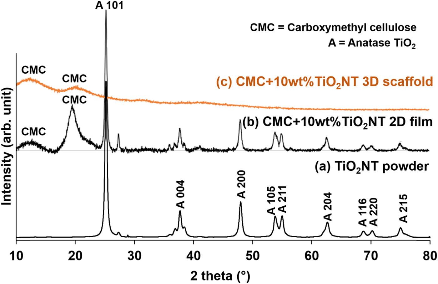
XRD patterns of (a) TiO2NT, (b) CMC + 10 wt% TiO2NT 2D film, and (c) CMC + 10 wt% TiO2NT 3D scaffold CMC scaffold.
The detected peaks at approximately 2θ = 12.54° and 19.64° were attributed to the CMC matrix [53]. Notably, no distinct diffraction peaks corresponding to TiO2 were observed in the scaffold sample, indicating the successful incorporation of TiO2NT into the CMC-based scaffold. This suggests that the scaffold material exhibits a disordered structural arrangement, likely influenced by differences in preparation and processing methods.
Thermogravimetric analysis (TGA) was employed to evaluate the thermal stability of TiO2NT powder, as well as CMC + 10 wt%TiO2NT 2D film and 3D scaffold samples. As illustrated in Figure 6, the synthesized TiO2NT exhibited exceptional thermal stability, with a minimal weight loss of only 1% observed up to 880°C. For both CMC + 10 wt%TiO2NT 2D film and 3D scaffold, the initial weight loss occurred between 30°C and 210°C. Based on the previous study, this weight loss is associated with the evaporation of absorbed moisture and volatile constituents [55,56], somehow the analysis of pre-dried sample could help to confirm this.

TGA curves of (a) TiO2NT, (b) CMC + 10 wt% TiO2NT 2D film, and (c) CMC + 10 wt% TiO2NT 3D scaffold CMC scaffold.
The primary degradation stage, spanning from 210°C to 280°C, is predominantly attributed to the thermal decomposition of the CMC polymer matrix [57]. Beyond 280°C, the weight loss continued gradually, extending up to 880°C, as the thermal degradation process approached completion [58,59]. The total weight loss for the CMC + 10 wt%TiO2NT 2D film and 3D scaffold was approximately 90%. The remaining 10% of the samples, which exhibited thermal stability, is attributed to the presence of TiO2NT, confirming the successful incorporation of 10 wt% TiO2NT filler into the CMC matrix.
Figure 7(a) presents the SEM images of CMC + 10 wt% TiO2NT 2D film sample following immersion in SBF for 7 days. The film surface exhibits the formation of cubic crystals, which are attributed to NaCl resulting from an imbalance in ion exchange processes [60]. The NaCl compounds were confirmed by EDX analysis as Na and Cl elements were detected (Figure 7(b)). The high concentration of TiO2NT in the 2D film likely disrupts the local ionic environment, causing an accumulation of Na⁺ and Cl⁻ ions on the film surface. These ions facilitate the crystallization of NaCl, which competes with and suppresses HAp nucleation [61]. The XRD diffractogram in Figure 7(c) confirms the formation of NaCl, with four distinct peaks observed at 27.47°, 31.70°, 45.50°, and 76.62°, corresponding to the 111, 200, 220, and 420 planes of the face-centered cubic structure of NaCl [62]. The high intensity and sharpness of these peaks indicate the highly crystalline nature of the produced NaCl, consistent with the SEM observations of cubic NaCl crystals. The accumulation of Na and Cl originates from the use of Na-containing precursors and chloride-based reagents during the immersion in SBF solution [63]. These ions have a stronger affinity toward surface hydroxyl groups and defect sites on the CMC + 10 wt% TiO2NT 2D film, leading to preferential surface adsorption. In contrast, other ionic species remained dissolved in the SBF solution and did not significantly participate in surface deposition.
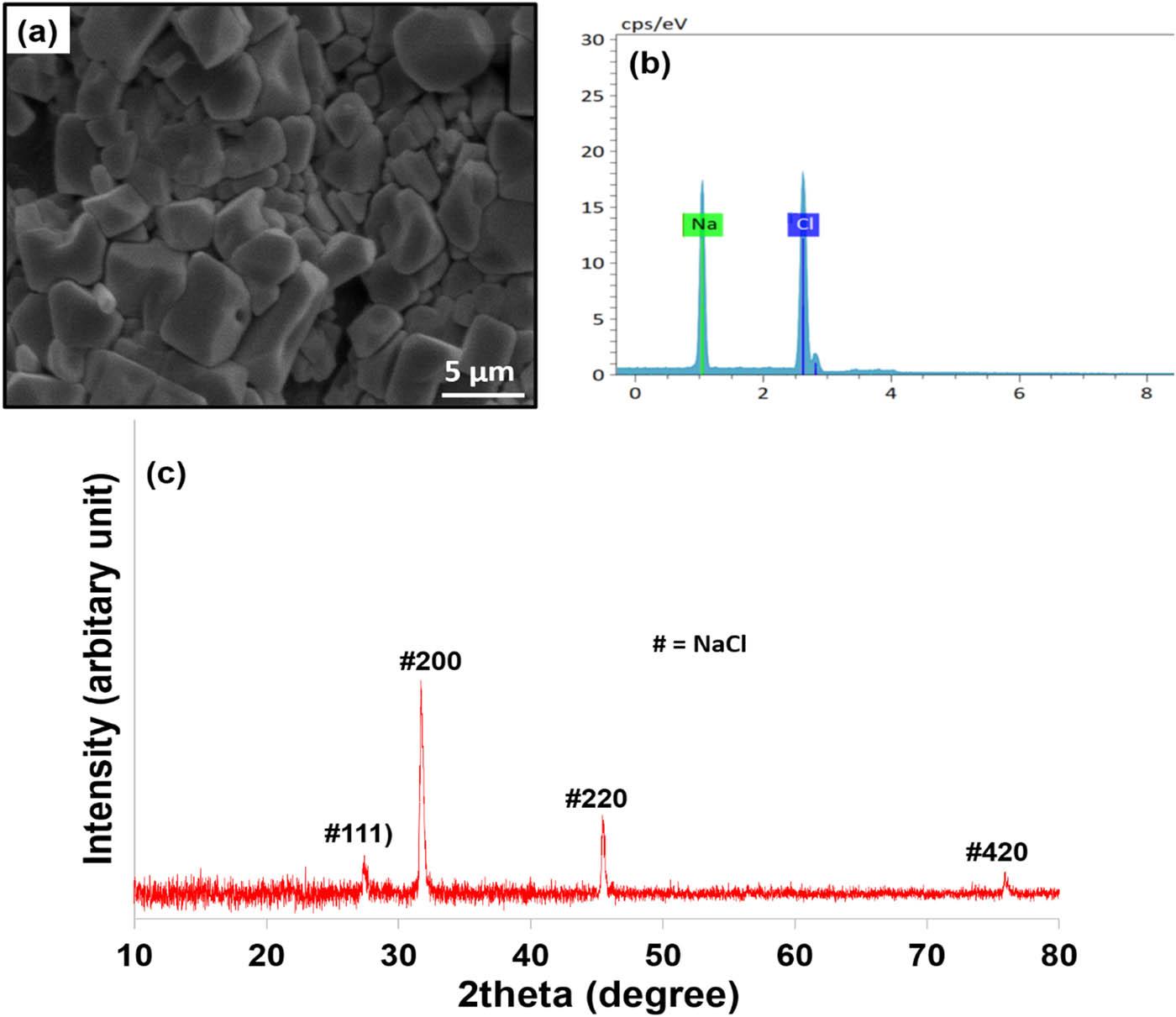
(a) SEM micrograph, (b) EDX spectra, and (c) XRD pattern of CMC + 10 wt% TiO2NT 2D film after immersion in SBF for 1 week.
On the other hand, following 1 week of immersion in SBF, the surface of the CMC + 10 wt% TiO2NT 3D scaffold, as observed in SEM images (Figure 8(a), exhibits a web-like structure with numerous small particles dispersed unevenly across it, along with distinct clusters of mineral deposits indicative of HAp formation. Somehow, the morphology of HAp obtained in this study is different with the typical HAp morphology reported in the literature [28]. This behavior can be attributed to the unique 3D porous architecture, surface chemistry, and ionic environment of our material. Unlike dense or planar substrates used in conventional SBF studies, the 3D scaffolds present a highly interconnected pore network and abundant surface functional groups, which alter local ion diffusion, nucleation density, and crystal growth kinetics [64]. As a result, HAp preferentially nucleates within the internal porous regions and grows in a more compact or fused morphology rather than forming the classical plate-like or needle-like apatite crystals typically observed on flat surfaces [65].
The EDX results in Figure 8(b) confirm the presence of calcium (Ca), phosphorus (P), and oxygen (O) elements, which are characteristic of HAp. The surface of the CMC + 10 wt% TiO2NT 3D scaffold significantly enhanced its ability to induce HAp-like layer formation after SBF immersion indicating improved in vitro mineralization behavior. Additionally, the presence of TiO2 in the nanocomposite 3D scaffold has been reported to facilitate the deposition of calcium–phosphate (Ca–P) minerals on its surface [66]. TiO2 absorbs moisture from the air, forming titanium hydroxide (Ti–OH) groups. These Ti–OH groups, along with charge separations on the TiO2 surface, influence the precipitation behavior of HAp. The Ti–OH groups act as nucleation sites for HAp formation in SBF [67,68]. At the SBF pH of 7.4, the Ti–OH groups interact with hydroxyl ions, forming a negatively charged surface with Ti–O functional groups [69]. This negatively charged surface attracts positively charged Ca²⁺ ions from the SBF, which subsequently attract negatively charged PO₄³⁻ ions. This alternating attraction of ions leads to the formation of an amorphous calcium phosphate layer, which eventually crystallizes into HAp [70]. XRD analysis further corroborates these findings, revealing peaks at 2θ values of 25.84°, 31.70°, 32.90°, 34.08°, 39.82°, 45.40°, 46.76°, and 49.56° (Figure 8(c). These peaks correspond to the 002, 211, 112, 300, 130, 222, 215, and 004 crystal planes of hexagonal HAp, as denoted by their Miller indices [71,72]. The difference in crystal phases observed between the 2D film and 3D scaffold samples arises from their distinct nucleation and growth environments [73]. In the 2D film, lower local diffusion rates favor the formation of the metastable phase, whereas in the 3D scaffold architecture, enhanced mass transport, higher local supersaturation, and multidirectional crystal growth promote transformation into the thermodynamically more stable phase. Furthermore, differences in drying dynamics and internal stress within the 3D porous framework contribute to phase rearrangement [74].

(a) SEM micrograph, (b) EDX spectra, and (c) XRD pattern of CMC + 10 wt% TiO2NT 3D scaffold after immersion in SBF for 1 week.
Figure 9 show the increment trend of degradation rate of CMC + 10 wt% TiO2NT 2D film and 3D scaffold within 24 h in phosphate buffer solution. The CMC + 10 wt% TiO2NT 3D scaffold exhibits a higher degradation rate with 94.28 ± 8.22% as compared to 72.88 ± 6.46% for 2D film sample after 24 h. This is due to their structural and functional differences of the samples. The high porosity and interconnected architecture of scaffolds provide a significantly larger surface area and allow for deeper penetration of water, enzymes, and other degrading agents, accelerating hydrolytic and enzymatic degradation [75]. The material in scaffolds is often distributed in fibers, making them more susceptible to breakdown compared to the denser, thicker, and continuous structure of films, which restricts the diffusion of degrading agents. Additionally, scaffolds are designed to degrade faster in biological environments to match tissue regeneration rates, facilitating cell infiltration and new tissue formation, whereas films are typically engineered for durability, with slower degradation suited for protective or controlled-release applications [76]. These factors collectively result in 3D scaffolds degrading at a much faster rate than 2D films, thus supporting the good performance of scaffold for biomedical application such as bone tissue regeneration. The rapid degradation rate observed in the present study warrants careful evaluation in the context of bone tissue engineering, where scaffold resorption must be synchronized with new tissue formation. Although excessively fast degradation may compromise mechanical stability, several studies have shown that moderately rapid-degrading biopolymer scaffolds can still be suitable for early-stage bone repair, especially when they promote ion exchange, surface reactivity, and cellular infiltration [77].

Biodegradation of CMC + 10 wt% TiO2NT 2D film and 3D scaffold within 24 h.
This study developed TiO2NT incorporated into CMC matrices to create 2D films and 3D scaffolds for bone tissue engineering. The CMC + 10 wt% TiO2NT 3D scaffold exhibited high porosity, swelling capacity, and controlled degradation, essential for cell infiltration and nutrient diffusion. Bioactivity assessments revealed HAp formation on the scaffold after immersion in SBF, confirming its potential for bone regeneration. In contrast, the 2D film formed NaCl crystals, underscoring the importance of structural design in biological performance. Comprehensive characterization using FTIR, XRD, SEM, and TGA confirmed the successful integration of TiO2NT into the CMC matrix, with the 3D scaffold displaying a porous, interconnected structure ideal for tissue regeneration. The scaffold’s-controlled degradation aligns with the requirements for gradual bone repair, making it a promising candidate for bone tissue engineering. The CMC + 10 wt% TiO2NT 3D scaffold show physicochemical and in vitro mineralization characteristics that warrant further biological evaluation for potential bone tissue regeneration applications. Somehow, future in vitro and in vivo studies are required to confirm biocompatibility and bone regeneration capability.
We thank Universiti Malaysia Terengganu for providing funding support for this project (UMT/PPP/IPRG/2024/55533).
This research was supported by UMT (UMT/PPP/IPRG/2024/55533).
Lei Ren: conceptualization, methodology. Tuo Jia: funding acquisition, project administration. Mahani Yusoff: resources, formal analysis. Nur Ain Atisya C.M. Khairuddin: software, visualization. Alina Irwana Muhamad A’srai: investigation, validation. Nur Adibah Roslan: data curation. Mohd Hasmizam Razali: supervision, writing-original draft preparation Zhiming Kang: writing – reviewing and editing.
Authors state no conflict of interest.
The data supporting the findings of this study are included within the article. Additional datasets are available from the corresponding author upon reasonable request.