

Figure 1
Pictorial representation of development of YTO nanoparticles.
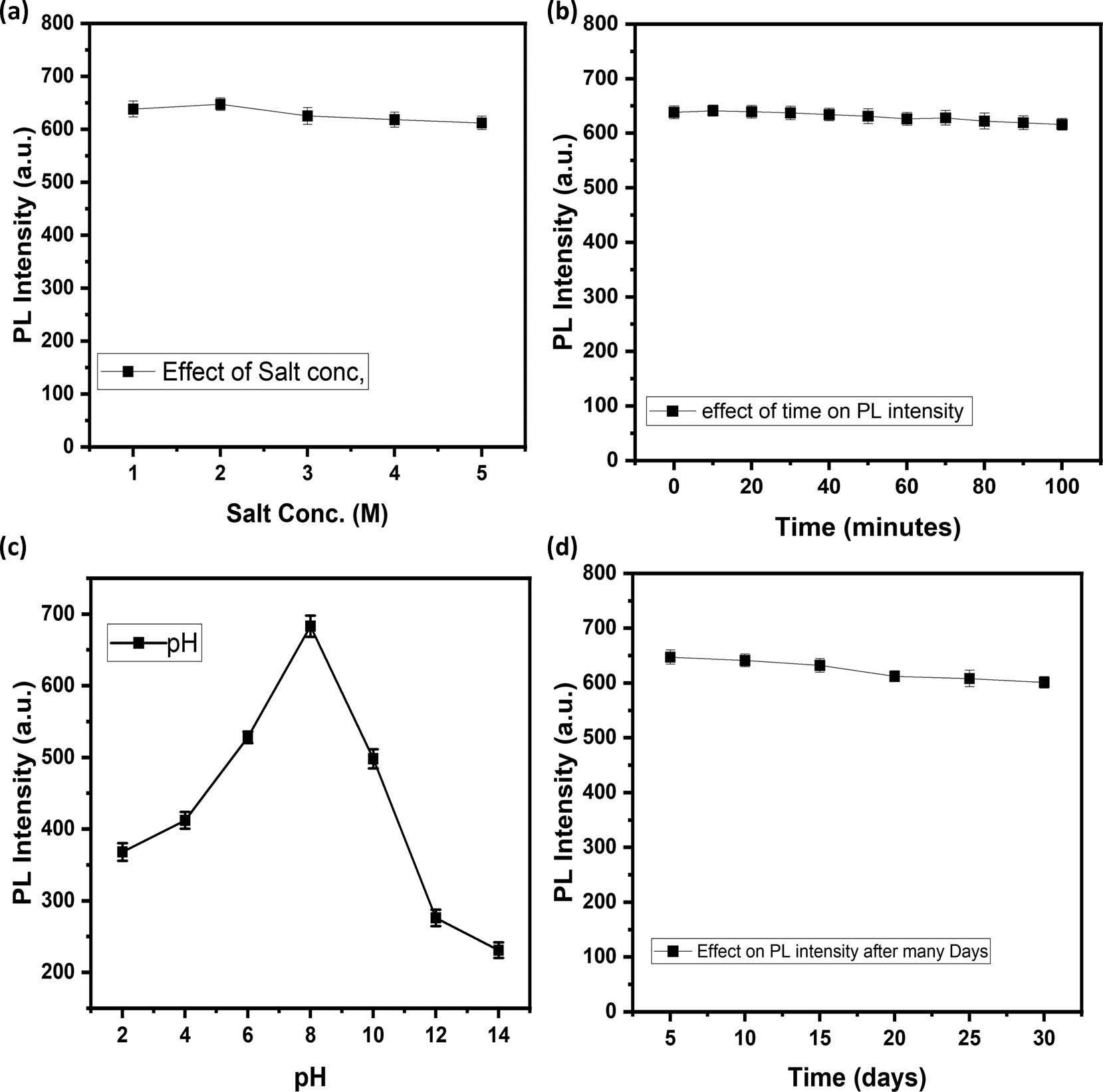
Figure 2
Effect of (a) factor of salt conc.; (b) factor of time (min); (c) pH; and (d) time (days) on the PL intensity of YTOs.
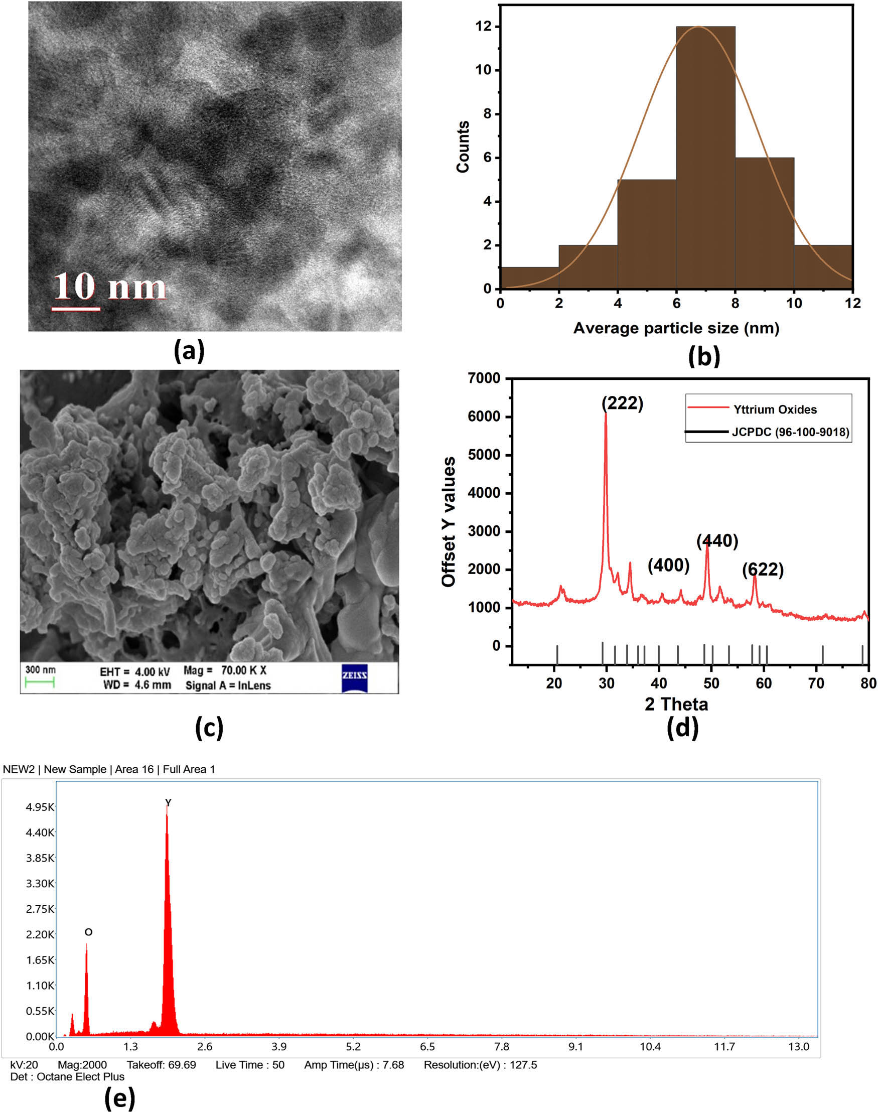
Figure 3
(a) HR-TEM image, (b) average particle size, (c) FE-SEM image, (d) XRD spectra, and (e) EDX spectra of YTOs.
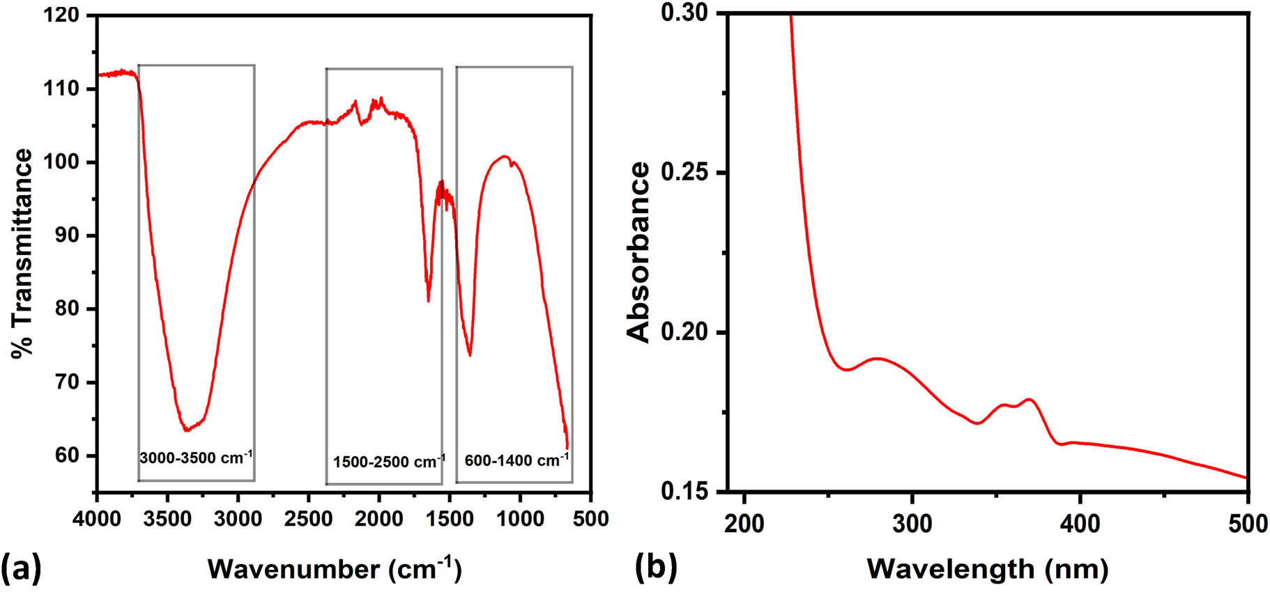
Figure 4
(a) FT-IR spectra and (b) UV-Vis absorption spectrum of YTO.
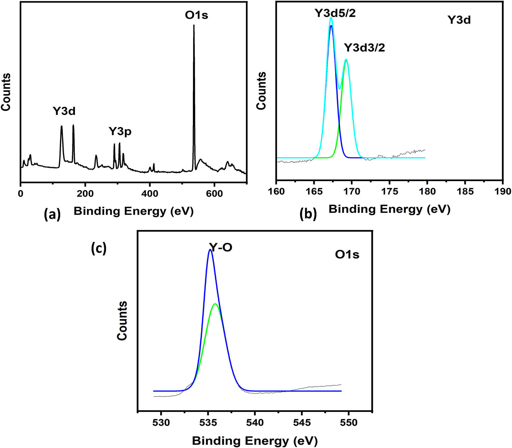
Figure 5
(a) Comprehensive XPS of YTO; (b) enhanced resolution spectra of Y3d; and (c) spectra of O1s.
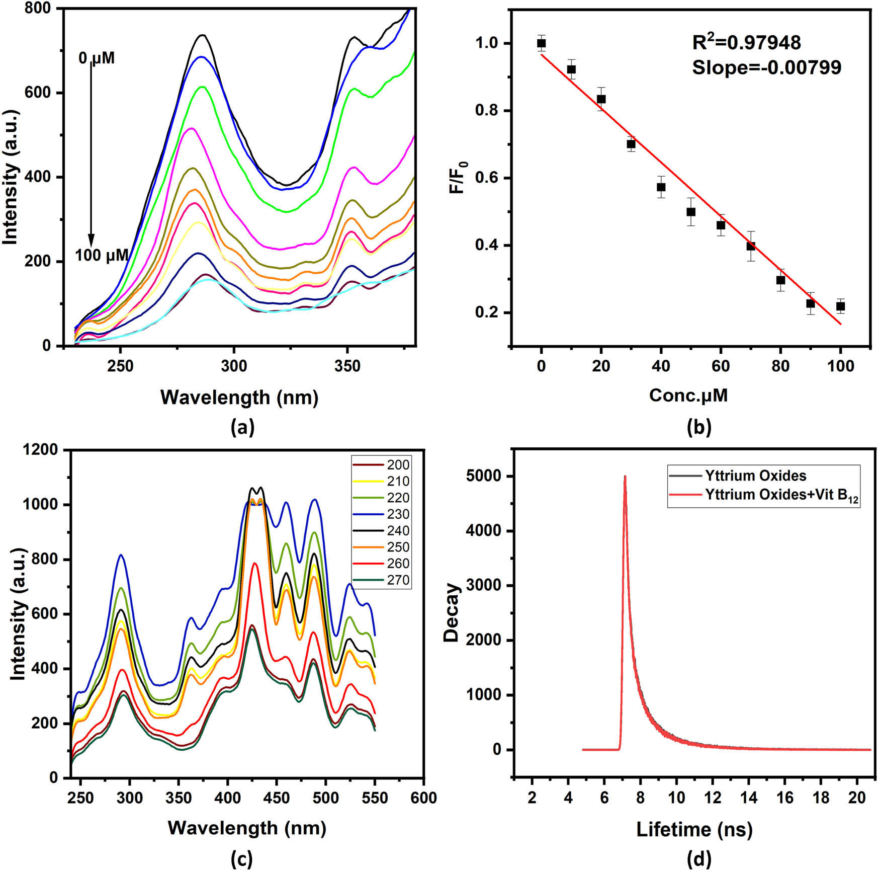
Figure 6
(a) PL spectra of YTO quenching with vitamin B12, (b) linear calibration of fluorescence intensity with vitamin B12, (c) excitation dependent emission spectra of YTO, and (d) fluorescence lifetime of pure YTOs nanoparticles and mixture with vitamin B12.
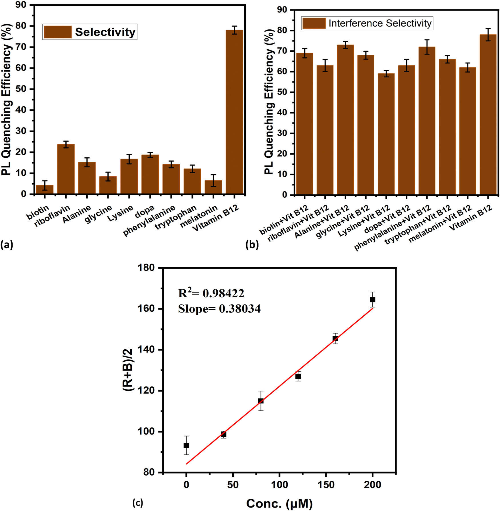
Figure 7
(a) Selectivity plot of YTO with various analytes, (b) interference selectivity plot of YTO with various analytes in the presence of vitamin B12, and (c) RBG-based detection of vitamin B12 with YTO.