Vitamin B12 is a cobalt-containing chemical complex that is required for human health. Vitamin B12, present in the active coenzyme forms methylcobalamin and adenosylcobalamin, plays a vital role in cellular development, growth, and metabolic regulation, while cyanocobalamin serves as a synthetic precursor commonly used in supplementation. Deficiency in this vitamin can result in pernicious anemia, weakness, nausea, and fatigue [1,2]. Vitamin B12 is an essential coenzyme that plays a crucial role in cellular growth and development. As the human body is unable to produce this vitamin, it needs to be supplemented through food items like dairy products, fish, eggs, meat, oysters, and poultry. Plant foods have very minimal amounts of vitamin B12, so vegetarians and those who do not consume red meat need supplementation [3,4]. Vitamin B12 is crucial for producing red blood cells, DNA synthesis, neurological function, and a healthy immune system. Deficiency may cause low energy levels, muscle weakness, and gastrointestinal disturbances, as well as neurological features of peripheral neuropathy and dementia. Biochemically, it leads to the rise in homocysteine and methylmalonic acid, which in turn add to systemic and neurological complications, thus having a profound impact on life expectancy [5,6]. Vitamin B12, produced only by microorganisms, is obtained mainly from animal-derived foods, while plant foods lack it unless fortified. Vitamin B12 is used as a cofactor by L-methylmalonyl-CoA mutase and methionine synthase, the latter of which converts homocysteine into the amino acid methionine. Deficiency is usually identified by a complete blood count and a vitamin B12 blood test.
For the detection of vitamin B12, several analytical techniques have been described, such as high-performance liquid chromatography, electrochemical detection, enzyme-linked immunosorbent assay, and microbiological tests [7,8,9]. Though satisfactory, these methods are hampered by being costly and complex in instrument requirements, labor, and trained personnel. Furthermore, the absence of a standardized procedure complicates routine analysis. New developments have proposed new alternatives; for instance, the use of a time-resolved fluorescent microsphere immunochromatographic assay allowed rapid and ultrasensitive detection of vitamin B12 in infant formula with high accuracy and stability [10]. In the same line, yellow-emissive carbon dots prepared through a one-pot hydrothermal process were employed as a label-free fluorescent probe with a low detection limit of 0.1 µM and successful application to real samples [11]. Other methods are the application of magnetic nanoparticles to selectively enrich and preconcentrate cyanocobalamin, which enhances sensitivity in matrices that are complex [12]. Optical methods commonly depend on noble metals (such as gold, silver, and platinum) or heavy-metal-doped quantum dots, posing cost and toxicity issues, yet fluorescence-based sensing with non-metal-doped quantum dots has attracted much interest due to its high sensitivity, low cost, simplicity in operation, and possibility of direct visual detection.
Nanoparticles are hybrid or heterogeneous materials designed at the nanometric level, possessing different properties from their bulk form. Their structures are more complex than those of micro-composites [13]. Because of their heterogeneity, the properties of nanoparticles are dependent on factors related to those in conventional composites, such as component properties, composition, structure, and interfacial interactions [14]. The nanoscale nature of these particles changes their atomic characteristics, resulting in improved functionalities. This change in nanoparticle characteristics has been beneficial in different applications (biomedical applications/sensors, optical filters, solar cells, etc.) [15,16,17,18].
Yttrium oxide (YTO) is a highly promising material for developing advanced architectures with applications in photocatalysis [19], sensing [20], biosensing [21], and hydrogen production [22]. Its widespread interest stems from its biocompatibility and non-toxicity, making it suitable for eco-friendly systems and biomedical applications [23,24]. The most common crystalline form of YTO is hexagonal, serving as the foundation for various morphologies, including hexagonal plates, cones (needles), and prisms (rods or tubes) [25]. These morphologies can be tailored by controlling preferential growth directions. Additionally, hydrothermal synthesis provides a straightforward method for fabricating diverse YTO nanostructures, with temperature and solution alkalinity playing crucial roles in influencing nanoparticle growth. It is an extensively used transition metal oxide and has high potential for future development because of its superior mechanical [26], chemical [27], and thermal stability [28]. It is an established host matrix for luminescent ions and has recently come into focus as an integral part of transition materials. YTO is an excellent raw material for chemical catalysis and optoelectronic devices, besides other applications in biological systems and photodynamic imaging. Additionally, it is commonly used as a dopant for rare-earth doping. Through the reduction in electron-hole pair combination, the creation of innovative active sites, and the enhancement of material properties through synergistic effects, YTO might be combined with additional non-metals, metals, lanthanides, and oxides to improve photocatalytic, optoelectronic, and antibacterial capabilities [17].
Literature on the application of YTO nanoparticles for the optical sensing of vitamin B12 has not been reported, as per our literature review. But some limited research has been done with similar oxide nanomaterials, like iron oxide, zinc oxide, and rare-earth-doped oxides, for sensing vitamins, in which they have shown highly positive optical and catalytic features. But as yet, no report is available specifically for YTO, demonstrating a definite lacuna in this regard. This gap, together with the need for in-depth research on the role of metal oxides in composite properties with sensing implications, motivated this study. The goal of this research was to synthesize and characterize YTO nanoparticles and examine how their concentration affects structural, vibrational, morphological, optical, and surface features. To achieve this, we used a set of sophisticated characterization tools: X-ray diffraction (XRD) to identify crystal structure and phase purity; transmission electron microscopy (TEM) and scanning electron microscopy (SEM) to investigate particle size, shape, and morphology; Fourier-transform infrared spectroscopy (FT-IR) to determine functional groups and surface interactions; X-ray photoelectron spectroscopy (XPS) to investigate surface composition and oxidation states; UV-Visible (UV-Vis) spectroscopy to investigate optical absorption; and photoluminescence (PL) spectroscopy to investigate emission properties for assessment of sensing performance. Consequently, the novelty of our work lies in the development and thorough characterization of a nanomaterial with potential applications in optical sensing.
Deionized water from the Millipore Milli-Q water purification system was used to create the solution, having a resistivity of 18.2 MΩ cm. Analytical-grade hydrochloric acid and sodium hydroxide, used in pH maintenance studies, were bought from Loba, India. Vitamin B12 and melatonin were acquired from Sigma Aldrich. A few other substances, including sodium chloride, glycine, lysine, biotin, riboflavin, dopa, yttrium nitrate, phenylalanine, ammonia, and tryptophan, were acquired from Loba. Distilled water was employed as the reference solution in the measurements.
0.725 g of yttrium nitrate was dissolved in 20 mL of distilled water using a magnetic stirrer and stirred constantly at 6,000 rpm for 2 h. When a clear solution was obtained, it was placed in a crucible and calcined at 500°C for 1 h, giving a white solid. The product was filtered and washed with ethanol and distilled water in 1:1 ratio, and then dried at 60°C, as shown in Figure 1.
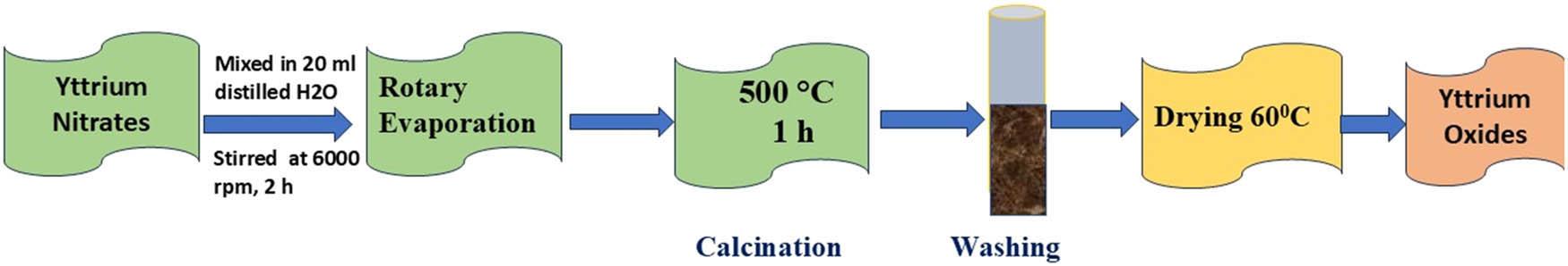
Pictorial representation of development of YTO nanoparticles.
The YTO nanoparticles were also characterized with FTIR, TEM, XRD, XPS, SEM, PL and UV-Vis spectroscopic methods. The size and surface particle morphology were determined by high-resolution TEM (HR-TEM, JEOL-2010 microscope running at 300 kV). Topography and thickness information were gathered with an Agilent 5500 SEM. The crystallinity of YTO was determined on an XRD (P-Analytical Empyrean), and chemical classes of metal oxides and their binding energies were detected with an XPS (Thermo Fisher Scientific EscaLab Xi+) of Physical Electronics. The UV-Vis absorption spectra were noted by a Lab-India UV-Vis spectrometer. The fluorescence spectra of YTOs were monitored by a fluorescence spectrophotometer (Hitachi FL-4600 spectrophotometer) in the range of 200–270 nm excitation wavelengths. Furthermore, surface functional groups on the material were detected through an FT-IR spectrophotometer (Agilent Cary 630).
A fluorescence spectrophotometer (with λ ex = 230 nm) was utilized to see the fluorescence spectra and intensities of YTO solution (10⁻3 M) after various quantities of vitamin B12 (10⁻4 M) were introduced into the solution, and distilled water was used as the solvent for all experiments in this study. The experiment was conducted three times for quality control. At room temperature, the vitamin B12 diagnostic procedure was performed. Various vitamins, amino acids, and metal ions were introduced to YTO solution under the same circumstances as vitamin B12 to further estimate the selectivity of the nano-sensing system for this nutrient.
In various challenging conditions, including UV lamp exposure, high-salinity solutions, pH fluctuations, and prolonged stress, the stability and exceptional properties of YTO were thoroughly examined to enhance its applicability. Notably, the fluorescence intensity of YTO remained consistent across a wide range of sodium chloride salt concentrations (1–5 M), demonstrating remarkable stability even in environments with high ionic strength (5 M, Figure 2a). This resilience suggests that YTO can effectively resist quenching from salts and other adverse factors, making it highly valuable for environmental and sensing applications. Furthermore, when subjected to UV lamp radiation, YTO exhibited outstanding photostability, with its PL intensity showing negligible variation even after 100 min of continuous exposure (Figure 2b). This property highlights its superior resistance to photobleaching, further reinforcing its potential for long-term use in demanding conditions. For sensor applications, it is essential to maintain steady fluorescence intensity to ensure accurate and reproducible observations. YTO is an appropriate selection for monitoring the environment tasks necessitating reliable and accurate detection of analytes or pollutants, as its fluorescence intensity stays steady at high ionic strength circumstances. Furthermore, fluorescence stability across numerous pH solutions was evaluated, with the results presented in Figure 2c. In acidic conditions (pH 2–5), fluorescence intensity was initially low. However, it peaked between pH 7 and 8 before declining at higher pH levels. This reduction in fluorescence intensity at elevated pH values can be credited to factors such as surface passivation layer degradation, nanoparticle aggregation, and other chemical changes [29,30].
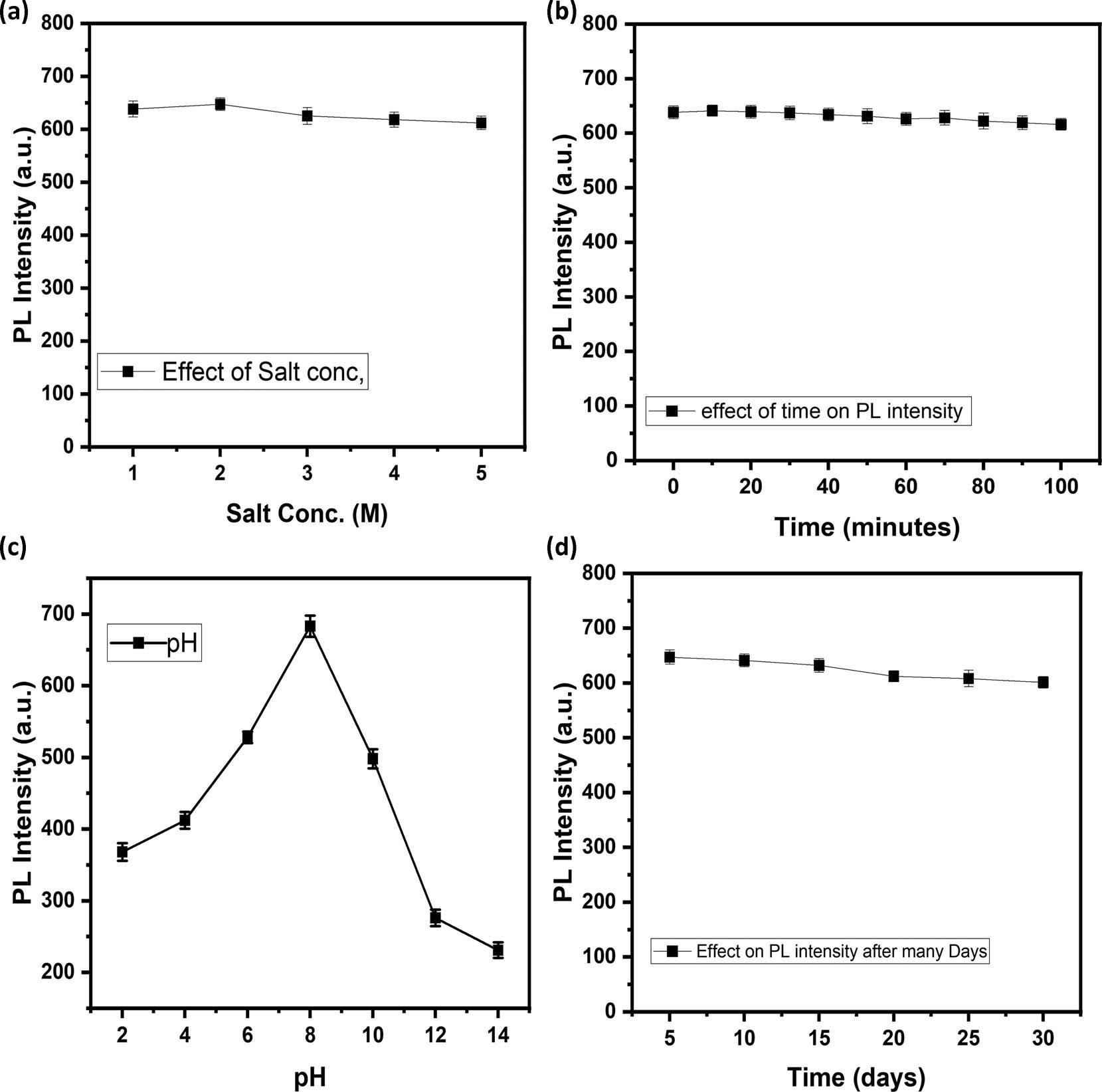
Effect of (a) factor of salt conc.; (b) factor of time (min); (c) pH; and (d) time (days) on the PL intensity of YTOs.
These findings highlight YTO’s significant potential for use in various fields, including sensing and environmental studies. Notably, the fluorescence peaks of YTO remained stable over a 1-month period (Figure 2d), indicating excellent long-term stability. Continuous sensing and long-term environmental monitoring are two examples of applications that require fluorescence performance that is consistent over lengthy periods of time. This stability is therefore crucial for these applications [31,32]. Additionally, Figure 6c presents the emission spectra of YTO at different excitation wavelengths fluctuating from 200 to 270 nm. The PL intensity at 285 nm initially increases with excitation from 200 to 230 nm, followed by a decline in the 240–270 nm range. The results show that the emission wavelength of YTO, which peaks at 230 nm, is unaffected by the excitation wavelength.
Fluorescence-based sensing experiments were performed to sense vitamin B12 with high sensitivity. Vitamin B12 concentration was shown to cause a linear dampening of YTO fluorescence intensity. The fluorescence quenching equation was used to compute the Stern–Volmer constant (K
sv) [33]:
TEM was used to study the structural and morphological characteristics of YTO nanoparticles. HR-TEM images showed clearly defined nanostructures with a fairly homogeneous size distribution and visible lattice fringes, attesting to the crystalline nature of the material (Figure 3a). The particles showed predominantly spherical or slightly irregular shapes, ranging in nanometer size [35]. The observation of lattice fringes in HR-TEM images also signified well-ordered atomic structures, implying high purity and phase stability. Apart from morphology, TEM analysis further indicated a propensity of some nanoparticles to self-assemble into small aggregates, a feature typically found in oxide nanoparticles because of high surface energy and van der Waals forces [36]. Such an aggregation can impact surface reactivity and optical properties, although in some cases, it can also lead to improved stability. The microstructural characteristics also indicate the existence of voids and interparticle voids, as consistent with mesoporous features commonly described for rare-earth oxides [37]. This porosity increases the surface area, thereby enhancing analyte interactions and improving performance in sensing and catalysis. Figure 3b illustrates the particle size distribution histogram of YTO nanoparticles, fitted with a log-normal distribution curve, exhibiting a mean particle size of 7.0 ± 1.8 nm and an R 2 value of 0.96, indicating a good fit.
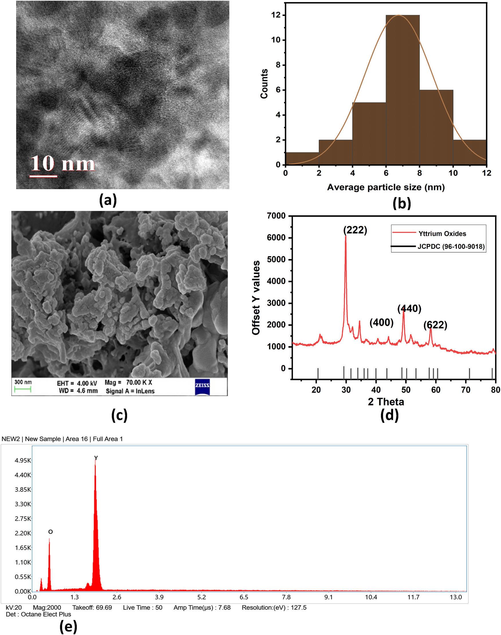
(a) HR-TEM image, (b) average particle size, (c) FE-SEM image, (d) XRD spectra, and (e) EDX spectra of YTOs.
SEM imaging (Figure 3c) validated the nanoscale nature of the particles, though particle size and shape variations were apparent, further corroborating the partial aggregation tendency. The EDX spectrum (Figure 3e) confirmed the elemental structure of the nanoparticles with robust signals for yttrium and oxygen, determining material purity. These findings collectively suggest that YTO nanoparticles have a crystalline, porous, and partially aggregated morphology, characteristics that make them more favorable as sensing, catalysis, and optoelectronic device materials.
XRD examination was conducted to ascertain phase formation and crystalline structure of YTO nanoparticles, as shown in Figure 3d. Sharp and distinct diffraction peaks were visible in the XRD spectra, indicating that the samples were extremely crystalline. The diffraction peaks were indexed to the cubic phase of YTOs. The most intense peaks appeared at characteristic 2θ values, corresponding to the (222), (400), (440), and (622) crystal planes, which are typical of cubic YTOs. Overall, the XRD results confirmed the successful synthesis of phase-pure YTO nanoparticles with high crystallinity [38].
The FTIR spectra of YTO nanoparticles, as depicted in Figure 4a, have three major absorption bands. The first band, in the range of 600–1,400 cm⁻1, is related to the stretching vibrations of the Y–O bond, validating the existence of YTO. The second band, within the range of 1,500–2,500 cm⁻1, is associated to the asymmetric stretching vibrations of the C–O band. This is probably due to the uptake of atmospheric CO2, even in the absence of precursors containing C–O during the synthesis. The O–H stretching vibrations of water molecules are responsible for the third band, which is located between 3,000 and 3,500 cm⁻1. This frequency range is ascribed to water absorption by either KBr pellets, which are used as a reference material, or by YTO nanoparticles from the ambient atmosphere [39]. With decreasing particle size, surface effects are more significant, resulting in increased adsorption characteristics. The UV–Vis absorption spectrum of the prepared YTO nanoparticles (Figure 4b) displays a sharp absorption edge around 280 nm, which can be assigned to the fundamental bandgap transition of YTOs (5.5–5.8 eV). The absorption intensity is gradually decreased with increasing wavelength with weak shoulder-like features present in the 300–400 nm range. These characteristics may be attributed to intrinsic defect states, which are mainly oxygen vacancies and surface-related energy levels, that create localized states within the bandgap and enable sub-bandgap absorption. The broad, weak band found in the range 350–380 nm could be caused by charge-transfer transitions of oxygen vacancy states. The fact that these defect-related absorptions are present confirms the nanoscale character of the material and the effect of surface chemistry on its optical behavior, which can be the key to its future applications in photocatalysis and optoelectronic devices [40–42].
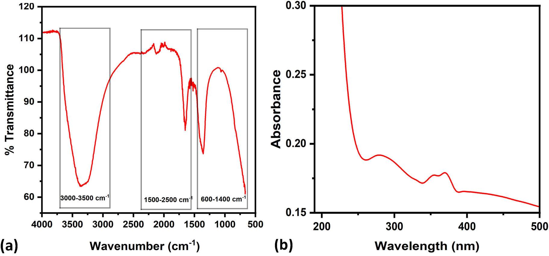
(a) FT-IR spectra and (b) UV-Vis absorption spectrum of YTO.
Figure 5a illustrates the full-scan XPS survey spectra of YTO nanoparticles. The deconvoluted Y3d spectra in Figure 5b reveal two main peaks at 167.4 and 170.5 eV corresponding to Y3d5/2 and Y3d3/2, respectively. Moreover, the O1s spectrum (Figure 5c) displays a peak of deconvolution at 535.7 eV corresponding to O–Y bonding, indicating that YTO was formed successfully [43,44].
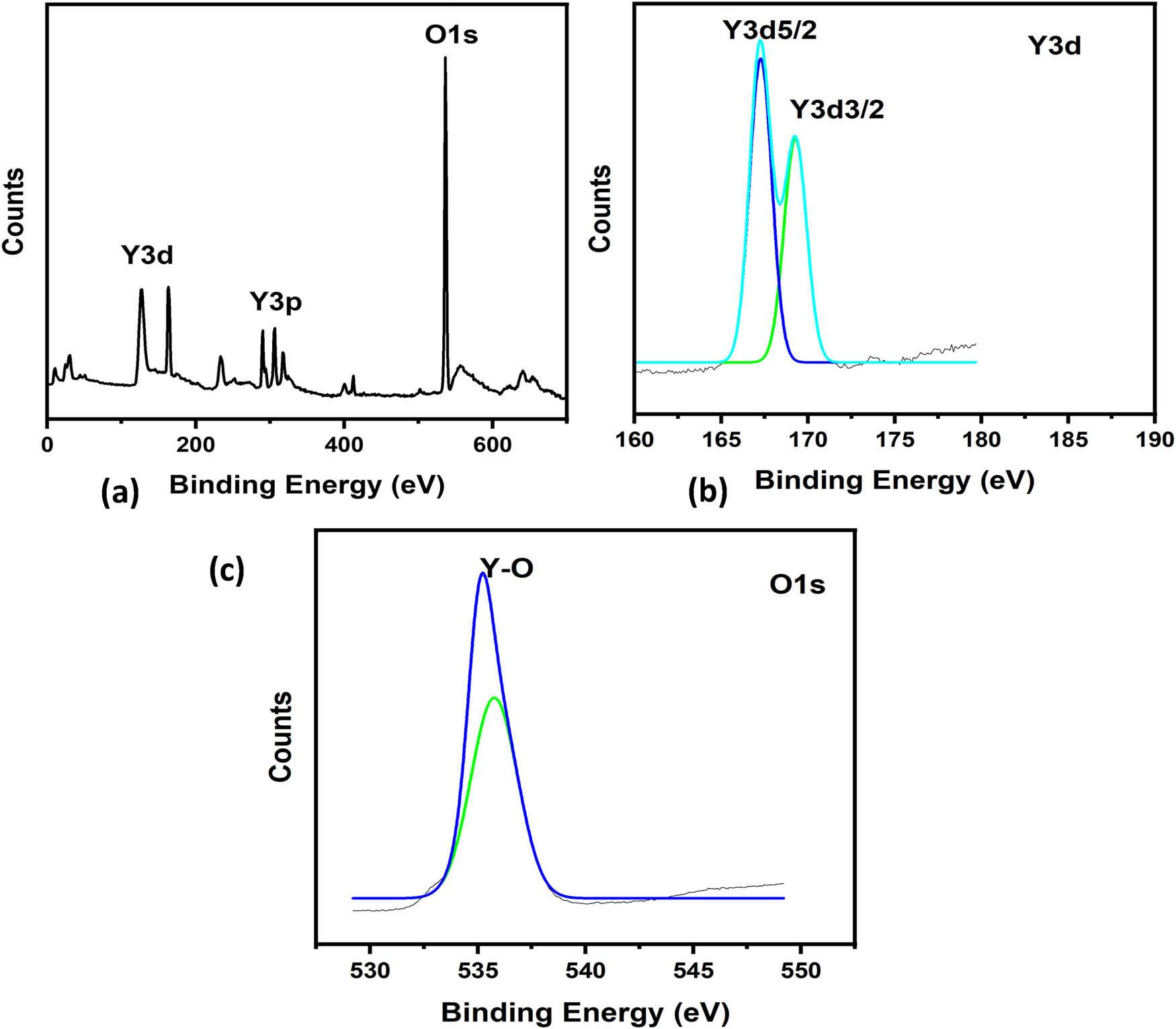
(a) Comprehensive XPS of YTO; (b) enhanced resolution spectra of Y3d; and (c) spectra of O1s.
Researchers looked at YTO ability for usage in fluorescence sensors due to its remarkable photostability and fluorescent properties. The investigation encompassed lysine, glycine, sodium chloride, alanine, riboflavin, biotin, yttrium nitrate, dopa, phenylalanine, tryptophan, and melatonin, which include amino acids, vitamins, and metal ions. Selectivity plots illustrate the material’s selectivity for vitamin B12, revealing that only vitamin B12 significantly reduces the fluorescence spectra of YTO. The fluorescence intensity of YTO at 285 nm gradually decrease as vitamin B12 concentrations (10–100 μM) rise, as seen in Figure 6a, demonstrating the nanosensor’s ability to reliably and precisely detect vitamin B12. A strong linear relationship was observed between the fluorescence ratio (F/F0) and vitamin B12 concentrations in the range of 10–100 μM. Figure 6b shows the titration procedure and the linear connection (R 2 = 0.97948) between YTO and vitamin B12. Moreover, it has been determined that the detection of vitamin B12 has a limit of detection (LOD) of 18.37 μM and a quantification limit of 55.66 μM, which is much higher than the capabilities of current methods. These results all support the excellent sensitivity and high selectivity of the “turning-off” fluorescence nanosensor for specific vitamin B12 identification in sample analysis. The LOD/ limit of quantification values achieved are at the micromolar level, which is above clinically significant nanomolar levels of vitamin B12. This work is therefore a proof-of-concept, and future efforts will concentrate on enhancing sensitivity by nanoparticle surface modification and experimental optimization.
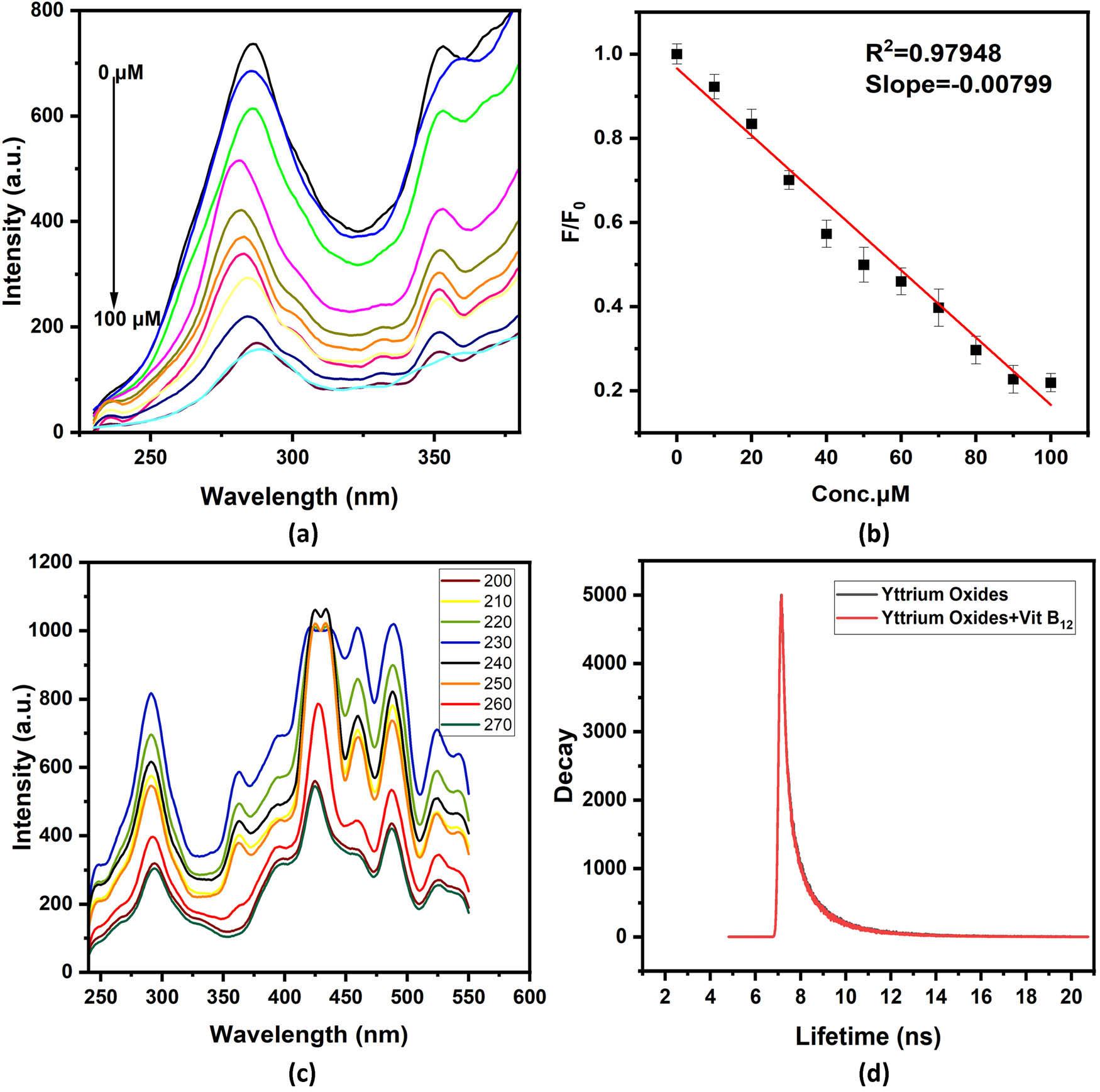
(a) PL spectra of YTO quenching with vitamin B12, (b) linear calibration of fluorescence intensity with vitamin B12, (c) excitation dependent emission spectra of YTO, and (d) fluorescence lifetime of pure YTOs nanoparticles and mixture with vitamin B12.
Fluorescence quenching is a decrease in the fluorescence emission intensity of probe-detecting molecules by ground-state complex formation, energy transfer, molecular interactions, and collision processes. Typical quenching processes include photoinduced electron transfer (PET) and fluorescence resonance energy transfer (FRET) [45,46]. PET is often associated with observable changes in fluorophore lifetime [47]. In this work, FRET can be eliminated since the positively charged YTO nanoparticles render the sub 10 nm donor-acceptor distances necessary for FRET improbable under mildly basic to neutral conditions (pH 7–8). The reduction in fluorescence observed is mainly due to the inner filter effect (IFE), an optical event brought about by the overlap of the quencher’s UV absorption spectrum with the fluorophore’s excitation or emission spectrum. In contrast to real quenching events involving molecular interactions, IFE is independent of temperature and gives a way of distinguishing it from dynamic quenching. Fluorescence lifetime measurements on the nanoparticles before and after mixing with vitamin B12 further corroborated the dominance of the IFE mechanism. The addition of vitamin B12 did not significantly alter the fluorescence lifetime, which remained at 8.4 ns before and 8.2 ns after. This indicates that the decrease in fluorescence intensity is due to the IFE rather than FRET, as shown in Figure 6d. Moreover, quenching can be static or dynamic: static quenching is the creation of a ground-state non-emissive complex, whereas dynamic quenching occurs as a result of collisional interactions. The Stern–Volmer plot (Figure 6b) is highly linear, reflecting that the observed quenching is largely static, with IFE being the major contributor [48,49].
The main factor influencing YTO selectivity is the specific identification of vitamin B12. The fluorescence response of YTO was compared with that of several different metal salts, vitamins, and amino acids to determine the degree of selective sensing activity. The PL intensity of several other analytes with comparable chemical structures, including glycine, alanine, lysine, sodium chloride, riboflavin, biotin, yttrium nitrate, dopa, tryptophan, phenylalanine, and melatonin, were tested to verify the selectivity of the proposed sensing probe. Figure 7a shows the histogram of the fluorescence intensity response. Figure 7b shows the interference selectivity plot of various analytes with vitamin B12. The selectivity analysis revealed that the fluorescence intensity of YTO was essentially unaffected by the presence of these species (100 μM).
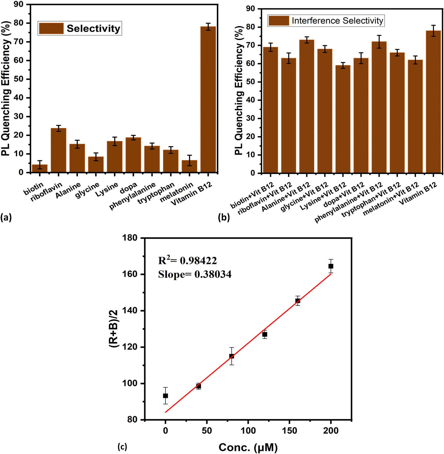
(a) Selectivity plot of YTO with various analytes, (b) interference selectivity plot of YTO with various analytes in the presence of vitamin B12, and (c) RBG-based detection of vitamin B12 with YTO.
Smartphone-based imaging sensors and portable devices provide better accuracy than the human eye’s visual detection [50,51]. For this research, an Android-based software was developed in-house to complement YTOs solution-based nanosensor. The sample was photographed by the smartphone, and the images were processed with RGB (red, green, and blue color space) software to identify color changes. To facilitate precise image acquisition, the camera was set around 6 cm away from the sample container without flash. The incremental addition of vitamin B12 to the solution of YTOs brought about an easily observed, naked-eye-visible color transition from light blue to dark blue. The lower LOD was made to be 51.64 μM, in the concentration range of 0–200 μM (Figure 7c). RGB profiling was subsequently applied to assess the degree of color transitions employing the camera within a smartphone with higher precision determination of vitamin B12 [52,53].
The concentration of vitamin B12 in human urine samples was measured with YTOs as fluorescence probes to check the reliability and usefulness of the fluorescence detection process. Spiking of the urine samples with known concentrations of vitamin B12 was employed for the assay validation [54,55]. Recovery of spiked vitamin B12 (98.06–102.15%) with relative standard deviation (0.853–2.245%) is shown in Table 1. These findings prove that the established fluorescence method is reliable and accurate for determining vitamin B12 in human urine samples. Given the excellent recovery and precision observed, this method has potential applications for other biological fluids, such as serum, which can be explored in future research.
Vitamin B12 real sample studies using YTOs.
| Sample | Spikey | Found | % Recovery | R.S.D |
|---|---|---|---|---|
| Urine | 20 | 20.23 | 101.15 | 1.235 |
| 40 | 40.86 | 102.15 | 1.527 | |
| 60 | 59.29 | 98.82 | 0.853 | |
| 80 | 78.45 | 98.06 | 2.245 |
This work successfully synthesized fluorescent YTO nanoparticle for the sensitive and selective detection of vitamin B12, leveraging its interaction with YTO for precise fluorometric measurements. Comprehensive spectroscopic and microscopic characterization confirmed the structural and optical properties of the nanoparticles. The wavelengths of 230 and 285 nm were found to be the most effective for excitation and emission, respectively. Key analytical parameters, including pH, incubation time, and NaCl concentration, were systematically optimized, yielding a linear calibration range of 10–100 μM. The method demonstrated reliability, with detection and quantification limits of 18.37 and 55.66 μM, respectively. Furthermore, an innovative RGB-based sensor was developed using these nanoparticles, enabling efficient visual detection of vitamin B12. The method was successfully applied to human urine samples, achieving high precision and satisfactory recoveries, underscoring its potential for real-world biomedical and analytical applications.
The authors extend their appreciation to Taif University, Saudi Arabia, for supporting this work through project number (TU-DSPP-2025-44).
This research was funded by Taif University, Saudi Arabia, Project No. (TU-DSPP-2025-44).
Salma Alshehri: Writing – Review & Editing, Formal analysis; Mohammad Shariq: Supervision, Writing – Original Draft, Writing – Review & Editing; Project administration; Wafa Al-Gethami: Project administration, Writing – Review & Editing, Formal analysis; Aisha H. Al-Moubaraki: Validation, Writing – Review & Editing, software; M. D. Alshahrani: Validation; Writing – Review & Editing, resources; Nouf Alharbi: Visualization, Writing – Review & Editing, Resources; Hind S. Alzahrani: Writing – Review & Editing; Noha Al-Qasmi: Validation.
The authors declare no competing financial or personal interests.
The data that support the findings of this study are available upon reasonable request from the authors.