
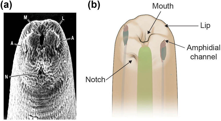
Figure 1
S. stercoralis iL3s have two tripartite labia. (a) Scanning electron micrograph shows the head of an S. stercoralis iL3. M, mouth; A, amphid openings; L, one of the lateral labia; N, the notch that attaches the mouth cavity to the cuticle. Image is reprinted from Fine et al., 1997 with permission; data are from Ashton et al., 1995 (Ashton et al., 1995, Fine et al., 1997). (b) Illustration of the head of an S. stercoralis iL3, showing the position of the amphidial channels, lips (raised bumps on the head), and notch relative to the mouth opening. One of the two amphidial channels and one of the six lips are labeled. The pharynx is shown in green.
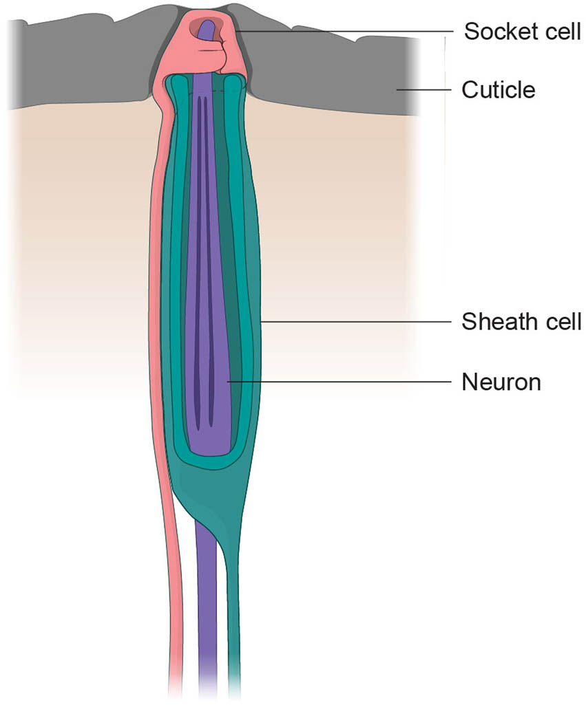
Figure 2
A typical nematode sensillum consists of a socket cell, a sheath cell, and neuronal processes. The illustration shows a typical nematode sensillum with a circular socket cell (pink), a sheath cell that forms the sensillar channel (teal), and the neuronal processes that are housed within the sensillum (purple). The image is adapted from Ashton & Schad, 1996 (Ashton et al., 1995, Ashton & Schad, 1996).
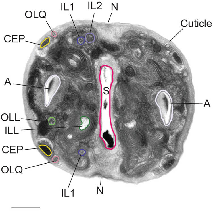
Figure 3
The putative mechanosensilla of S. stercoralis. Transmission electron micrograph (TEM) of the head of an S. stercoralis iL3. The labial neurons (IL1, IL2, OLL, ILL, and OLQ), cephalic neurons (CEP), amphid sensilla (A), and cuticle in the iL3 head are labeled. The notch (N), which attaches the mouth cavity to the cuticle, and the stoma (S), which is the mouth opening, are also labeled. A subset of the cephalic and labial neurons, the amphid, and the stoma are outlined. Scale bar = 0.5 µm. TEM image is reprinted from Fine et al. 1997 with permission; data are from Ashton et al., 1995 (Ashton et al., 1995, Fine et al., 1997).
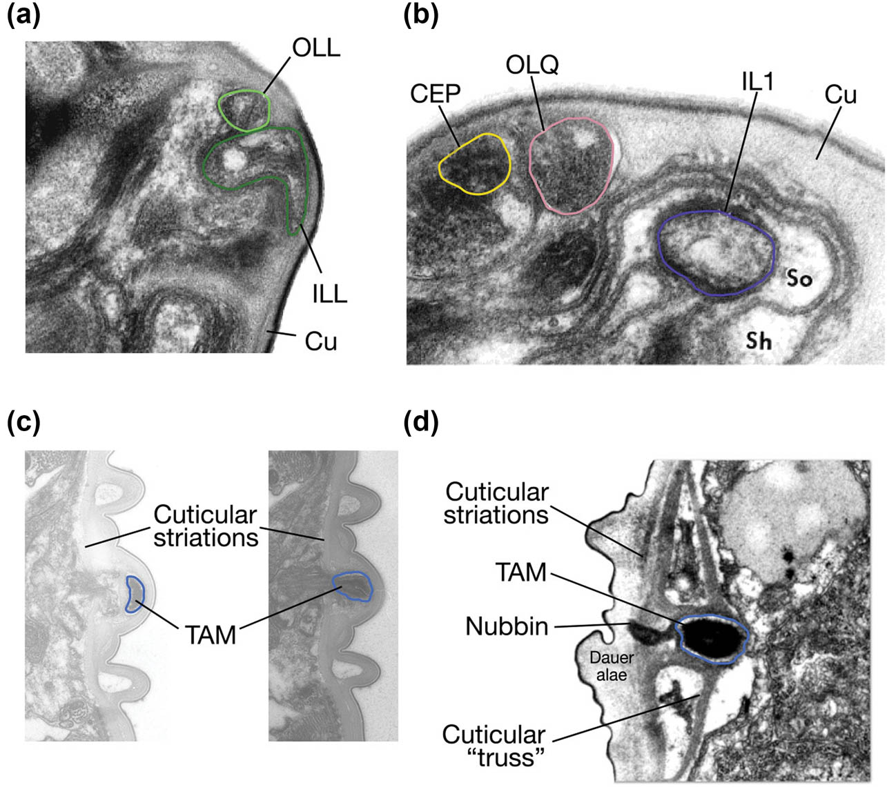
Figure 4
The sensory endings of most labial, cephalic, and deirid neurons abut or are embedded within the cuticle. (a) Transmission electron micrograph shows that the flattened foot-shaped endings of the S. stercoralis inner lateral labial neuron (ILL) and the sensory endings of the S. stercoralis outer lateral labial neuron (OLL) are located next to each other, underneath the cuticle. The ILL and OLL endings are outlined using the same color-coding as in Figure 3. The cuticle (Cu) is indicated. Adapted from Fine et al., 1997 with permission; data are from Ashton et al., 1995 (Ashton et al., 1995, Fine et al., 1997). (b) Transmission electron micrograph shows that both Sst-OLQ (light pink) and Sst-CEP (yellow) terminate underneath the cuticle, in close proximity to each other. The IL1 neuron (dark purple), the cuticle (Cu), the sheath cell (Sh), and the socket cell (So) are also labeled. Adapted from Fine et al., 1997 with permission; data are from Ashton et al., 1995 (Ashton et al., 1995, Fine et al., 1997). (c) Transmission electron micrographs show the sensory endings of the Sst-ADE neuron embedded in the iL3 cuticle, above the cuticular striations, in two distinct sections. The Sst-ADE dendrite is outlined in blue, and the cuticular striations and electron-dense tubule-associated material (TAM) found in the Sst-ADE cilia are labeled. Negatives: Sste_GAS_198.1_015894-1 (left) and Sste_GAS_198.1_015897 (right). Image credit: Nathan Schroeder; images originally acquired by G. Schad. (d) Transmission electron micrograph shows the sensory ending of the Cel-ADE neuron, which is outlined in blue, in a C. elegans dauer larva. Unlike in S. stercoralis iL3s, in C. elegans dauers the sensory ending of Cel-ADE is enclosed in a cuticular truss and lies mostly below the striated section of the cuticle. The sensory ending of Cel-ADE also has electron-dense TAM. Reproduced from Wolkow and Hall, 2011 (Wolkow & Hall, 2011).
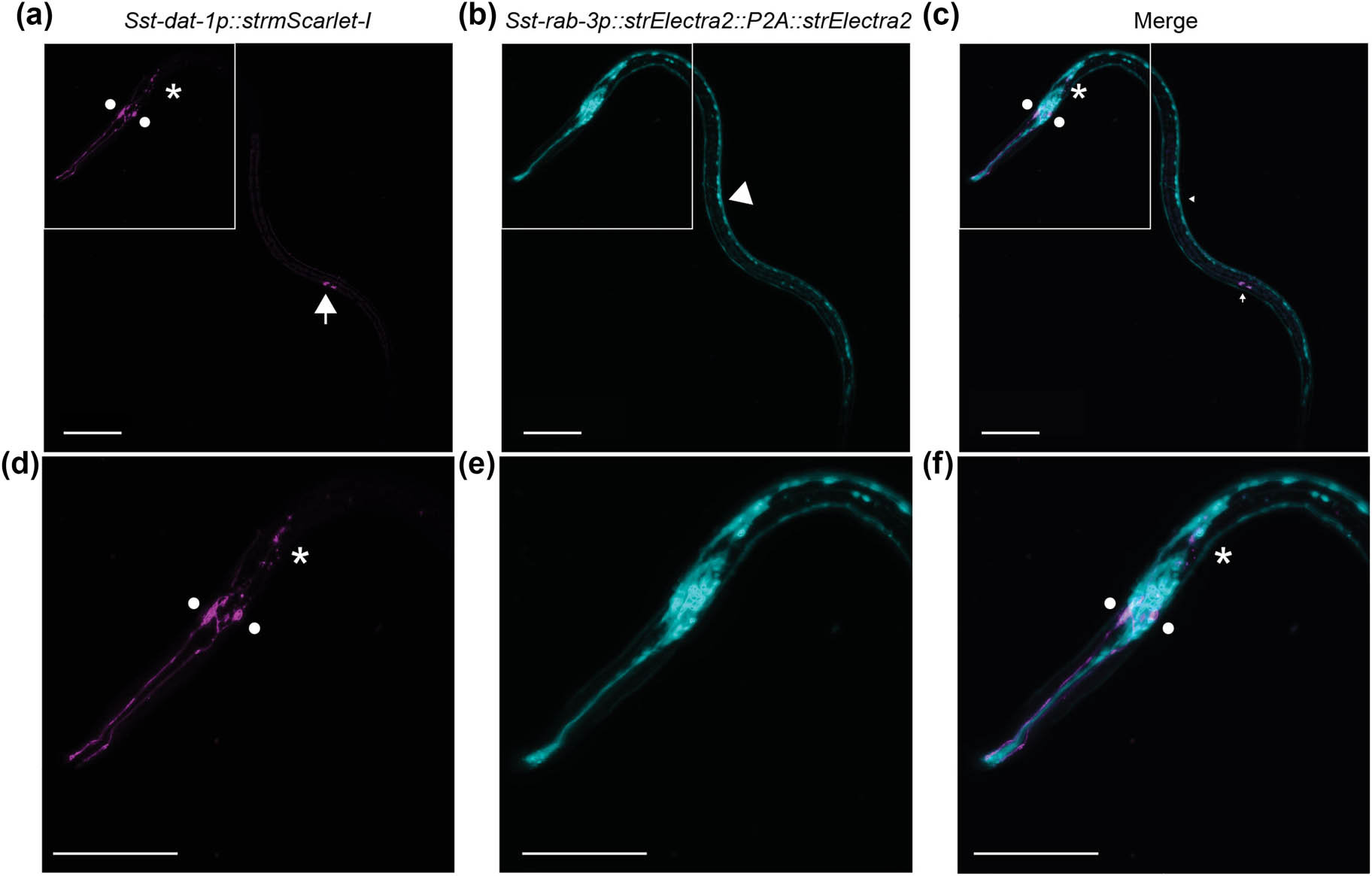
Figure 5
Detection of S. stercoralis CEP, ADE and PDE neurons using a transcriptional reporter for Sst-dat-1. (a) Confocal micrograph shows the labeling of the Sst-CEP, Sst-ADE, and Sst-PDE neurons in an iL3 using the transcriptional reporter for the S. stercoralis dat-1 gene, which is predicted to encode the S. stercoralis dopamine reuptake transporter (Patel et al., 2025). (b) A transcriptional reporter for the pan-neuronal marker gene Sst-rab-3 labels most neurons in the head and along the body of the iL3. (c) The merged image shows the positions of the dopaminergic neurons relative to other neurons in the iL3. (d–f) Images show the same worm as in (a–c), with the head magnified and zoomed in. For all images, the circles, asterisk, and arrow label the putative Sst-CEP, Sst-ADE, and Sst-PDE neurons, respectively. The white arrowhead labels the ventral nerve cord. The head of the iL3 is to the left, and the dorsal side of the iL3 is facing down. Scale bar = 50 µm. Reproduced from Patel et al., 2025 (Patel et al., 2025).
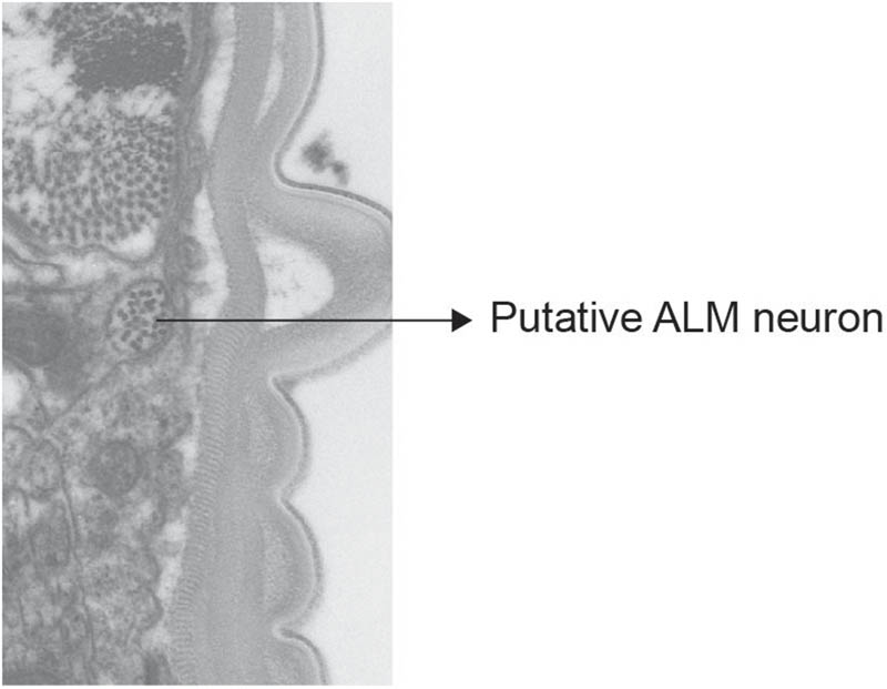
Figure 6
Putative touch receptor neurons have been detected in S. stercoralis. A transmission electron micrograph shows the detection of the putative Sst-ALM neuron. Similar to the Cel-ALM neuron processes, the Sst-ALM processes are located medial to the subdorsal body-wall muscle and are enriched in microtubules. Negative: Sste_GAS_198.1_018714. Image credit: Nathan Schroeder; images originally acquired by G. Schad.
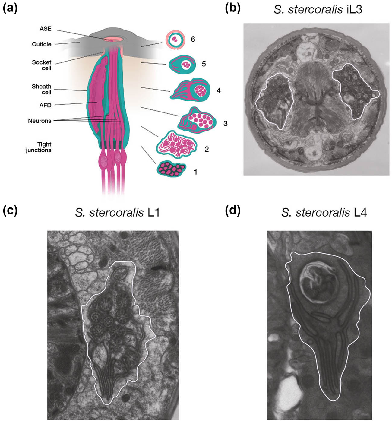
Figure 7
The amphid sensilla of S. stercoralis. (a) Longitudinal representation of an amphid sensillum with transverse sections at various levels. The first transverse section is taken just above the base of the amphid and shows the region where all thirteen amphid neurons form tight junctions with each other. All the amphid neurons are enclosed within the sheath cell. Shortly thereafter, as shown in Section 2, twelve of the thirteen amphid neurons (all but Sst-AUA) send out lateral, fin-like projections that intertwine. The central process of Sst-AFD, along with the dendritic processes of eleven amphid neurons, is visible in Section 3. Section 4 shows the dramatic, branched morphology of the Sst-AFD process, which is embedded in the sheath cell and wraps around the other amphid neurons. Section 5 shows ciliated processes of fewer amphid neurons, enclosed in the sheath cell. The process of Sst-AFD is less branched in this section. The final section shows the ciliated dendrites of two neurons, one of which is Sst-ASE, enclosed in the sheath and socket cells. Also note the enlarged and bulbous tip of the Sst-ASE neuron at the top of the sensillum, located very close to the cuticular opening. Image adapted from Ashton & Schad, 1996; data are from Ashton et al., 1995 (Ashton et al., 1995, Ashton & Schad, 1996). (b) Transmission electron micrograph shows the amphid neurons of an S. stercoralis iL3. The lateral, fin-like projections can be clearly seen on the right side of the section. The left side of the section shows the large central process of Sst-AFD as well as some of its lamellae. Negative: Sste_GAS_198.1act_012865. Image credit: Nathan Schroeder; images originally acquired by G. Schad. (c) Transmission electron micrograph shows the amphid neurons of an S. stercoralis L1-stage larva. The lamellar structure of the Sst-AFD dendrite is visible. Negative: Sste_GAS_L1_11_014700. Image credit: Nathan Schroeder; images originally acquired by G. Schad. (d) Transmission electron micrograph shows the amphid neurons of an S. stercoralis L4-stage larva. The lamellar structure of Sst-AFD is also visible at this stage. Negative: Sste_GAS_L4_FI_020528-1. Image credit: Nathan Schroeder; images originally acquired by G. Schad. For (b–d), the amphid is outlined in white.
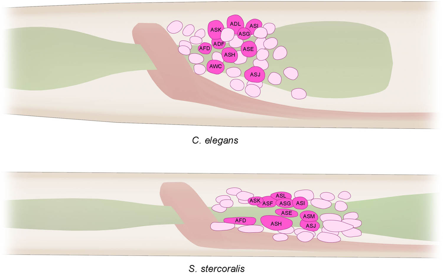
Figure 8
Positions of the cell bodies of a subset of amphid neurons in a C. elegans L1 larva and an S. stercoralis iL3. The schematic shows the relative positions of the amphid cell bodies that lie posterior to the nerve ring (peach), within the lateral ganglia, in C. elegans and S. stercoralis. The pharynx is shown in green. Adapted from Ashton et al., 1995 with permission (Ashton et al., 1995). Cell body locations are approximate, and only a subset of the head neuron cell bodies is shown.
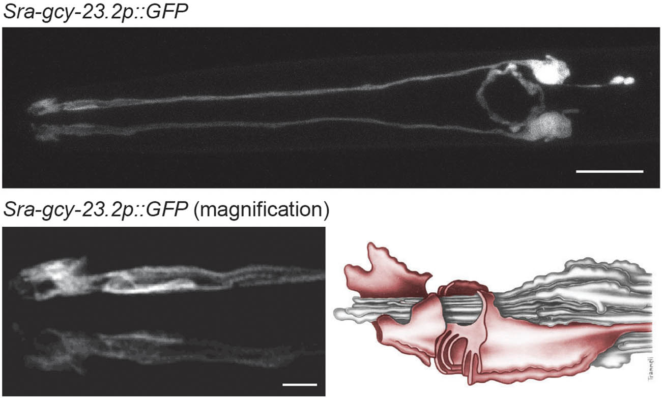
Figure 9
Detection of the S. stercoralis AFD neurons using a transcriptional reporter for a Strongyloides thermoreceptor gene. Top, confocal micrograph shows the morphology and position of the Sst-AFD neurons, as labeled by a transcriptional reporter for the Strongyloides ratti Sra-gcy-23.2 gene, which encodes a putative thermoreceptor (Bryant et al., 2022). Scale bar = 10 µm. Lower left, a magnified view of the dendritic endings of the AFD neurons. Scale bar = 2 µm. Lower right, a reconstruction of the amphid neurons modified from Ashton et al., 1995 and Lopez et al., (2000) with permission; figure reproduced from Bryant and Hallem, 2018 with permission (Ashton et al., 1995, Lopez et al., 2000, Bryant et al., 2018). Entire image reproduced from Bryant et al., 2022 (Bryant et al., 2022).
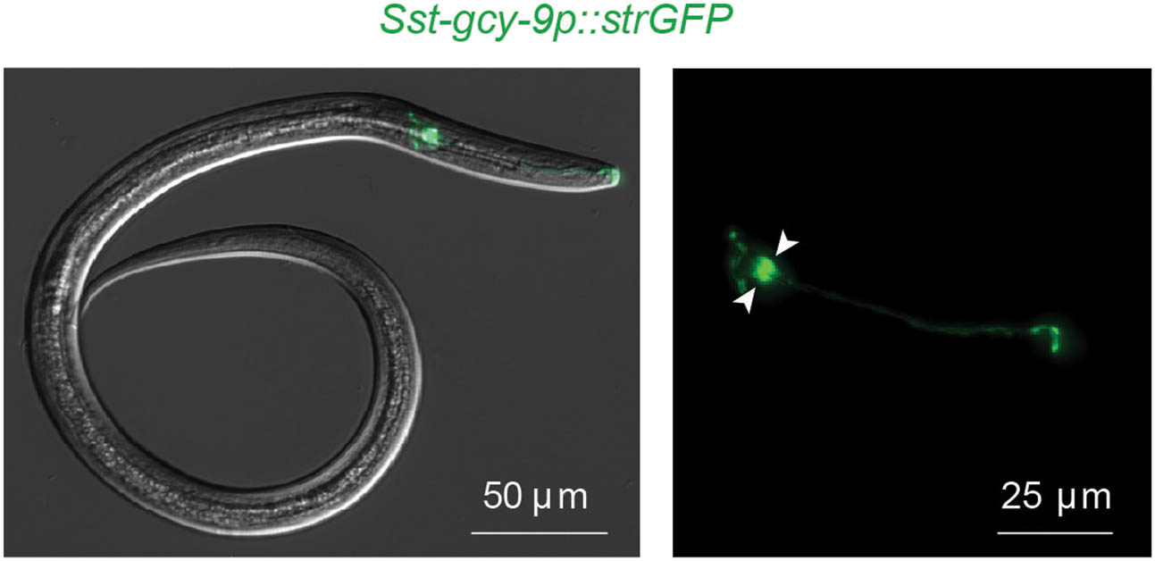
Figure 10
S. stercoralis has a single pair of BAG sensory neurons in the head. A fluorescence micrograph shows the S. stercoralis BAG sensory neurons of an iL3, as labeled by a transcriptional reporter for the Sst-gcy-9 gene, which encodes a receptor guanylate cyclase that is necessary for the detection of carbon dioxide (Banerjee et al., 2025). Arrowheads indicate the cell bodies of the paired Sst-BAG neurons. Scale bar = 50 µm (left image) and 25 µm (right image). Reproduced from Banerjee et al., 2025 (Banerjee et al., 2025).