
The nervous system of the human-parasitic nematode Strongyloides stercoralis is presumed to be largely similar to that of the free-living nematode Caenorhabditis elegans. This assumption is based on the general conservation of sensory neuroanatomy and function between S. stercoralis and C. elegans (Ashton et al., 1995, Ashton & Schad, 1996, Fine et al., 1997, Ashton et al., 1998, Lopez et al., 2000, Forbes et al., 2004, Ashton et al., 2007, Schafer, 2016, Bryant et al., 2022, Banerjee et al., 2025, Patel et al., 2025). S. stercoralis likely has both a distinct somatic nervous system, of which the sensory neurons are best characterized, and a pharyngeal nervous system, of which nothing is known. Similar to C. elegans, S. stercoralis has a circumpharyngeal nerve ring (Ashton et al., 1995, Fine et al., 1997, Schafer, 2016), which is a dense bundle of neural processes that is often called the worm brain. Additionally, structures resembling the dorsal and ventral nerve cords have been detected in S. stercoralis iL3s (Patel et al., 2025).
The existing knowledge of the structure of the S. stercoralis nervous system comes from serial-section electron microscopy reconstructions of the amphid, labial, and cephalic sensilla of the head of infective third-stage larvae (iL3s) (Ashton et al., 1995, Fine et al., 1997). These sensilla house sensory neurons that innervate the two lips of S. stercoralis, which are each tripartite in structure and composed of large medial lobes and smaller dorsal and ventral lobes (Figure 1). The amphid sensilla are considered the primary chemosensory organs of S. stercoralis because they are open to the external environment (Ashton et al., 1995, Ashton & Schad, 1996, Ashton et al., 1999). In contrast, the labial and cephalic sensilla are likely mechanosensory because these organs do not open to the outside but instead house sensory processes that terminate in close association with the cuticle (Fine et al., 1997). As in C. elegans, the relative positions and morphologies of the sensilla in S. stercoralis are largely invariant among individuals that were studied (Ashton et al., 1995, Fine et al., 1997).
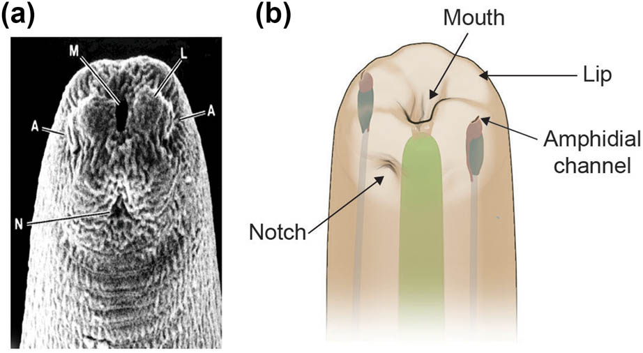
Figure 1
S. stercoralis iL3s have two tripartite labia. (a) Scanning electron micrograph shows the head of an S. stercoralis iL3. M, mouth; A, amphid openings; L, one of the lateral labia; N, the notch that attaches the mouth cavity to the cuticle. Image is reprinted from Fine et al., 1997 with permission; data are from Ashton et al., 1995 (Ashton et al., 1995, Fine et al., 1997). (b) Illustration of the head of an S. stercoralis iL3, showing the position of the amphidial channels, lips (raised bumps on the head), and notch relative to the mouth opening. One of the two amphidial channels and one of the six lips are labeled. The pharynx is shown in green.
The original nomenclature used for S. stercoralis neurons was similar but not identical to that used for C. elegans. Each S. stercoralis amphid neuron was designated by a three-letter code, where the “A” in the first position indicates that the neuron is part of the amphid, the “S” in the second position indicates that the neuron extends a single ciliated dendritic ending, and the letter designation in the third position indicates a letter that was given to each of the thirteen amphid neurons in S. stercoralis (Ashton et al., 1995). It should be noted that there are no dual-ciliated amphid neurons in S. stercoralis (Ashton et al., 1995), unlike in C. elegans (Altun & Hall, 2011); thus, “S” is the second letter in the name of most S. stercoralis amphid neurons. The exception to this rule is the Sst-AFD neuron (originally called the ALD neuron, where the “L” designation was given to indicate the lamellar shape of its dendritic process), which was named Sst-AFD because it is the S. stercoralis homolog of the C. elegans thermosensory AFD neuron (Ashton et al., 1995, Bryant et al., 2022). Homology of individual neurons between S. stercoralis and C. elegans has been deduced partly on the basis of conservation of the relative positions of neuronal cell bodies (i.e., positional homology) between the two species and partly on the basis of conservation of function. When positional homologs of C. elegans amphid neurons could be clearly identified in S. stercoralis, the letter in the third position of the neuron name was conserved between the two species. For example, the Sst-ASE neuron is the S. stercoralis positional homolog of the Cel-ASE neuron, while the Sst-ASL neuron (“S” because it has a single dendritic process) is the positional homolog of the Cel-ADL neuron (“D” because it has two dendritic processes) (Ashton et al., 1995). In the future, cell lineage studies in S. stercoralis would further bolster the identification of homologous neurons between this nematode and C. elegans.
The inner labial, outer labial, and cephalic neurons of S. stercoralis have also been named after their putative C. elegans homologs, whenever such homologs were identified (Fine et al., 1997). Thus, the nomenclature for these neurons in S. stercoralis follows that of C. elegans. The exceptions to this case are the inner lateral labial neurons of S. stercoralis, which are quite different from the inner lateral labial neurons of C. elegans although they share similar anatomical positions in both species (Fine et al., 1997); the exact differences between the S. stercoralis and C. elegans inner lateral labial neurons are described below. As such, the inner lateral labial neurons in S. stercoralis have been named the Sst-ILL neurons (Fine et al., 1997), whereas the inner lateral labial neurons of C. elegans are called the Cel-IL1 and Cel-IL2 neurons. As more sensory neurons are functionally characterized in S. stercoralis, it may be beneficial to rename the S. stercoralis neurons with the same name as their C. elegans homologs to avoid confusion and facilitate comparative analysis across species.
1. Neurons
Only the morphologies of the sensory neurons in the amphid, labial, and cephalic sensilla of iL3s have been studied (Ashton et al., 1995, Fine et al., 1997). These neurons are all bipolar, with a single dendritic process and a single axon. The dendritic processes of these neurons have either simple, rounded tips or complex, multilayered endings, as described in detail below.
1.1. Nervous system development
The development of the nervous system of S. stercoralis has not yet been characterized.
1.2. Neuron categories
The different categories of neurons in S. stercoralis include sensory neurons, interneurons, polymodal neurons, and motor neurons. Of these, only the sensory neurons have been morphologically and/or functionally characterized thus far (Ashton et al., 1995, Fine et al., 1997, Ashton et al., 1998, Lopez et al., 2000, Forbes et al., 2004, Nolan et al., 2004, Ashton et al., 2007, Bryant et al., 2022, Banerjee et al., 2025, Patel et al., 2025).
1.3. Motor neurons and the motor circuit
Nothing is known about the motor neurons and motor circuits of S. stercoralis.
1.4. Sensory neurons
The sensory neurons in the head of S. stercoralis iL3s are the most thoroughly characterized of all the neuronal categories. Reconstructions of serial section electron micrographs of the head of the worm have shown the presence of 13 neurons in each of the two amphid sensilla and a total of twenty labial and cephalic neurons that are contained within 16 putative mechanosensilla (Ashton et al., 1995, Ashton & Schad, 1996, Fine et al., 1997). As described below, the relative positions of many of these neurons are largely conserved between S. stercoralis and C. elegans, but some neurons have alterations in morphology, which might relate to differences in function between the two species.
1.4.1. Mechanosensation
Sixteen putative mechanosensilla have been identified at the anterior tip of S. stercoralis iL3s via serial-section transmission electron microscopy (Fine et al., 1997). The mechanosensilla share the following components that are typical of a nematode sensillum: the dendritic processes of one or more neurons that traverse the lumen of the sensillar channel; a sheath cell, which forms the sensillar channel through which neurons send their processes; and a socket cell, which abuts the anterior end of the sheath cell, connecting the sensillar channel to the nematode body wall and creating a circular opening through which dendrites interface with the cuticle (in the case of mechanosensilla), the external environment (in the case of the amphid sensilla), or both (Figure 2) (Ward et al., 1975, Wright, 1980).
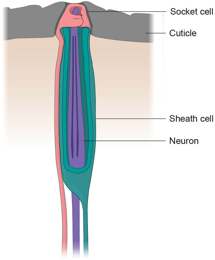
Figure 2
A typical nematode sensillum consists of a socket cell, a sheath cell, and neuronal processes. The illustration shows a typical nematode sensillum with a circular socket cell (pink), a sheath cell that forms the sensillar channel (teal), and the neuronal processes that are housed within the sensillum (purple). The image is adapted from Ashton & Schad, 1996 (Ashton et al., 1995, Ashton & Schad, 1996).
Like C. elegans, S. stercoralis has six inner labial sensilla, six outer labial sensilla, and four cephalic sensilla (Figure 3; Fine et al., 1997). The dendrites of the labial and cephalic neurons of S. stercoralis iL3s are not exposed to the external environment, and as such, it has been postulated that their function is primarily mechanosensory (Fine et al., 1997). As in C. elegans, the dendritic processes of the labial and cephalic sensilla of S. stercoralis are cilium-like and contain microtubule doublets (Fine et al., 1997).
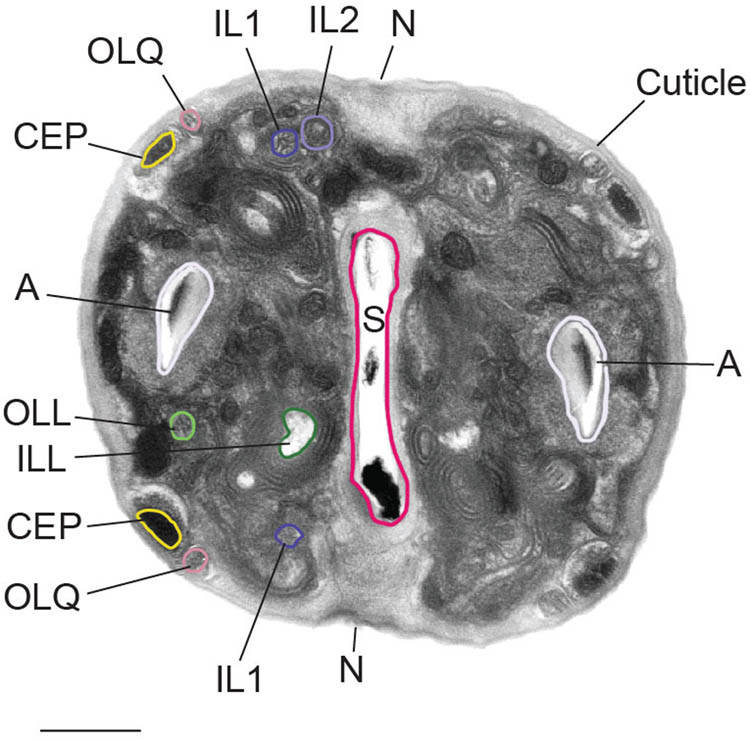
Figure 3
The putative mechanosensilla of S. stercoralis. Transmission electron micrograph (TEM) of the head of an S. stercoralis iL3. The labial neurons (IL1, IL2, OLL, ILL, and OLQ), cephalic neurons (CEP), amphid sensilla (A), and cuticle in the iL3 head are labeled. The notch (N), which attaches the mouth cavity to the cuticle, and the stoma (S), which is the mouth opening, are also labeled. A subset of the cephalic and labial neurons, the amphid, and the stoma are outlined. Scale bar = 0.5 µm. TEM image is reprinted from Fine et al. 1997 with permission; data are from Ashton et al., 1995 (Ashton et al., 1995, Fine et al., 1997).
The lips of S. stercoralis are innervated by both inner labial and outer labial sensilla (Fine et al., 1997). Two inner labial sensilla on the dorsal side, two inner labial sensilla on the ventral side, and one inner labial sensillum on each of the two lateral sides of the worm together comprise the complement of inner labial sensilla in S. stercoralis. The dorsal and ventral inner labial sensilla extend into the dorsal and ventral lobes of the nematode lips, while the lateral inner labial sensilla extend into the medial lobes of the lips. Each dorsal and ventral inner labial sensillum is innervated by the dendritic processes of two neurons, which have been named the S. stercoralis IL1 and IL2 neurons because of resemblance to the C. elegans IL1 and IL2 neurons. In iL3s, the Sst-IL1 neurons send dendritic processes that terminate just under the cuticle, whereas the dendritic processes of Sst-IL2 neurons terminate slightly posterior to those of Sst-IL1 and do not appear to directly contact the cuticle (Fine et al., 1997). In contrast, both the Cel-IL1 and Cel-IL2 neurons of C. elegans dauers terminate at roughly the same point and directly contact the dauer cuticle (Wolkow & Hall, 2011).
The inner lateral labial neurons (named the S. stercoralis ILL neurons) innervate the inner lateral labial sensilla, which extend into the medial lobes of the lips. It should be noted that only a single pair of inner labial neurons innervate the inner lateral labial sensilla of S. stercoralis iL3s, as opposed to two pairs of neurons that innervate these sensilla in C. elegans dauer and non-dauer stages (Fine et al., 1997, Altun & Hall, 2010, Wolkow & Hall, 2011). Interestingly, the Sst-ILL dendrites terminate in a flattened, foot-like shape immediately underneath the iL3 cuticle (Figure 4a; Fine et al., 1997); the foot-shaped sensory ending is unique to Sst-ILL and not detected in either Sst-IL1, Sst-IL2, or the inner labial neurons of C. elegans dauers (Wolkow & Hall, 2011).
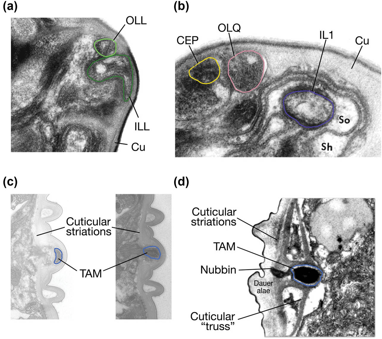
Figure 4
The sensory endings of most labial, cephalic, and deirid neurons abut or are embedded within the cuticle. (a) Transmission electron micrograph shows that the flattened foot-shaped endings of the S. stercoralis inner lateral labial neuron (ILL) and the sensory endings of the S. stercoralis outer lateral labial neuron (OLL) are located next to each other, underneath the cuticle. The ILL and OLL endings are outlined using the same color-coding as in Figure 3. The cuticle (Cu) is indicated. Adapted from Fine et al., 1997 with permission; data are from Ashton et al., 1995 (Ashton et al., 1995, Fine et al., 1997). (b) Transmission electron micrograph shows that both Sst-OLQ (light pink) and Sst-CEP (yellow) terminate underneath the cuticle, in close proximity to each other. The IL1 neuron (dark purple), the cuticle (Cu), the sheath cell (Sh), and the socket cell (So) are also labeled. Adapted from Fine et al., 1997 with permission; data are from Ashton et al., 1995 (Ashton et al., 1995, Fine et al., 1997). (c) Transmission electron micrographs show the sensory endings of the Sst-ADE neuron embedded in the iL3 cuticle, above the cuticular striations, in two distinct sections. The Sst-ADE dendrite is outlined in blue, and the cuticular striations and electron-dense tubule-associated material (TAM) found in the Sst-ADE cilia are labeled. Negatives: Sste_GAS_198.1_015894-1 (left) and Sste_GAS_198.1_015897 (right). Image credit: Nathan Schroeder; images originally acquired by G. Schad. (d) Transmission electron micrograph shows the sensory ending of the Cel-ADE neuron, which is outlined in blue, in a C. elegans dauer larva. Unlike in S. stercoralis iL3s, in C. elegans dauers the sensory ending of Cel-ADE is enclosed in a cuticular truss and lies mostly below the striated section of the cuticle. The sensory ending of Cel-ADE also has electron-dense TAM. Reproduced from Wolkow and Hall, 2011 (Wolkow & Hall, 2011).
S. stercoralis iL3s also have six outer labial sensilla, including two that are each innervated by a single Sst-OLL neuron and four that are each innervated by a single Sst-OLQ neuron. The knob-shaped tips of the Sst-OLL processes are located beneath the cuticle of the medial lobes of the lips (Figure 4a), whereas the processes of Sst-OLQ terminate as expanded tips beneath the cuticle of the dorsal and ventral lobes of the iL3 lips (Figure 4b). The types of mechanical stimuli (e.g., gentle touch, harsh touch, texture, or vibration) that activate the labial neurons, as well as their roles in parasitic behaviors such as host seeking, host attachment, skin penetration, and tissue migration, remain unknown.
Other putative mechanosensory neurons in the head of S. stercoralis include the four cephalic neurons, which are named the S. stercoralis CEP neurons, and the two anterior deirid neurons, which are named the S. stercoralis ADE neurons (Figures 4 and 5; Fine et al., 1997, Patel et al., 2025). The four Sst-CEP neurons each send processes through one of four cephalic sensilla, and the tips of these processes terminate as large, oblong-shaped structures that are flattened on the end abutting the cuticle (Figure 4b; Fine et al., 1997). These neurons innervate the dorsal and ventral lobes of the lips, with dendritic endings that lie in close proximity to those of Sst-OLQ (Fine et al., 1997). Like Cel-ADE, the sensory endings of Sst-ADE contain electron-dense material, which is likely necessary for the mechanosensory function of these neurons (Figure 4c, d). The Sst-ADE neurons in iL3s have mushroom-shaped sensory endings that are embedded underneath the alae and above the striated section of the cuticle (Figure 4c). In contrast, most of the electron-dense sensory endings of the Cel-ADE neurons in dauers are enclosed in a cuticular truss, with only a small nubbin sent into the alae (Figure 4d); neither cuticular trusses nor nubbins have been detected in S. stercoralis iL3s (Figure 4c, d). The functional significance of these differences remains unclear. A recent study showed that the Sst-CEP neurons, the Sst-ADE neurons, and/or the S. stercoralis posterior deirid neurons (Sst-PDE) are necessary for the behaviors performed by infective larvae during skin penetration (Patel et al., 2025). When S. stercoralis iL3s are placed on skin, they repeatedly push down on the skin with their heads, crawl on the skin, puncture the skin, and burrow into the skin until penetration is complete. Silencing the Sst-CEP, Sst-ADE, and Sst-PDE neurons using the histamine-gated chloride channel HisCl1 causes a reduction in pushing behavior and puncture attempts and ultimately inhibits skin penetration (Patel et al., 2025). These neurons are likely dopaminergic, similar to their C. elegans homologs, and act via conserved dopamine signaling pathways to drive skin penetration (Patel et al., 2025). Further studies will elucidate the downstream neural circuits that mediate the effect of the S. stercoralis dopaminergic neurons on skin penetration.
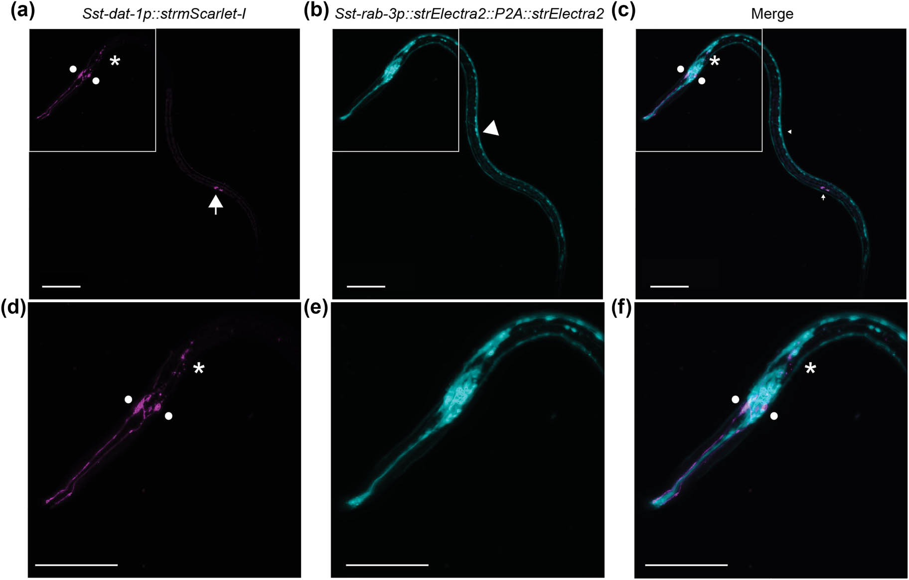
Figure 5
Detection of S. stercoralis CEP, ADE and PDE neurons using a transcriptional reporter for Sst-dat-1. (a) Confocal micrograph shows the labeling of the Sst-CEP, Sst-ADE, and Sst-PDE neurons in an iL3 using the transcriptional reporter for the S. stercoralis dat-1 gene, which is predicted to encode the S. stercoralis dopamine reuptake transporter (Patel et al., 2025). (b) A transcriptional reporter for the pan-neuronal marker gene Sst-rab-3 labels most neurons in the head and along the body of the iL3. (c) The merged image shows the positions of the dopaminergic neurons relative to other neurons in the iL3. (d–f) Images show the same worm as in (a–c), with the head magnified and zoomed in. For all images, the circles, asterisk, and arrow label the putative Sst-CEP, Sst-ADE, and Sst-PDE neurons, respectively. The white arrowhead labels the ventral nerve cord. The head of the iL3 is to the left, and the dorsal side of the iL3 is facing down. Scale bar = 50 µm. Reproduced from Patel et al., 2025 (Patel et al., 2025).
Because of the largely conserved sensory neuroanatomy between S. stercoralis and C. elegans (Ashton et al., 1995, Ashton & Schad, 1996, Fine et al., 1997, Schafer, 2016, Bryant et al., 2022, Banerjee et al., 2025, Patel et al., 2025), it is presumed that S. stercoralis also has homologs of C. elegans mechanosensory neurons that are not mentioned earlier. Indeed, a neuron that resembles the gentle touch receptor neuron Cel-ALM (Chalfie & Sulston, 1981, Chalfie et al., 1985) has been detected in S. stercoralis (Figure 6). However, additional homologous neurons, as well as the mechanosensory behaviors that these neurons mediate in S. stercoralis, are as-yet unknown.
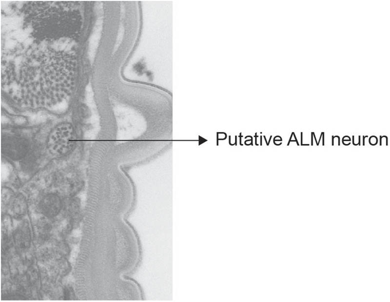
Figure 6
Putative touch receptor neurons have been detected in S. stercoralis. A transmission electron micrograph shows the detection of the putative Sst-ALM neuron. Similar to the Cel-ALM neuron processes, the Sst-ALM processes are located medial to the subdorsal body-wall muscle and are enriched in microtubules. Negative: Sste_GAS_198.1_018714. Image credit: Nathan Schroeder; images originally acquired by G. Schad.
1.4.2. Nociception
Nociceptive responses of S. stercoralis iL3s to two stimuli have been reported thus far: high concentrations of sodium chloride (NaCl) and acute increases in the concentration of ambient carbon dioxide (CO2) (Forbes et al., 2003, Forbes et al., 2004, Castelletto et al., 2014, Ruiz et al., 2017, Banerjee et al., 2025). S. stercoralis iL3s are repelled by high concentrations of NaCl (∼2.85 M NaCl), and this repulsion is dependent on the Sst-ASH neurons (Forbes et al., 2003, Forbes et al., 2004). Notably, the Sst-ASH neurons are only required for migration away from high concentrations of NaCl but are dispensable for migration toward preferred NaCl concentrations (Forbes et al., 2004). The S. stercoralis ASH neurons are part of the amphid sensilla, the morphology of which is described in detail below.
When ambient levels of CO2 increase suddenly from the trace levels found in air to 2.5%, S. stercoralis iL3s slow their crawling speed and increase their rate of reversals (Banerjee et al., 2025). Additionally, S. stercoralis iL3s migrate away from a CO2 source (Castelletto et al., 2014, Ruiz et al., 2017, Banerjee et al., 2025). These behavioral responses to CO2 are dependent on the S. stercoralis BAG neurons and the S. stercoralis receptor guanylate cyclase Sst-GCY-9, a putative receptor for CO2 (Banerjee et al., 2025). The morphology of the BAG neurons is also described below.
1.4.3. Chemosensation
The two amphid sensilla of S. stercoralis are considered the primary chemosensory organs (Ashton et al., 1995, Ashton & Schad, 1996, Ashton et al., 1999). These sensilla house neuronal processes exposed to the external environment, with openings that lie immediately lateral and posterior to the large, medial lobe of the lips. Each sensillum contains the components of a typical nematode sensillum, as described earlier, including a socket cell, a sheath cell, and neuronal processes (Ward et al., 1975, Wright, 1980). The S. stercoralis amphid neurons have ciliated dendritic processes endowed with microtubule doublets (Ashton et al., 1995), similar to C. elegans (Ward et al., 1975, Altun & Hall, 2010). In S. stercoralis, 13 neurons have been assigned to each amphid sensillum (Ashton et al., 1995), as compared with C. elegans, which has 12 neurons in each amphid (Ward et al., 1975, Altun & Hall, 2010). However, the so-called additional amphid neuron of S. stercoralis is likely the homolog of the C. elegans AUA neuron (Castelletto et al., 2024). The AUA neuron is not considered an amphid neuron in C. elegans and two other species of free-living nematodes, Pristionchus pacificus and Acrobeles complexus, because its dendritic processes terminate near the nerve ring and do not enter the amphid sensilla in these species (White et al., 1986, Bumbarger et al., 2009, Hong et al., 2019). However, the dendritic processes of the putative Sst-AUA neurons do enter the S. stercoralis amphid sensilla (Figure 7a; Ashton et al., 1995), and it remains to be seen whether this neuron has sensory function.
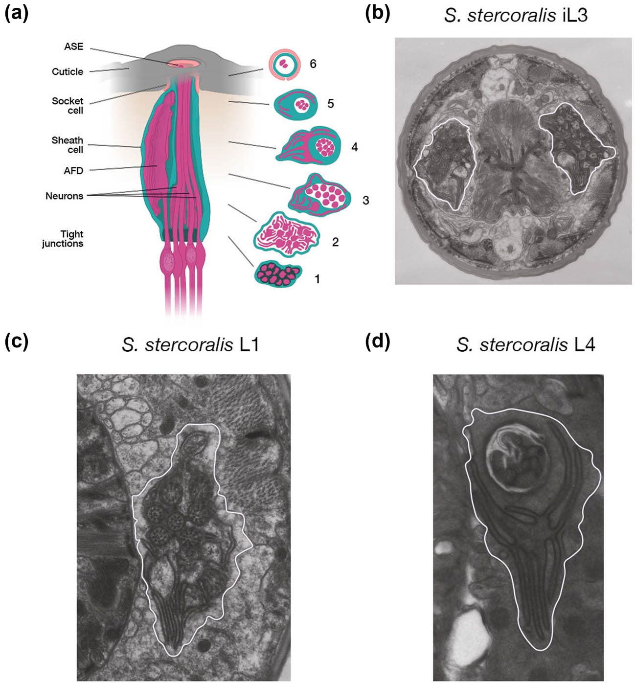
Figure 7
The amphid sensilla of S. stercoralis. (a) Longitudinal representation of an amphid sensillum with transverse sections at various levels. The first transverse section is taken just above the base of the amphid and shows the region where all thirteen amphid neurons form tight junctions with each other. All the amphid neurons are enclosed within the sheath cell. Shortly thereafter, as shown in Section 2, twelve of the thirteen amphid neurons (all but Sst-AUA) send out lateral, fin-like projections that intertwine. The central process of Sst-AFD, along with the dendritic processes of eleven amphid neurons, is visible in Section 3. Section 4 shows the dramatic, branched morphology of the Sst-AFD process, which is embedded in the sheath cell and wraps around the other amphid neurons. Section 5 shows ciliated processes of fewer amphid neurons, enclosed in the sheath cell. The process of Sst-AFD is less branched in this section. The final section shows the ciliated dendrites of two neurons, one of which is Sst-ASE, enclosed in the sheath and socket cells. Also note the enlarged and bulbous tip of the Sst-ASE neuron at the top of the sensillum, located very close to the cuticular opening. Image adapted from Ashton & Schad, 1996; data are from Ashton et al., 1995 (Ashton et al., 1995, Ashton & Schad, 1996). (b) Transmission electron micrograph shows the amphid neurons of an S. stercoralis iL3. The lateral, fin-like projections can be clearly seen on the right side of the section. The left side of the section shows the large central process of Sst-AFD as well as some of its lamellae. Negative: Sste_GAS_198.1act_012865. Image credit: Nathan Schroeder; images originally acquired by G. Schad. (c) Transmission electron micrograph shows the amphid neurons of an S. stercoralis L1-stage larva. The lamellar structure of the Sst-AFD dendrite is visible. Negative: Sste_GAS_L1_11_014700. Image credit: Nathan Schroeder; images originally acquired by G. Schad. (d) Transmission electron micrograph shows the amphid neurons of an S. stercoralis L4-stage larva. The lamellar structure of Sst-AFD is also visible at this stage. Negative: Sste_GAS_L4_FI_020528-1. Image credit: Nathan Schroeder; images originally acquired by G. Schad. For (b–d), the amphid is outlined in white.
The thirteen pairs of amphid neurons of S. stercoralis iL3s enter each amphidial channel at the base, at which point they form tight junctions with each other, as well as with the sheath cell (Figure 7a) (Ashton et al., 1995). Shortly thereafter, the process of the putative Sst-AUA neuron terminates (Ashton et al., 1995). Slightly anterior to the region with the tight junctions, the dendrites of the twelve remaining amphid neurons send lateral, fin-like projections that enmesh with each other and form an intricate web (Figure 7a, b) (Ashton et al., 1995). It has been postulated that these fin-like projections might improve the sensitivity of the amphid neurons to chemical stimuli by increasing the surface area available for contact with such stimuli (Ashton et al., 1995). Fin-like projections have not been reported in C. elegans (Ward et al., 1975, Ware et al., 1975), hookworms (Ashton et al., 1995), or the plant-parasitic nematode Meloidogyne incognita (Wergin & Endo, 1976). However, similar structures have been reported in the bird-parasitic nematode Heterakis gallinarum (Wright, 1977) and the filarial nematode Dipetalonema viteae (McLaren, 1972), and it remains to be seen how prevalent they are among other species of nematodes.
Anterior to the level of the fin-like projections, the processes of the Sst-AFD neurons separate from the others, embed within the sheath cell, and continue further anterior while wrapping around the other amphid neurons (Ashton et al., 1995). The morphology and thermosensory function of the Sst-AFD neurons (Ashton et al., 1995, Lopez et al., 2000, Nolan et al., 2004, Bryant et al., 2022) are discussed in detail below. The remaining eleven amphid neurons send processes that span almost the entire amphidial channel in S. stercoralis iL3s; each of these neurons sends a single process (Figure 7a) (Ashton et al., 1995, Ashton & Schad, 1996, Ashton et al., 1999).
The amphidial processes in S. stercoralis iL3s differ substantially from the more complex arrangement found in C. elegans dauers. In C. elegans dauers, the processes of four amphid neuron pairs (Cel-AFD, Cel-AWC, Cel-AWA, and Cel-AWB) do not traverse the amphid channel but instead embed within the amphid sheath cells. Of the 8 remaining amphid neuron pairs, only six (Cel-ADF, Cel-ADL, Cel-ASE, Cel-ASH, Cel-ASJ, and Cel-ASK) send processes that span the entire amphidial channel. Of these six pairs of neurons, two pairs (Cel-ADF and Cel-ADL) send two processes each, whereas the other four pairs (Cel-ASE, Cel-ASH, Cel-ASJ, and Cel-ASK) send a single process each (Albert & Riddle, 1983, Wolkow & Hall, 2011). Finally, the two remaining amphid neurons – Cel-ASI and Cel-ASG – send processes into the amphidial channel that do not extend all the way to the opening (Albert & Riddle, 1983).
In S. stercoralis, a single neuron pair, which has been named S. stercoralis ASE based on positional and functional homology to the C. elegans ASE neurons (Ashton et al., 1995, Forbes et al., 2004), sends its process the furthest, ending just ∼0.25 µm from the amphidial opening (Ashton et al., 1995). Thus, Sst-ASE is in a prime location to receive and detect signals from the external milieu. Moreover, the dendritic tip of the Sst-ASE neuron is bulbous and enlarged relative to the processes of the other 10 neurons, which have smaller, rounded tips (Figure 7a). In contrast to the species-specific differences in the morphology of amphidial neuron processes, the cell bodies of many of the amphid neurons of S. stercoralis are found in roughly similar positions to those of their C. elegans homologs (Figure 8).
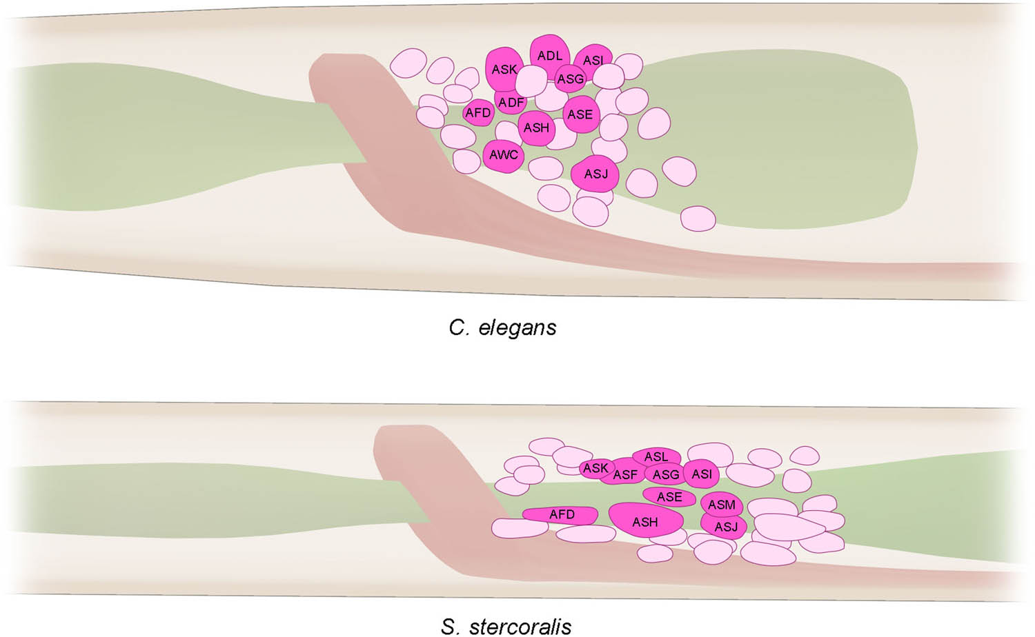
Figure 8
Positions of the cell bodies of a subset of amphid neurons in a C. elegans L1 larva and an S. stercoralis iL3. The schematic shows the relative positions of the amphid cell bodies that lie posterior to the nerve ring (peach), within the lateral ganglia, in C. elegans and S. stercoralis. The pharynx is shown in green. Adapted from Ashton et al., 1995 with permission (Ashton et al., 1995). Cell body locations are approximate, and only a subset of the head neuron cell bodies is shown.
S. stercoralis iL3s are robustly attracted to an array of chemical compounds that are normally found in human sweat, skin, and skin-associated microbes (Forbes et al., 2003, Forbes et al., 2004, Koga et al., 2005, Safer et al., 2007, Castelletto et al., 2014, Gang et al., 2020). This attraction likely enables soil-dwelling iL3s to seek out and make contact with a suitable host. It has been postulated that olfactory and gustatory neurons that reside within the amphid sensilla mediate the detection of human-specific olfactory and gustatory cues by S. stercoralis iL3s. Consistent with this hypothesis, the Sst-ASE neurons are required for the attraction of S. stercoralis iL3s to concentrations of sodium chloride that are normally found in human sweat (Forbes et al., 2004).
Upon contact with a human host, the S. stercoralis iL3s, which are a non-feeding, developmentally arrested life stage (Lok, 2007), penetrate through the skin and enter the body (Schad, 1989). Feeding and development resume within the host via a process termed activation. Incubation of iL3s in host-like conditions (i.e., in Dulbecco’s Modified Eagle Medium (DMEM), 37°C, 5% carbon dioxide) is sufficient for activation, suggesting that intra-host cues, including chemical cues and heat, drive the resumption of iL3 development (Ashton et al., 2007, Stoltzfus et al., 2012, Stoltzfus et al., 2014, Gang et al., 2020). The amphid sensilla likely detect some of these intra-host chemical cues and thereby spur the resumption of development; indeed, it has been shown that laser-mediated ablation of the amphidial Sst-ASJ neurons reduces the efficiency of activation in vitro (Ashton et al., 2007).
Through activation and development inside the host, S. stercoralis infective larvae become parasitic adults that live and reproduce in the gastrointestinal tract (Dionisio et al., 2000, Rivasi et al., 2006, Buonfrate et al., 2023). The larval progeny of these parasitic adults may then either remain within the host and perpetuate the infection or exit the host in feces (Schad, 1989, Buonfrate et al., 2023). The larvae then develop through one of two trajectories: (1) they directly become iL3s that must infect a host for development to continue; or (2) they become free-living adults, which reproduce outside of the host to yield progeny that all become iL3s. The amphid neurons control the number of worms that follow either developmental trajectory, as laser-mediated ablation of both the Sst-ASF (the likely homolog of Cel-ADF) and Sst-ASI amphid neurons in L1 hatchlings leads to a preponderance of iL3s, rather than free-living adults, in the first extra-host generation (Ashton et al., 1998). Whether the amphid neurons sense particular chemicals in the intra-host milieu or extra-host environment and thereby nudge development toward either the infective or the free-living life stage remains to be seen.
The amphid sensilla of many nematode species may also have excretory/secretory function, beyond acting as detectors of chemosensory cues. For example, it has been shown that the amphid sensilla of D. viteae, the rodent hookworm Nippostrongylus brasiliensis, and the human hookworm Necator americanus have a system of ducts within them that likely have secretory function (McLaren, 1972, McLaren, 1974, Wright, 1975, Ashton & Schad, 1996). These ducts have all been detected in the adult life stage of these species. Interestingly, extracts of the amphid sensilla of adult Ancylostoma caninum, which are dog hookworms, have an anticoagulatory effect on blood (Thorson, 1956, Eiff, 1966). Although ducts have not been reported in the amphid sensilla of S. stercoralis iL3s (Ashton et al., 1995), they might exist in S. stercoralis parasitic adults. Moreover, even in the absence of duct-like structures, it remains an enticing possibility that other components of the amphid sensilla (e.g., the sheath cell or the neurons) secrete molecules that help iL3s penetrate skin, migrate within the host body, and/or establish an infection. Consistent with this idea, it has been shown that some C. elegans amphid neurons secrete extracellular vesicles (Wang et al., 2024).
1.4.4. Thermosensation
The AFD neurons (originally referred to as the ALD neurons) of S. stercoralis are the primary thermosensory neurons (Lopez et al., 2000, Nolan et al., 2004, Bryant et al., 2022). There are two Sst-AFD neurons, with one sending processes through each amphid sensillum (Figures 7, 9) (Ashton et al., 1995, Ashton & Schad, 1996, Ashton et al., 1999). As with the other amphid neurons, the dendritic processes of the Sst-AFD neurons enter at the base of each amphidial channel by poking through the sheath cell (Figure 7a). Near the base of each channel, at the same level where lateral, fin-like projections are detected from the eleven other S. stercoralis amphid neurons, the Sst-AFD dendrites also send out lateral, fin-like projections (Figure 7a, b) (Ashton et al., 1995, Ashton & Schad, 1996). Shortly thereafter, the process of Sst-AFD adopts a multi-layered, lamellar structure that interdigitates with the sheath cell and wraps around the amphidial channel whilst being embedded in the sheath cell (Figures 7, 9) (Ashton et al., 1995, Ashton & Schad, 1996). Thus, the processes of the S. stercoralis AFD neurons briefly enter each amphidial channel before they embed within the sheath cell, unlike the processes of C. elegans AFD, which do not enter the amphidial channel and are instead always embedded within the sheath cell (Ashton et al., 1995, Ashton & Schad, 1996, Altun & Hall, 2010, Wolkow & Hall, 2011).
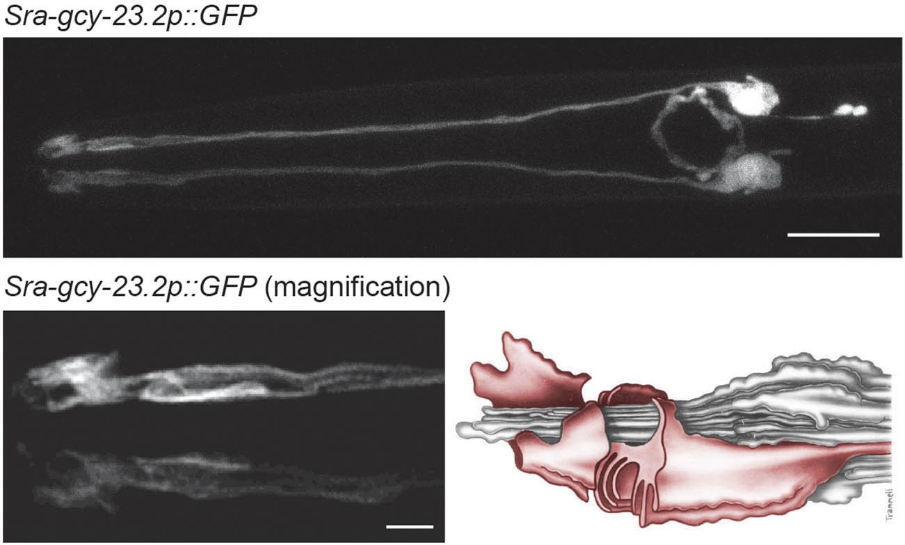
Figure 9
Detection of the S. stercoralis AFD neurons using a transcriptional reporter for a Strongyloides thermoreceptor gene. Top, confocal micrograph shows the morphology and position of the Sst-AFD neurons, as labeled by a transcriptional reporter for the Strongyloides ratti Sra-gcy-23.2 gene, which encodes a putative thermoreceptor (Bryant et al., 2022). Scale bar = 10 µm. Lower left, a magnified view of the dendritic endings of the AFD neurons. Scale bar = 2 µm. Lower right, a reconstruction of the amphid neurons modified from Ashton et al., 1995 and Lopez et al., (2000) with permission; figure reproduced from Bryant and Hallem, 2018 with permission (Ashton et al., 1995, Lopez et al., 2000, Bryant et al., 2018). Entire image reproduced from Bryant et al., 2022 (Bryant et al., 2022).
The multi-layered lamellar structure of Sst-AFD is very atypical and has not been described in any other nematode species to date (McLaren, 1972, McLaren, 1974, Ward et al., 1975, Wright, 1975, Wergin & Endo, 1976, Wright, 1977, Ashton et al., 1995, Ashton & Schad, 1996, Li et al., 2001, Hoholm et al., 2005, Hong et al., 2019). Moreover, the lamellar structure has been detected in S. stercoralis L1-stage larvae, iL3s, and adults, suggesting that it is a feature of this nematode species and not a developmental variation (Figure 7b–d).
S. stercoralis iL3s that are placed in a thermal gradient migrate up the gradient toward warmer temperatures (Lopez et al., 2000, Bryant et al., 2018). This heat-seeking behavior likely helps soil-dwelling iL3s find and infect a host. The heat-seeking behavior of iL3s is mediated by the Sst-AFD neurons, as either laser-mediated ablation or silencing of these neurons using the histamine-gated chloride channel HisCl1 (Pokala et al., 2014) disrupts this behavior (Lopez et al., 2000, Bryant et al., 2022). Sst-AFD senses increases in ambient temperature (Bryant et al., 2022). Interestingly, neural activity of Sst-AFD increases nearly linearly within a large temperature range, which likely enables S. stercoralis iL3s to track temperature gradients ranging from ambient to human body temperature (Bryant et al., 2022). Temperature sensitivity in Sst-AFD is mediated by three receptor guanylate cyclases, Sst-GCY-23.1, Sst-GCY-23.2, and Sst-GCY-23.3 (Bryant et al., 2022). Further studies are needed to illuminate how thermoreceptors and intracellular signaling pathways work together to shape the dynamics of Sst-AFD neural activity upon exposure to both acute and prolonged temperature changes (Bryant et al., 2022).
The development of S. stercoralis L1-stage larvae to the infective vs. free-living life stage, which was described earlier, is also influenced by ambient temperature. Incubation at temperatures that are close to human body temperature promotes development to the infective life stage, and laser-mediated killing of the Sst-AFD neurons partially suppresses this effect of high temperature on development (Nolan et al., 2004). Thus, the AFD neurons of S. stercoralis influence both behavior and development.
1.4.5. Light sensation
Nothing is currently known about whether S. stercoralis detects and responds to light.
1.4.6. Oxygen and carbon dioxide sensation
The BAG sensory neurons in S. stercoralis detect carbon dioxide (CO2) and drive the ensuing behavioral response (Banerjee et al., 2025). The ultrastructure of the Sst-BAG sensory neurons has yet to be described. However, it is known that S. stercoralis iL3s have a single pair of BAG sensory neurons in the head that send processes to the tip of the nose (Figure 10) (Banerjee et al., 2025). As in C. elegans, the receptor guanylate cyclase Sst-GCY-9 is necessary both for the detection of CO2 by the Sst-BAG neurons and for the behavioral response of S. stercoralis to CO2 (Hallem et al., 2011, Banerjee et al., 2025). Interestingly, the response of S. stercoralis to CO2 changes as it progresses through its life cycle: the L1- and L2-stage progeny of parasitic adults, and the free-living adults, are neutral to CO2; the iL3s are strongly repelled by CO2; and activated iL3s are strongly attracted to CO2 (Banerjee et al., 2025). It has been posited that the switch to CO2 attraction in activated iL3s, which are an intra-host life stage, might enable worms to navigate within the host body and ultimately reach areas with a higher concentration of CO2, such as the intestine (Banerjee et al., 2025). Future work will elucidate the changes in the CO2-sensing circuit between iL3s and activated iL3s that cause changes in CO2 valence.
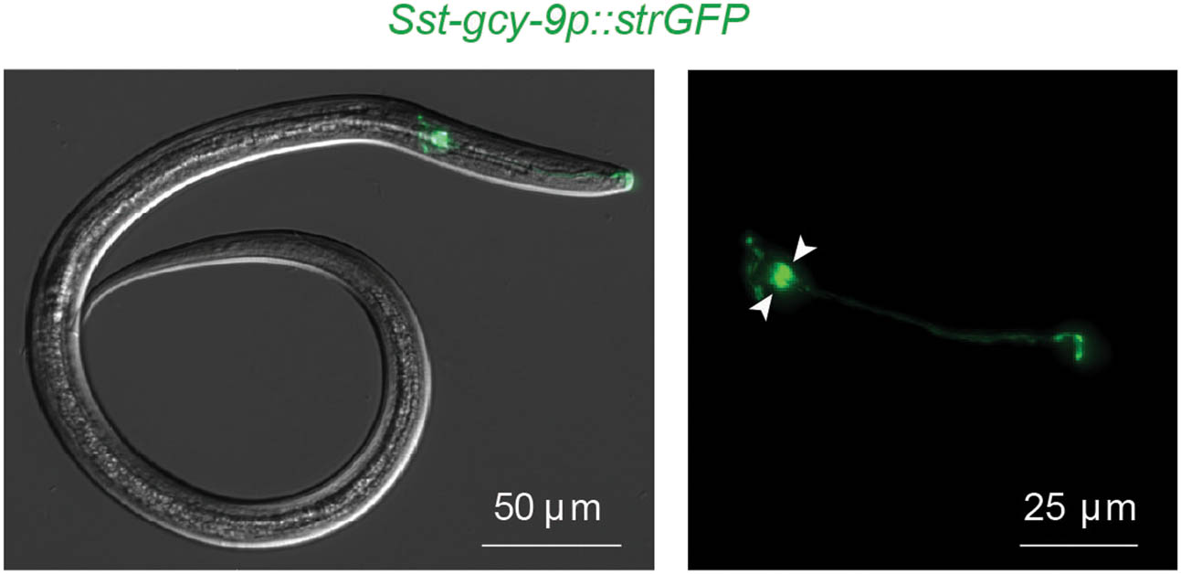
Figure 10
S. stercoralis has a single pair of BAG sensory neurons in the head. A fluorescence micrograph shows the S. stercoralis BAG sensory neurons of an iL3, as labeled by a transcriptional reporter for the Sst-gcy-9 gene, which encodes a receptor guanylate cyclase that is necessary for the detection of carbon dioxide (Banerjee et al., 2025). Arrowheads indicate the cell bodies of the paired Sst-BAG neurons. Scale bar = 50 µm (left image) and 25 µm (right image). Reproduced from Banerjee et al., 2025 (Banerjee et al., 2025).
The response of S. stercoralis to oxygen and the S. stercoralis neurons that drive this behavioral response have yet to be described.
1.5. Interneurons
The interneurons of S. stercoralis have not yet been characterized.
1.6. Polymodal neurons
Polymodal neurons in S. stercoralis have not yet been identified. In C. elegans, the ASH neurons function as polymodal avoidance neurons, mediating responses to both chemical and mechanical noxious stimuli (Yassin et al., 2001, Altun & Hall, 2010, Wolkow & Hall, 2011, Ferkey et al., 2021). The S. stercoralis ASH neurons mediate repulsion from high concentrations of sodium chloride (Forbes et al., 2004), as described earlier, but whether they also mediate averse mechanosensory responses has not yet been investigated.
1.7. Ganglia
Very little is known about the ganglia of S. stercoralis beyond the fact that the cell bodies of several amphid, labial, and cephalic neurons reside in the lateral ganglia (Ashton et al., 1995, Fine et al., 1997).
1.8. Process bundles
The structures of the neuronal process bundles in S. stercoralis have yet to be fully characterized. One interesting difference has been identified between S. stercoralis and C. elegans amphid bundles: whereas the neurons in the amphid bundle of S. stercoralis iL3s all form tight junctions with each other and with the sheath cell at the same level, the amphid neurons in C. elegans dauer and non-dauer stages form tight junctions with the sheath cell at different levels (Figure 7) (Ward et al., 1975, Albert & Riddle, 1983, Ashton et al., 1995). The functional significance of this difference remains unclear.
1.9. Commissures
The commissures of S. stercoralis remain to be studied.
1.10. Synapses
Synapses in S. stercoralis remain to be studied.
1.11. Changes in nervous system structure across the life cycle
In C. elegans, it is now well-established that the nervous system is remodeled between non-dauer and dauer stages. For example, the Cel-IL2 neurons in the L2 stage are chemosensory and have sensory endings that are exposed to the external environment (Yassin et al., 2001, Altun & Hall, 2010, Wolkow & Hall, 2011, Ferkey et al., 2021). In dauers, these sensory endings are retracted and, unlike in the L2 stage, are coated with electron-dense material, which is predicted to interfere with the chemosensory function of these neurons (Albert & Riddle, 1983, Wolkow & Hall, 2011, Yim et al., 2024). In addition, there is substantial remodeling of the connectome in C. elegans dauers, which likely supports dauer-specific behaviors (Yim et al., 2024). For example, the Cel-IL2 neurons form synapses with the Cel-RIG neurons in C. elegans dauers, and as a result, the RIG neurons are a critical component of the nictation circuit in dauers (Yim et al., 2024). Similar to the processes of the Cel-IL2 neurons, the ciliated endings of the Cel-ASI and Cel-ASG neurons are retracted in dauers relative to the L2 stage (Albert & Riddle, 1983, Wolkow & Hall, 2011). Moreover, like the Cel-RIG neurons, the Cel-ASG neurons play a dauer-specific role in nictation behavior (Yim et al., 2024). In S. stercoralis, sensory neuron morphology and position have so far only been characterized in detail at the iL3 stage. In future studies, it will be interesting to determine how much the structure of the nervous system changes across the parasite life cycle and how these changes drive life-stage-specific behaviors.
Acknowledgements
We thank Chris Crocker for assistance in generating figures and Laura Herndon for formatting the WormAtlas version of this article.
Funding information
This work was supported by NIH F32AI174816 (R.P.), NIH R24OD010943 (N.E.S), and NIH R01DC021489 (E.A.H.).
Author contributions
Ruhi Patel: conceptualization, writing – original draft. Michelle L. Castelletto: conceptualization, writing – review & editing. Nathan E. Schroeder: conceptualization, writing – review & editing. Elissa A. Hallem: conceptualization, funding acquisition, supervision, writing – review & editing.
Conflict of interest statement
Authors state no conflict of interest.