
The genus Coslenchus was erected by Siddiqi (1978) with C. costatus (De Man, 1921) Siddiqi, 1978, as its type species. According to Geraert (2008), Coslenchus comprises 38 valid species. Recently, two other species were described from Iran (Hosseinvand et al., 2019, 2020). Eighteen species of Coslenchus have been reported from different provinces in Iran (Karegar, 2018; Hosseinvand et al., 2019, 2020).
Features such as the number of longitudinal cuticular ridges, the number of incisures in the lateral field, smooth or annulated lip region, body and stylet length, the presence or absence of a post-vulval uterine sac, and the shape of the tail terminus are among the most important diagnostic characters for the identification of Coslenchus species (Geraert, 2008).
Identification of Coslenchus species based only on morphology and morphometric data is challenging and not always reliable due to very small differences among species in terms of morphological characteristics. Therefore, molecular data are required to separate closely related species. Additionally, the family Tylenchidae Örley, 1880, contains cryptic species. Some of the nominal species can actually be species complexes (Qing and Bert, 2019). The present scarcity of molecular data for numerous Coslenchus species presents a considerable impediment to accurate species identification within the genus.
In this study, an unknown species of the genus Coslenchus was recovered from Iran. The aim of the present study is to provide morphological and morphometric data for the recovered new species and to determine its molecular phylogenetic affinities with other species of Coslenchus and other taxa in the family Tylenchidae using three markers.
1. Materials and methods
1.1. Nematode extraction and morphological observations
Several soil samples were collected from the rhizosphere of soapwort (Acanthophyllum squarrosum Boiss.) in Birjand city, South Khorasan province, Eastern Iran. The centrifugal-flotation technique (Jenkins, 1964) was used to extract the nematodes from the soil samples. The collected specimens were killed in a hot 4% formaldehyde solution and transferred to anhydrous glycerin, according to De Grisse (1969). Observations and measurements were conducted using a Leitz SM-LUX light microscope with a drawing tube. Some specimens were photographed using an Olympus BX51 light microscope with a Tucsen Michrome 20 digital camera. For cross-sections, the nematodes were first fixed and processed in glycerin and then transferred into a small streak of glycerin jelly on a glass slide, cut into thin sections with a sharp eye knife, and arranged in an upright position with a wire nematode pick. The position of the specimens on the slide was marked with a permanent marking pen on the underside (Perry et al., 2021).
1.2. DNA extraction, polymerase chain reaction (PCR), and sequencing
For molecular analyses, single female specimens were picked out, examined in a drop of distilled water on a temporary slide under a light microscope, and transferred to 7 μl of worm lysis buffer (500 mM KCl, 100 mM Tris-Cl pH 8, 15 mM MgCl2, 10 mM dithiothreitol (DTT), 4.5% Tween 20) on a clean slide, and then crushed using a coverslip. The suspension was collected by adding 7 μl of lysis buffer. Two microliters of proteinase K (600 μg/ml) were added, and the tubes were frozen at –70°C for at least 10 min and then incubated at 65°C (1 h) and 95°C (10 min) consecutively. After incubation, the tubes were centrifuged for 2 min at 14,000 rpm. The DNA samples were stored at –20°C until used as a PCR template (Maafi et al., 2003). Primers for amplification of SSU rDNA were forward primer SSUF22 (5′-TCCAAGGAAGGCAGCAGGC-3′) and reverse primer SSUR13 (5′-GGGCATCACAGACCTGTTA-3′) (Dorris et al., 2002). Primers for LSU rDNA D2–D3 amplification were forward primer D2Ab (5′-ACAAGTACCGTGAGGGAAAGT-3′) and reverse primer D3B (5′-TCGGAAGGAACCAGCTACTA-3′) (De Ley et al., 1999). Primers for amplification of ITS rDNA were forward primer rDNA1 (5′-TTGATTACGTCCCTGCCCTTT-3′) and reverse primer rDNA1.58S (5′-ACGAGCCGAGTGATCCACCG-3′) (Subbotin et al., 2000). To amplify the abovementioned loci, the PCRs were performed as described by Azimi and Abdolkhani (2023). Amplification success was evaluated by electrophoresis on a 1% agarose gel. The PCR products were subjected to sequencing using an Applied Biosystems 3500 (ABI) sequencer, Pishgam Corporation, Tehran, Iran. The newly obtained sequences of the new species were deposited into the GenBank database (accession numbers PZ025862-PZ025865 for SSU rDNA, PZ025867, PZ025868 for LSU rDNA D2–D3, and PZ036249, PZ036250 for ITS rDNA).
1.3. Phylogenetic analyses
The newly obtained sequences of the SSU, D2–D3 fragments of LSU, and ITS rDNA were compared with those of other nematode species available in the GenBank database using the BLAST homology search program, and the selected sequences to reconstruct each phylogeny were retrieved. The sequences were aligned by ClustalX version 2 using the default parameters (Larkin et al., 2007). The outgroup taxa were chosen according to previous studies (Pedram et al., 2018; Azimi, 2021; Azimi and Abdolkhani, 2023; Jumaah and Azimi, 2024). The editing of the three alignments was performed manually in the MEGA7 program (Kumar et al., 2016). The base substitution model was selected using MrModeltest 2 (Nylander, 2004) based on the Akaike information criterion. A general time-reversible model, including gamma distribution of rate heterogeneity among sites and estimates of invariant sites (GTR + G + I), was used in the three phylogenies.
Bayesian analyses were performed to infer the phylogenetic trees using MrBayes v3.1.2 (Ronquist and Huelsenbeck, 2003), running the chains for four million generations. After discarding burn-in samples and evaluating convergence, the remaining samples were retained for further analyses. The Markov chain Monte Carlo method within the Bayesian framework was used to determine the equilibrium distribution and help estimate the posterior probabilities of the phylogenetic trees (Larget and Simon, 1999) using the 50% majority rule. Bayesian posterior probability (BPP) values higher than 0.50 are given on appropriate clades. The output files of the phylogenetic program were visualized using Dendroscope v3.2.8 (Huson and Scornavacca, 2012), and trees were digitally drawn in CorelDRAW software version 23.
2. Results
2.1. Description of Coslenchus iranicus n. sp.
The line drawings and photomicrographs of Coslenchus iranicus n. sp. are presented in Figs 1 and 2. The morphometrics of Coslenchus iranicus n. sp. are shown in Table 1.
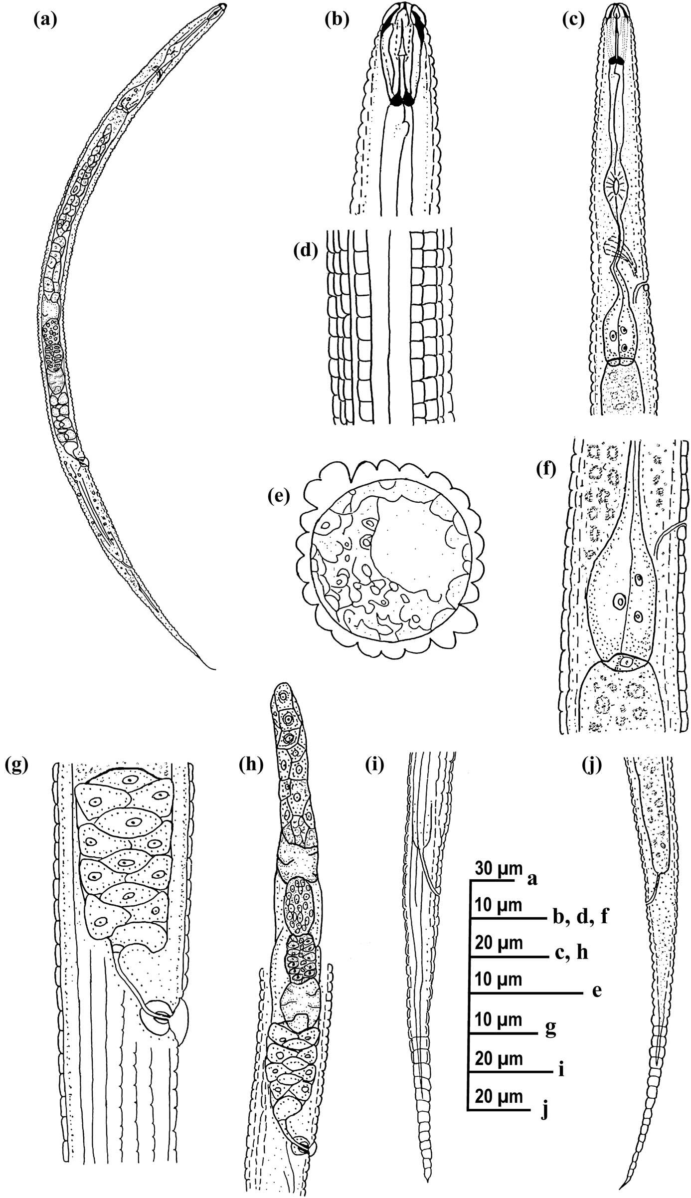
Figure 1
Line drawings of Coslenchus iranicus n. sp. from Iran. (a) Entire body; (b) anterior body region; (c) pharyngeal region; (d) lateral field at mid-body; (e) mid-body cross-section; (f) terminal bulb; (g and h) part of reproductive system; and (i and j) posterior body region.
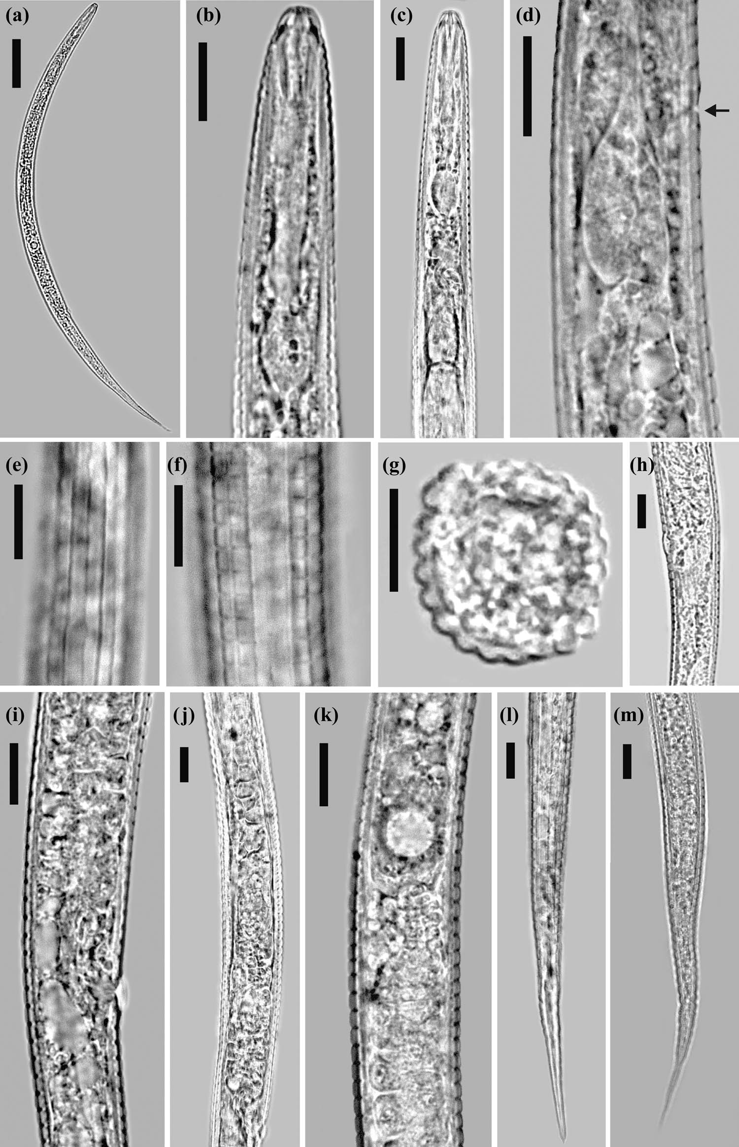
Figure 2
Light photomicrographs of Coslenchus iranicus n. sp. from Iran. (a) Entire body; (b and c) anterior body region; (d) terminal bulb (the arrow indicates the excretory pore); (e) lateral field at mid-body; (f) cuticle showing longitudinal ridges; (g) mid-body cross-section; (h and i) vulval region; (j and k) part of reproductive system; and (l and m) posterior body region. Scale bars: (a) = 50 μm; (b–m) = 10 μm.
Table 1
Morphometrics of Coslenchus iranicus n. sp. from South Khorasan province, Iran.
| Character | Holotype female | Paratype females |
|---|---|---|
| n | 1 | 11 |
| L | 501.5 | 484.7 ± 24.6 (447–525) |
| a | 26.7 | 27.8 ± 1.3 (25.8–29.7) |
| b | 5.0 | 5.4 ± 0.6 (5–7) |
| c | 5.6 | 5.8 ± 0.5 (5.3–6.9) |
| c′ | 9.2 | 8.5 ± 1.1 (6.9–10.8) |
| V | 66.6 | 67.2 ± 1.1 (66.0–69.8) |
| V′ | 81.1 | 81.3 ± 0.6 (80.6–82.2) |
| Stylet length | 11.8 | 11.1 ± 0.9 (9.4–12.0) |
| m | 45.8 | 42.5 ± 5.1 (34.5–52.0) |
| DGO | 1.8 | 2.1 ± 0.2 (1.8–2.5) |
| Annuli width | 2.5 | 2.2 ± 0.2 (1.9–2.5) |
| MB | 49 | 49.9 ± 1.1 (48.6–51.5) |
| Pharynx length | 100 | 90.4 ± 6.8 (74.7–100.0) |
| Anterior end to excretory pore | 71.8 | 71.7 ± 5.1 (57–77) |
| Rst | 9 | 7.5 ± 0.9 (6–9) |
| Rex | 33 | 33.1 ± 2.7 (25–35) |
| Roes | 45 | 41.8 ± 2.7 (35–45) |
| Rv | 141 | 137.2 ± 4.2 (130–142) |
| Ran | 171 | 168.2 ± 4.5 (161–173) |
| Rvan | 30 | 31.0 ± 1.2 (30–34) |
| Lip region-vulva | 334 | 325.6 ± 14.7 (303–350) |
| Lip region-anus | 411.8 | 400.6 ± 19.0 (368–412) |
| Maximum body width | 18.8 | 17.8 ± 0.9 (16.3–19.0) |
| Vulval body width | 15.7 | 15.3 ± 0.6 (14.3–15.8) |
| Vulva-anus | 77.8 | 75.0 ± 4.9 (65.5–83.0) |
| Tail length | 89.7 | 84.1 ± 8.5 (66.3–93.4) |
| Tail/vulva-anus | 1.2 | 1.1 ± 0.1 (0.9–1.2) |
Note: All measurements are in μm and in the form mean value ± SD (range).
2.1.1. Female
Body is slightly arcuate ventrally when heat-relaxed. Cuticle annuli is prominent, 1.9–2.5 μm wide at mid-body. Cuticle has 18 longitudinal ridges excluding lateral field with no variation. Lateral field about one-third of the corresponding body diameter, with three incisures. Lip region is unstriated, separated from body contour by a shallow depression, 2.8–3.0 μm high and 5.3–6.2 μm wide. Cephalic framework is distinct, basal plate extends posteriorly for about two annuli. Stylet is developed, its conus 38–47% of total length, knobs rounded, 3.0–3.3 μm across. Dorsal gland orifice is adjacent to the stylet base. Pharynx is composed of a slender procorpus, ovoid median pharyngeal bulb has distinct valve, 5–7 µm wide and 9–11 µm long; narrow slender isthmus; and pyriform terminal bulb, 7.0-9.7 µm wide and 14.5–18.0 µm long. Nerve ring encircles the middle of isthmus, 56.5 (46–65) μm from anterior end. Excretory pore is at the level of the anterior end of the terminal bulb. Hemizonid is just posterior to excretory pore. Intestine is simple. Reproductive system is monodelphic-prodelphic, and is composed of an outstretched ovary, oocytes mostly arranged in a single row (except germinal zone), spermatheca mostly bilobed, filled with rounded sperm, vagina with swollen walls, oblique, 4.5–5.1 µm long, post-vulval uterine sac absent, vulva sunken in body with large vulval flaps (5.0–5.8 µm), about 2–2.5 times the annuli width. Tail is regularly tapering, mostly with pointed terminus, while few have a finely rounded tip.
2.1.2. Male
Not found.
2.2. Type of host and locality
This population was recovered from the rhizosphere of soapwort collected from Birjand city in South Khorasan province, Eastern Iran. The GPS information of the sampling site is 32°45′48.6″N, 59°22′31.4″E.
2.3. Etymology
The specific epithet refers to the name of the country where it was found.
2.4. Type material
The holotype female, nine paratype females, and five juveniles were deposited in the nematology laboratory of the Department of Plant Protection, Shahid Chamran University of Ahvaz, Ahvaz, Iran. Two paratype females and one juvenile were deposited at the Wageningen Nematode Collection, Wageningen, The Netherlands. The Life Science Identifier code (LSID) for this publication is http://zoobank.org/urn:lsid:zoobank.org:pub:F5694A19-32FE-4D8A-844F-A7BA9DA3F19F.
2.5. Diagnosis and relationships
Coslenchus iranicus n. sp. is mainly characterized by its cuticle with 18 longitudinal ridges except for lateral lines, lateral field with three incisures, lip region unstriated, spermatheca filled with sperm, post-vulval uterine sac absent, vulva with large vulval flaps, and tail mostly with a pointed terminus, though few specimens have a finely rounded tip.
Based on general characterization, C. iranicus n. sp. is closely similar to C. areolatus (Egunjobi, 1967) Siddiqi, 1978, C. franklinae Siddiqi, 1981, C. japonicus Mizukubo & Minagawa, 1984, C. leiocephalus Brzeski, 1998, and C. maritus Andrássy, 1991.
It differs from C. areolatus by the spermatheca filled with sperm (vs empty), tail mostly with a pointed terminus (vs always finely rounded), lip region unstriated (vs with three indistinct annuli), and post-vulval uterine sac absent (vs present). From C. franklinae, it differs by having three incisures in the lateral field (vs four incisures), and lip region unstriated and separated from the body contour by a shallow depression (vs with three annuli and continuous with the body contour). From C. japonicus, with less longitudinal ridges (18 vs 18–22), three incisures in the lateral field (vs three and four incisures), an unstriated lip region that separated from body contour by a shallow depression (vs with three annuli and continuous with body contour), a higher C ratio (5.3–6.9 vs 4.4–4.9), and a lower T/VA ratio (0.9–1.2 vs 1.3–1.6). From C. leiocephalus, with less longitudinal ridges (18 vs 18–22), spermatheca filled with sperm (vs empty), epiptygma absent (vs present), and tail mostly with pointed terminus, in a few with the finely rounded tip (vs pointed tip or hair-like). From C. maritus, by shorter stylet (9.4–12.0 vs 12–13), three incisures in the lateral field (vs four incisures), lip region unstriated (vs with four to five annuli), post-vulval uterine sac absent (vs present), higher MB ratio (48.6–51.5 vs 48), and higher V ratio (66.0–69.8 vs 64–66).
2.6. Molecular characterization and phylogenetic relationships
2.6.1. Partial SSU rDNA phylogeny
To determine the phylogenetic relationships of Coslenchus iranicus n. sp. with other nematode species using SSU sequences, two 1,091 nt and two 1,118 nt long identically aligned sequences of SSU rDNA with accession numbers PZ025862, PZ025863, PZ025864 and PZ025865 were used. The four sequences differed only in length. The BLAST search using these sequences revealed that they have 98.63 and 98.66% identity, respectively (resulting from 14 mismatches and one gap), with the SSU sequence of C. polonicus Brzeski, 1982 (KJ869314). A total of 58 SSU sequences of the family Tylenchidae were used in SSU phylogeny. The phylogenetic tree inferred using this dataset is presented in Fig. 3. The SSU sequences of the new species formed a clade with the SSU sequence of C. polonicus with relatively high support (BPP = 0.82).
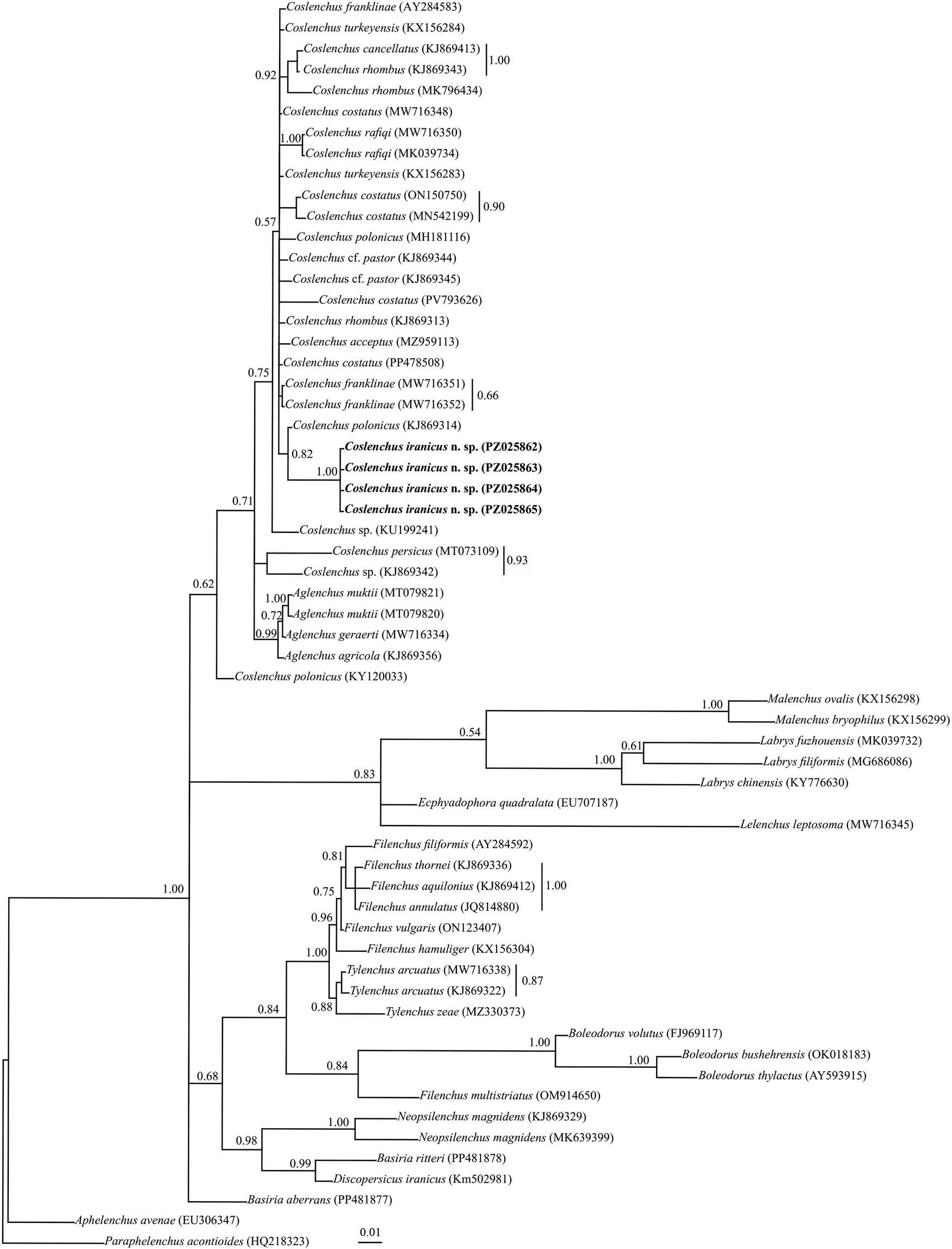
Figure 3
Bayesian 50% majority rule consensus tree inferred from analysis of the SSU rDNA sequences of Coslenchus iranicus n. sp. under the GTR + G + I model. BPP values more than 0.50 are given for appropriate clades. New sequences are indicated in bold.
2.6.2. D2–D3 fragment of LSU rDNA phylogeny
To reconstruct the LSU rDNA tree, two identical 709 nt long partial sequences of the D2–D3 region with accession numbers PZ025867 and PZ025868 were used. A BLAST search using these sequences revealed that they have 95.19% identity (resulting from 27 mismatches and three gaps) with the LSU sequence of C. leiocephalus (KM817176). A total of 48 LSU sequences of the family Tylenchidae were used in the LSU phylogeny. The phylogenetic tree inferred using this dataset is presented in Fig. 4. The clade including the newly generated LSU sequences of the new species is in a sister relationship with the LSU sequence of C. leiocephalus with maximal support (BPP = 1.00).
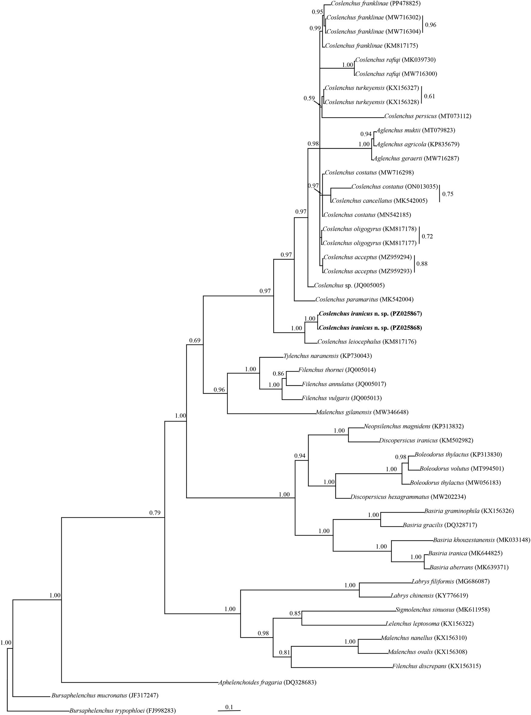
Figure 4
Bayesian 50% majority rule consensus tree inferred from analysis of the D2–D3 domains of the LSU rDNA sequences of Coslenchus iranicus n. sp. under the GTR + G + I model. BPP values more than 0.50 are given for appropriate clades. New sequences are indicated in bold.
2.6.3. Partial ITS rDNA phylogeny
Two 724 nt long identically aligned sequences of ITS rDNA (PZ036249 and PZ036250) were used in ITS phylogeny. A BLAST search using the ITS sequences of the new species revealed they have 94.54% identity with the corresponding locus of C. acceptus (MZ959285 and MZ959286). Sequence variations between these sequences were 86 mismatches and six gaps. There are only four sequences of ITS rDNA for the genus Coslenchus in the GenBank database. Twenty-seven sequences of the family Tylenchidae were used for ITS phylogeny. The phylogenetic tree inferred using this dataset is presented in Fig. 5. The ITS sequences of the new species formed a clade with the ITS sequences of two other species of the genus with maximal support (BPP = 1.00).
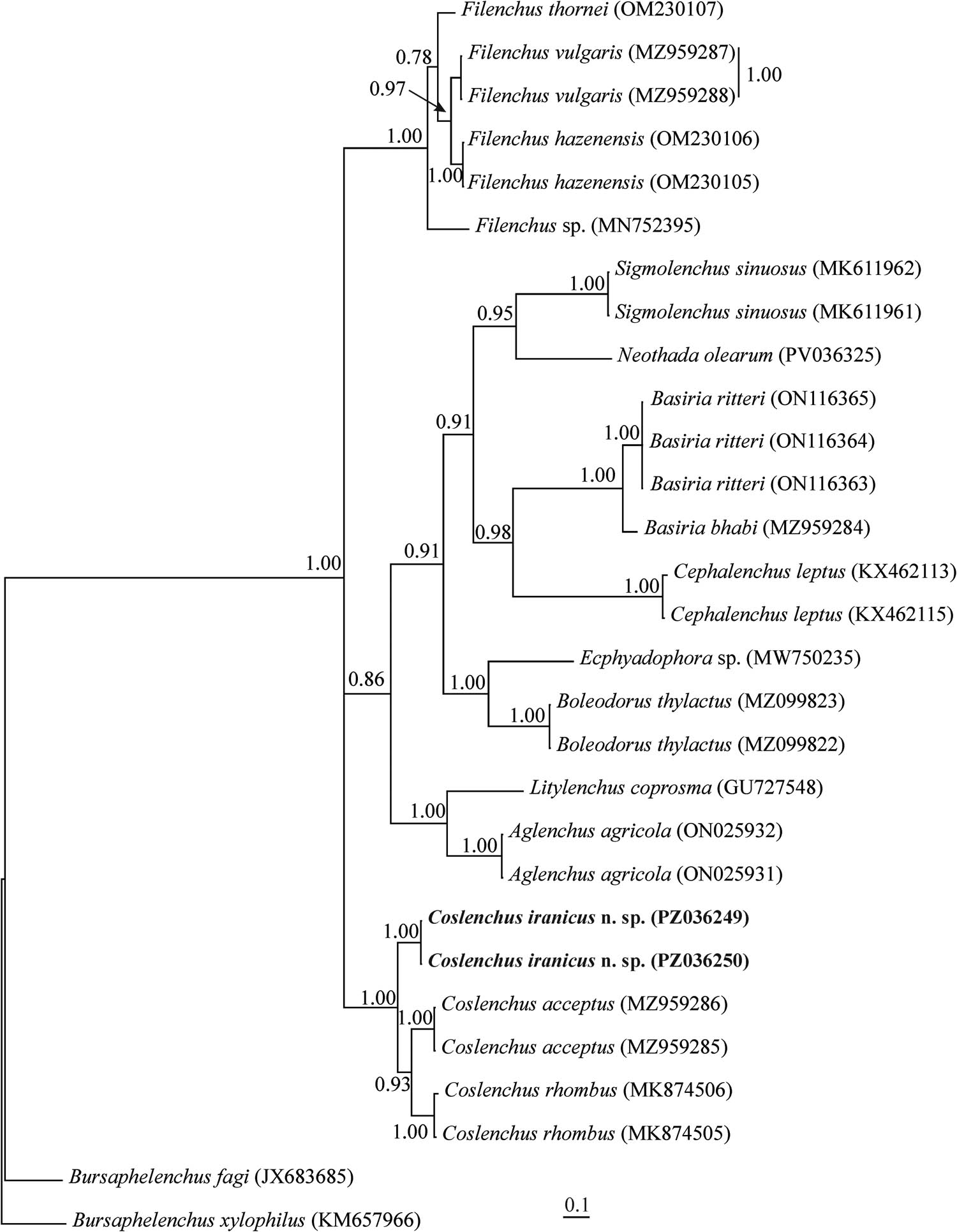
Figure 5
Bayesian 50% majority rule consensus tree inferred from analysis of the ITS rDNA sequence of Coslenchus iranicus n. sp. under the GTR + G + I model. BPP values more than 0.50 are given for appropriate clades. New sequences are indicated in bold.
3. Discussion
The objectives of this study were to characterize a new species of the genus Coslenchus from Iran and to investigate the molecular phylogenetic affinities of the new species with other Coslenchus species. According to the SSU rDNA phylogenetic tree, the new species formed a clade with C. polonicus. In LSU rDNA phylogeny, the new species has a sister relationship with the LSU sequence of C. leiocephalus.
Former phylogenetic analyses based on LSU D2–D3 showed that Coslenchus spp. is a monophyletic taxon (Panahandeh et al., 2016). However, they emphasized that the phylogeny of Tylenchidae needs deep sampling of representatives of the genera and multi-locus phylogenetic analyses. The results of the present study based on LSU D2–D3 and SSU rDNA phylogeny showed it is polyphyletic.
The larger number of sequences used in the phylogenetic analysis of the present study was effective in obtaining this result, such that in the ITS rDNA phylogeny, in which only sequences of two species available in GenBank and the new species studied were used in the analysis, the genus Coslenchus appeared monophyletic.
Of the 40 valid species of the genus, molecular data in GenBank are available only for about 10 species in SSU and LSU D2–D3, and in ITS rDNA for only 2 species. On the other hand, morphological data for some sequenced specimens are not available. The importance of obtaining molecular data from different populations of various species of the genus has been emphasized and will help provide a better resolution of the phylogeny of the genus in the family Tylenchidae.
Acknowledgments
The authors thank the Research Council of Shahid Chamran University of Ahvaz, Iran (Grant no. SCU.AP1404.638) for financial support.
Funding information
Authors state that there is no funding information.
Author contributions
Nasrin Noras Mofrad: Sampling, methodology, writing. Sedighe Azimi: Methodology, writing, review and editing, data curation, project administration, investigation, funding acquisition.
Conflict of interest statement
Authors state no conflict of interest.