
Figure 1
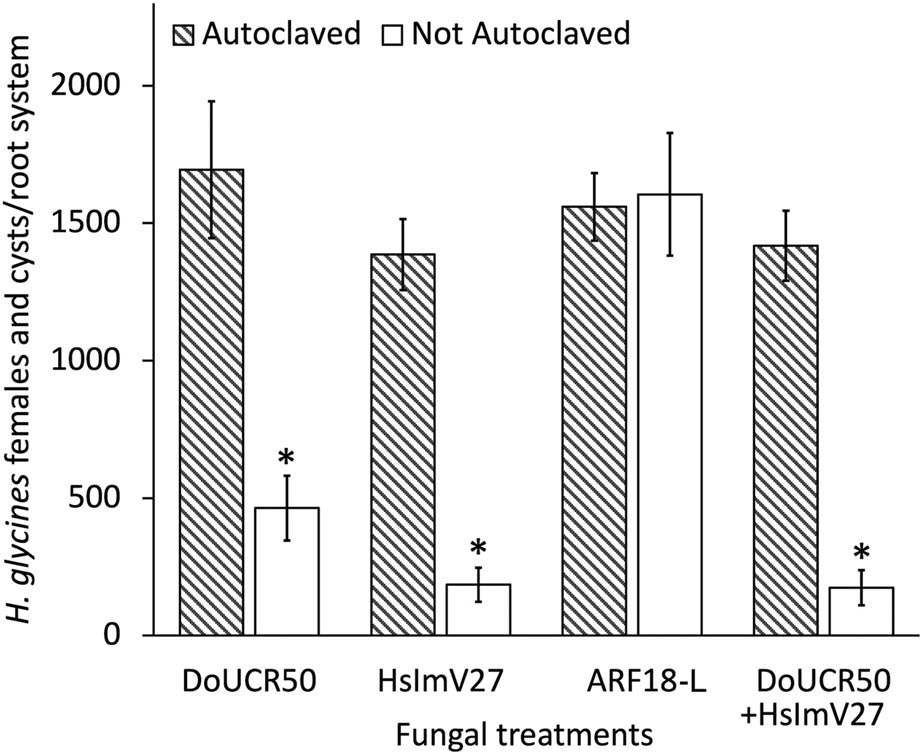
Nematode suppressive activity of Hyalorbilia spp. DoUCR50, HsImV27, and ARF-L were evaluated against Heterodera glycines on soybean (Glycine max cv. Williams 82) in soil-based greenhouse experiments. Autoclaved and not autoclaved fungal peat cultures were incorporated into autoclaved sandy-loam soil at a final concentration of 500 CFU/cm3 of soil. 3-week-old seedlings growing in cones with 200 cm3 of fungus-amended soil were inoculated with 250 H. glycines J2. Cysts and females were enumerated after approximately 2 months (1,260 DD). There was no statistically significant difference between the medians of the two data sets (Wilcoxon signed-rank test, P ≤ 0.05). The data were combined and analyzed using Kruskal–Wallis and Dunn’s multiple comparisons tests (P ≤ 0.05). Bars indicate standard error (n = 8). For each strain, * denotes a significant difference in nematode numbers at P ≤ 0.05.
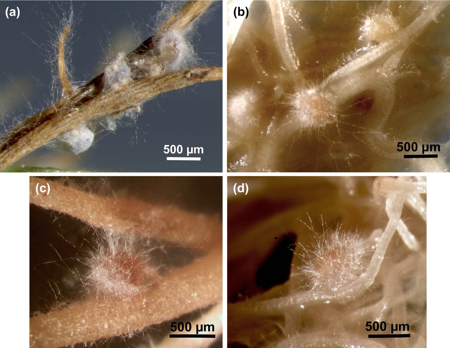
Figure 2
Parasitism of H. glycines and H. schachtii by HsImV27 and DoUCR50 in tissue culture. Females of H. glycines on soybean (a and c) and H. schachtii on cabbage (b and d) were inoculated with HsImV27 (a and b) or DoUCR50 (c and d) by placing a block of water agar culture of the fungus adjacent to the females as their posterior end broke through the epidermis of the roots. The blocks were removed after 3 days, and fungal parasitism was observed with a stereomicroscope. Photos were taken 10 days after fungal inoculation. Images a and b were captured using a Zeiss stereomicroscope (SteREO Discovery.V8) equipped with a camera (Zeiss Axiocam 305) and controlled by Zeiss ZEN software (3.11). The images are an extended depth of focus projection using the First/Last feature in ZEN. Images c and d were captured using a Leica stereomicroscope equipped with a Canon EOS Rebel T1i/500D digital camera controlled by Canon EOS Utility version 2.6.0. Scale bars represent 500 µm.
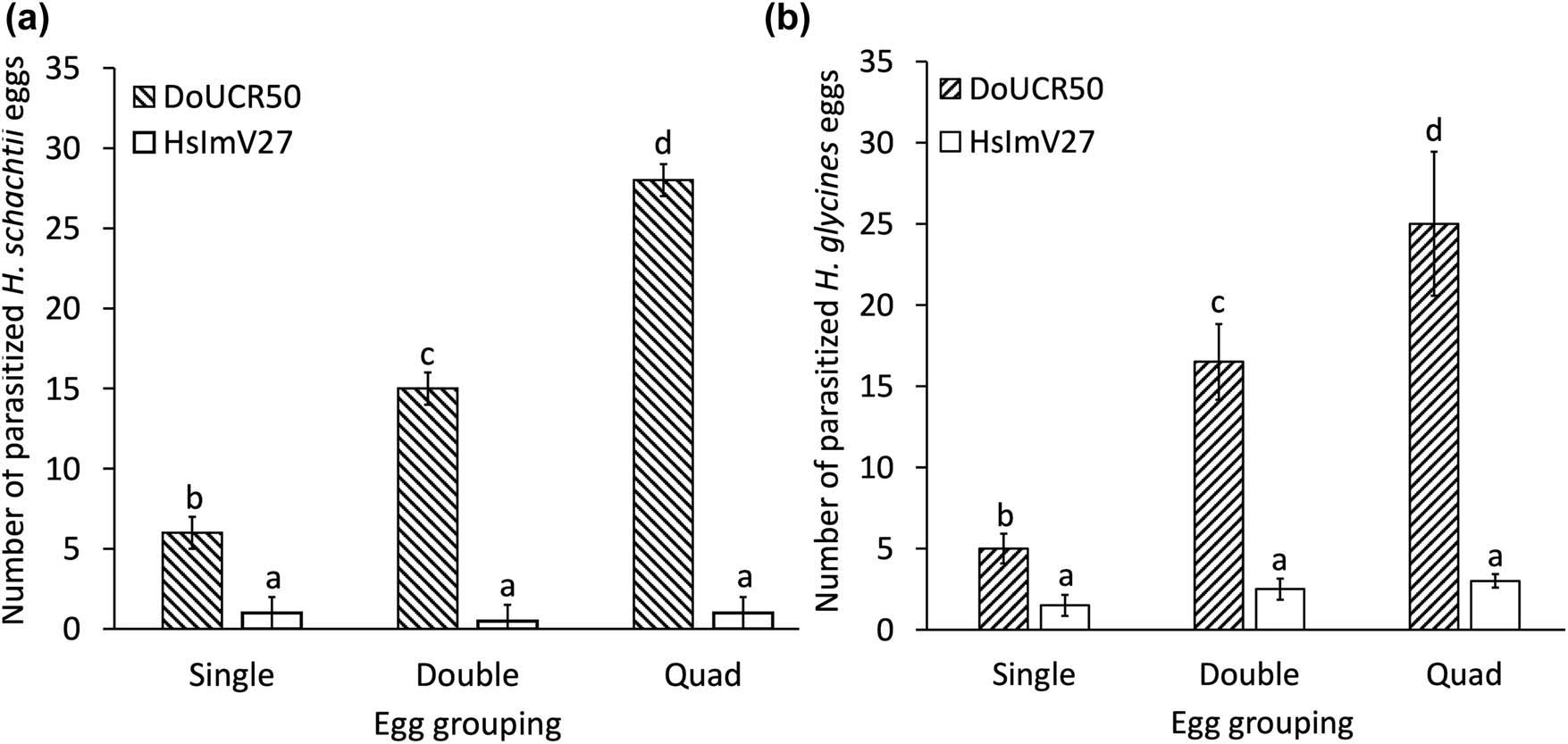
Figure 3
Effect of Heterodera schachtii and H. glycines egg group size on susceptibility to parasitism by Hyalorbilia sp. DoUCR50 and HsImV27. Forty undifferentiated eggs of H. schachtii (a) or H. glycines (b) were arranged on water agar cultures of the fungi in groups of 1 (Single), 2 (Double), or 4 (Quad). The total number of parasitized eggs in each group of 40 was determined after 6 days of incubation. The results of four experiments were combined, analyzed with ANOVA, and mean values compared using Fisher’s LSD (P ≤ 0.05). For each figure, a and b, different letters indicate a significant difference in the number of parasitized eggs among treatments (P ≤ 0.05). Bars represent standard error.