
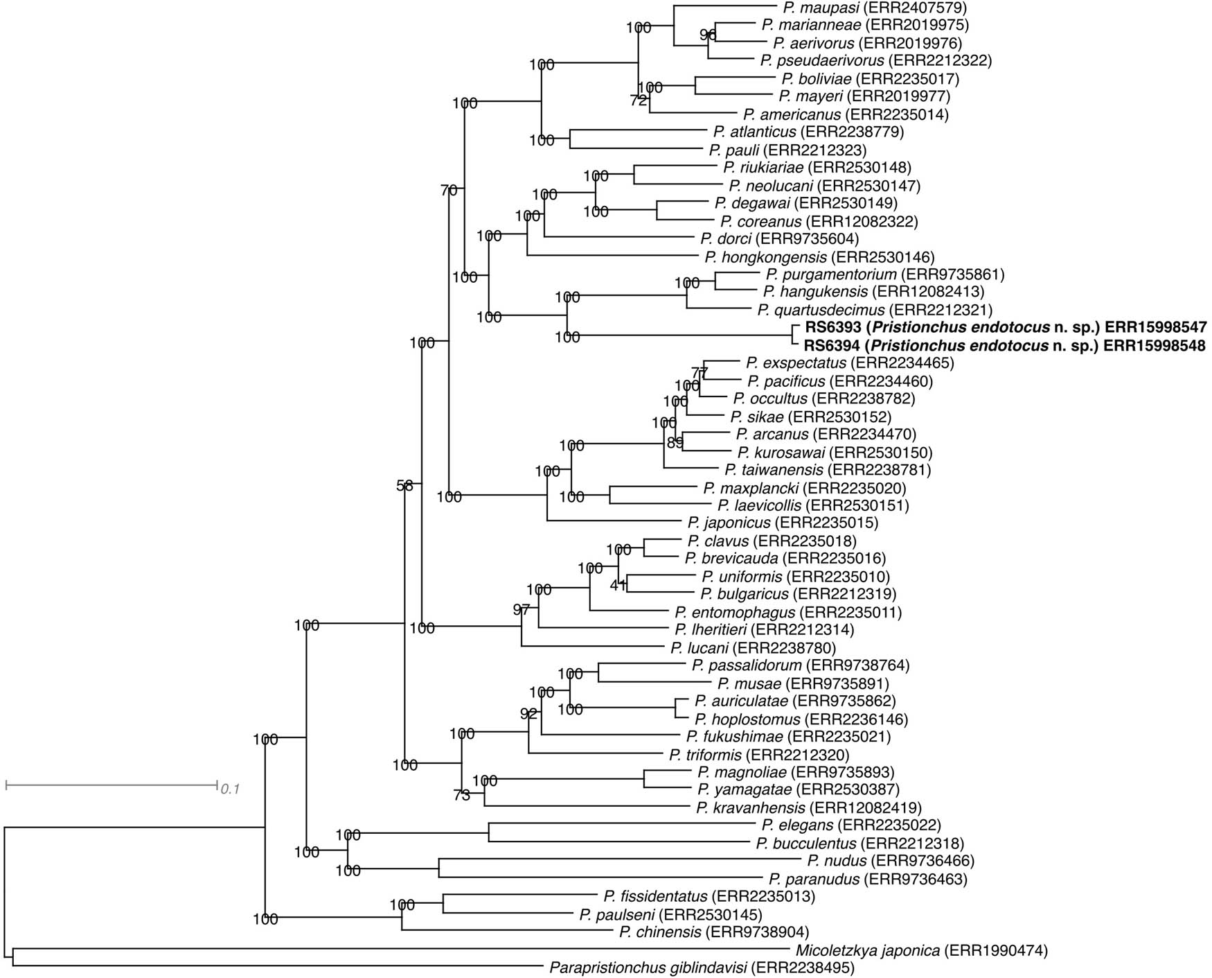
Figure 1
Molecular phylogeny of the genus Pristionchus as inferred from a maximum likelihood tree reconstructed from a concatenated protein alignment, with Micoletzkya japonica and Parapristionchus giblindavisi as outgroups. The identifiers after the species names indicate the Run accessions for the corresponding transcriptomic data at the European Nucleotide Archive.
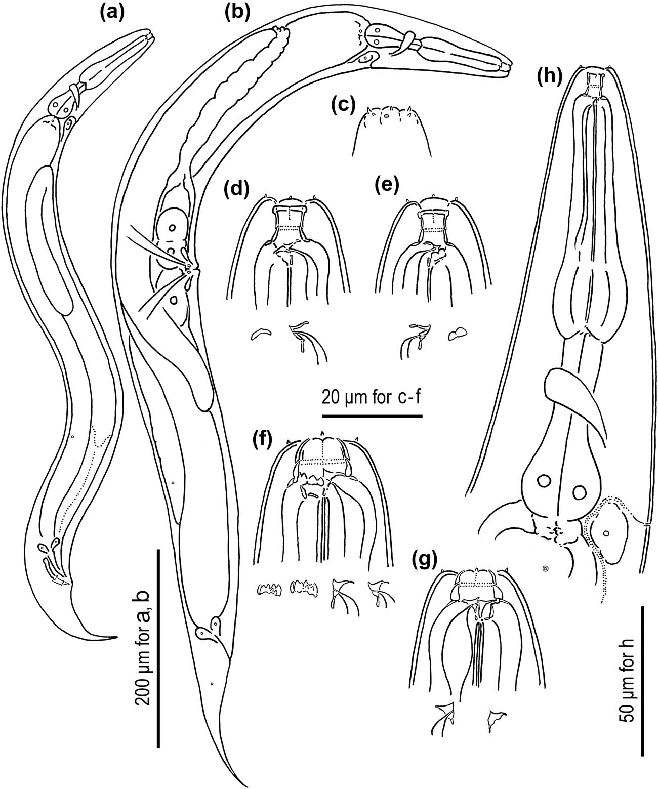
Figure 2
Adult male and hermaphrodite of Pristionchus endotocus n. sp. (a) male; (b) hermaphrodite; (c) lip region of male; (d, e) stomatal region of stenostomatous hermaphrodite where tooth and ridges are drawn separately; (f, g) stomatal region of eurystomatous hermaphrodite where tooth and ridges are drawn separately; (h) anterior part of the body of hermaphrodite. All subfigures are in the right lateral view except for d (left lateral view).
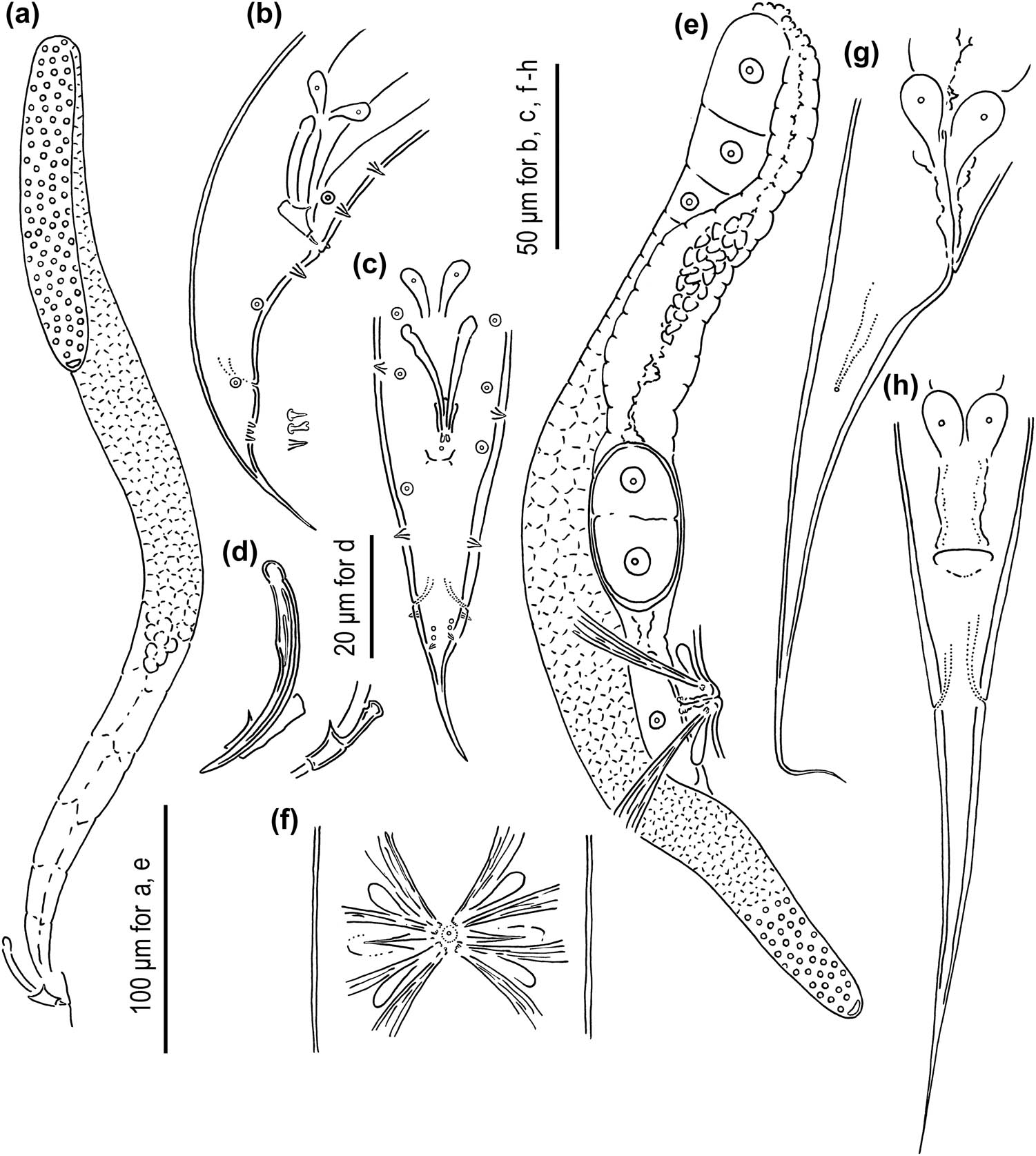
Figure 3
Adult male and hermaphrodite of Pristionchus endotocus n. sp. (a) Whole testis; (b, c) male tail; (d) spicule and gubernaculum; (e) whole part of anterior ovotestis; (f) vulval region of hermaphrodite; (g, h) tail region of hermaphrodite. a, b, e, and g are in the right lateral view; c, f, and h are in the ventral view; and d is in the left lateral view.
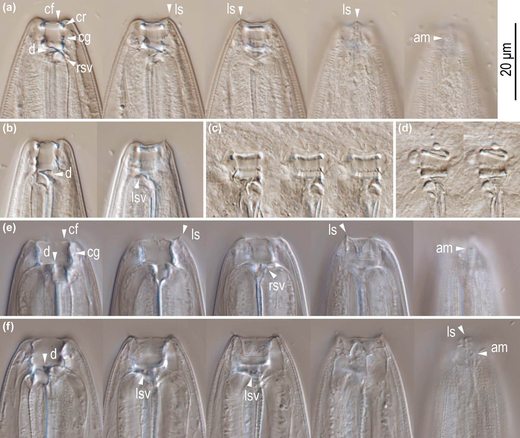
Figure 4
Stomatal morphology of Pristionchus endotocus n. sp. hermaphrodites. (a) Right lateral view of stenostomatous form in five different focal planes; (b) left lateral view of stenostomatous form in two different focal planes; (c) right lateral view of squashed stenostomatous form in three different focal planes; (d) left lateral view of squashed stenostomatous form in two different focal planes; (e) left lateral view of eurystomatous form in five different focal planes; (f) right lateral view of eurystomatous form in five different focal planes. Labels are as follows: am = amphid; cf = cheilostomatal flap; cg = cheilostom-gymnostom margin; cr = cheilostomatal rim; d = dorsal tooth; ls = labial sensilla; lsv = left subventral ridges; rsv = right subventral ridges.
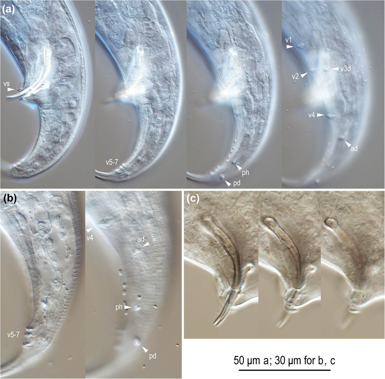
Figure 5
Left lateral view of male tail characters of Pristionchus endotocus n. sp. (a) Whole tail in five different focal planes; (b) posterior part of the tail in two different focal planes; (c) spicule and gubernaculum in three different focal planes. Labels are as follows: ad, pd, and v + number = genital papillae following the terminology in Sudhaus and Fürst von Lieven (2003); ph = phasmid; vs = ventral single papilla.
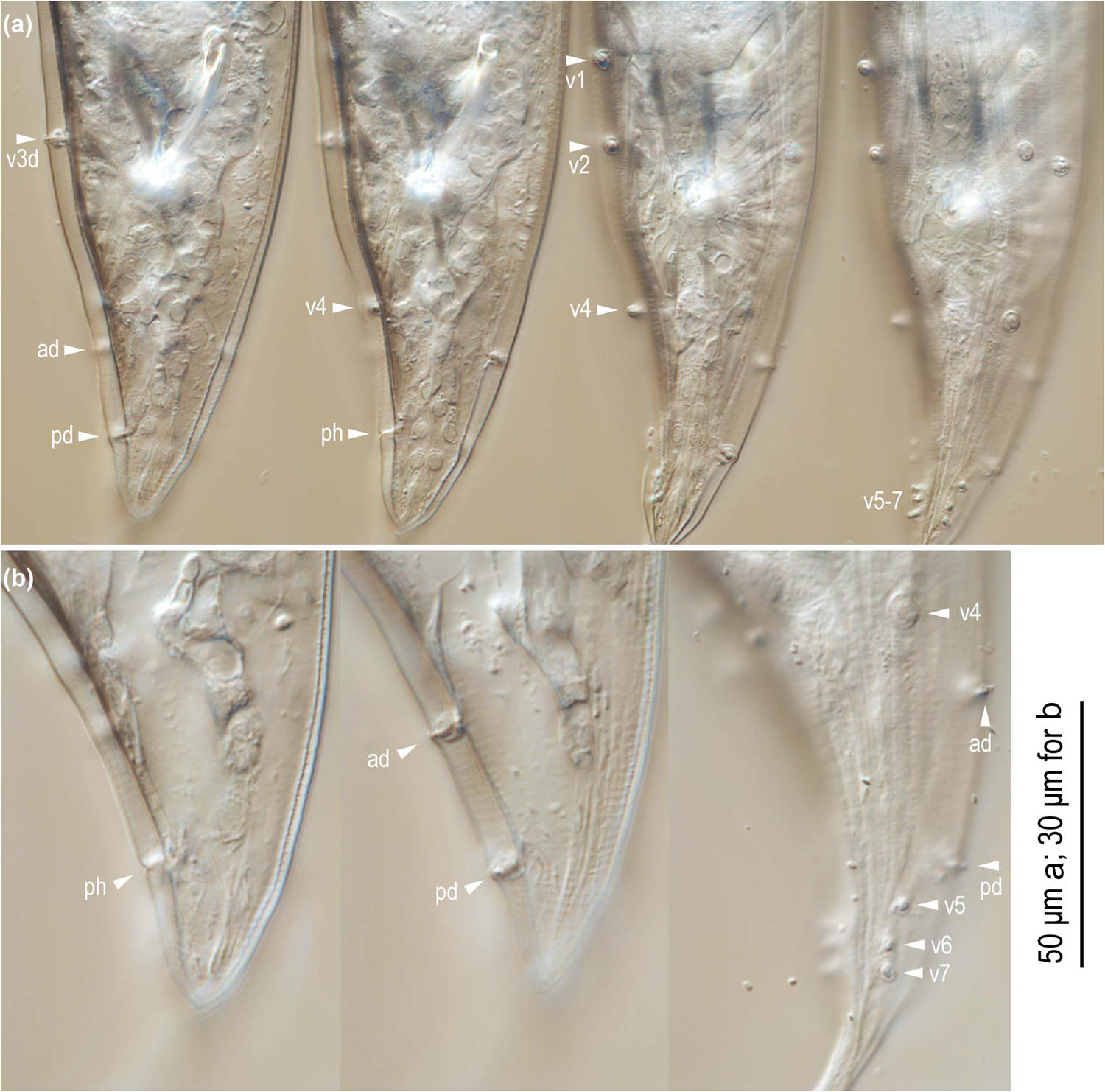
Figure 6
Ventral view of male tail characters of Pristionchus endotocus n. sp. (a) Whole tail in four different focal planes; (b) posterior part of the tail in three different focal planes. Labels are as follows: ad, pd, and v + number = genital papillae following the terminology presentd by Sudhaus and Fürst von Lieven (2003); ph = phasmid; vs = ventral single papilla.
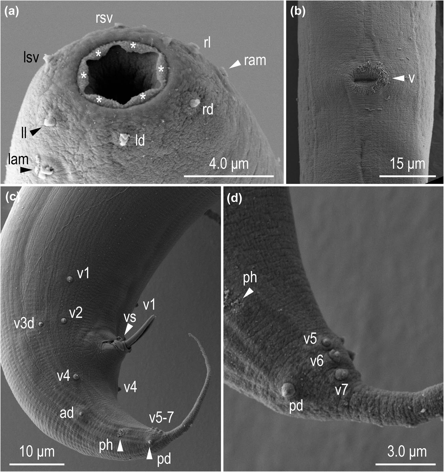
Figure 7
Scanning electron micrographs of Pristionchus endotocus n. sp. (a) Stomatal opening of stenostomatous hermaphrodite; (b) Vulval region of hermaphrodite; (c) Whole tail of male; (d) Tail tip region of male. Labels are as follows: ad, pd, and v + number = genital papillae following the terminology in Sudhaus and Fürst von Lieven (2003); asterisk = cheilostomatal flap-like extension; lam = left amphid; ld = left dorsal labial sensillum; ll = left lateral labial sensillum; lsv = left subventral labial sensillum; ram = right amphid; rd = right dorsal labial sensillum; rl = right lateral labial sensillum; rsv = right subventral labial sensillum; ph = phasmid; v = vulva; vs = ventral single papilla.
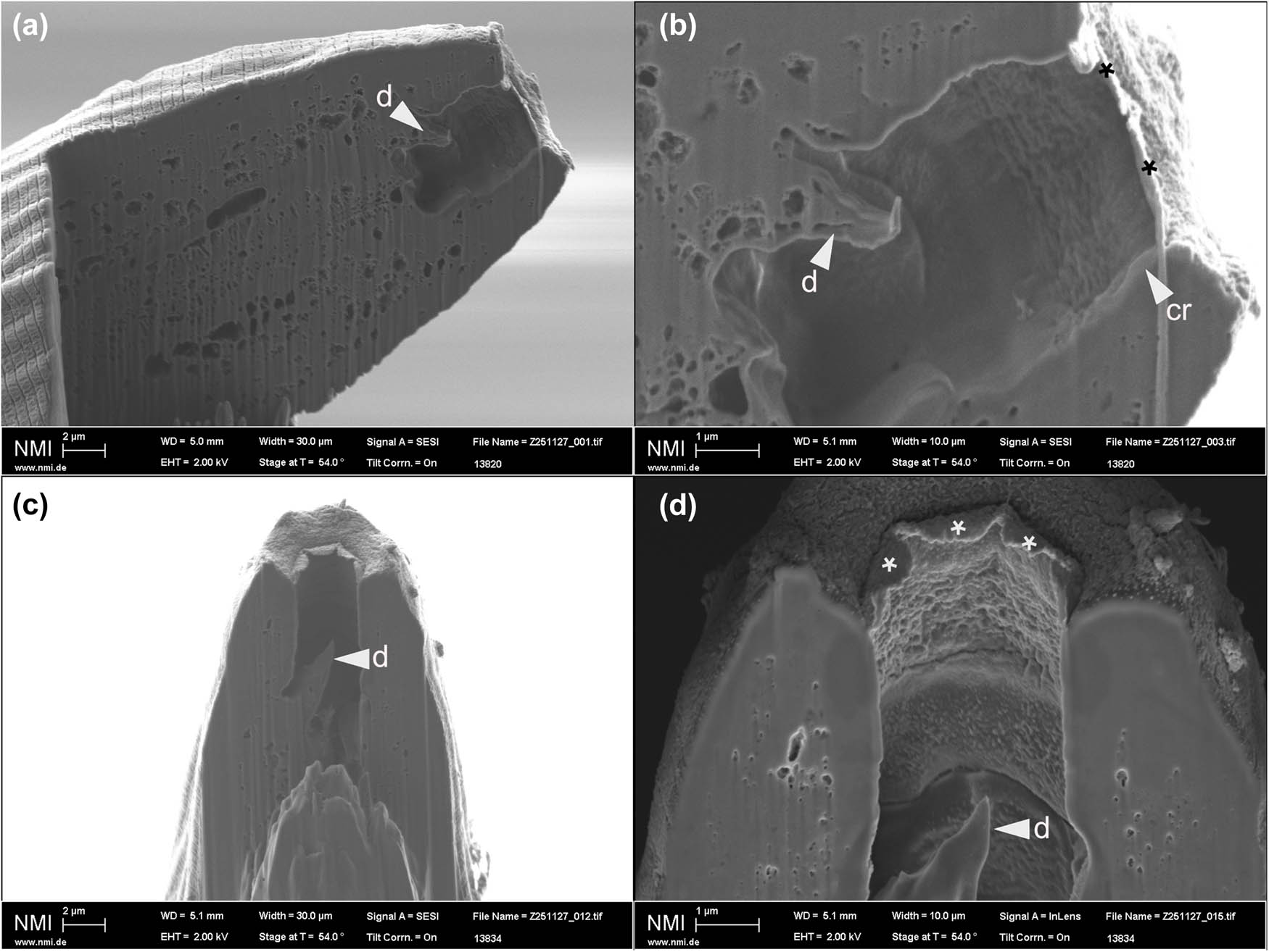
Figure 8
Focused ion beam-scanning electron micrographs of Pristionchus endotocus n. sp. (a, c) Sagittal section of the stomatal opening of stenostomatous hermaphrodites; (b, d) close-up views showing the fusion of cheilostomatal plates. Labels are as follows: asterisk = cheilostomatal flap-like extension; d = dorsal tooth; cr = rim-like thickened anterior end of cheilostom.
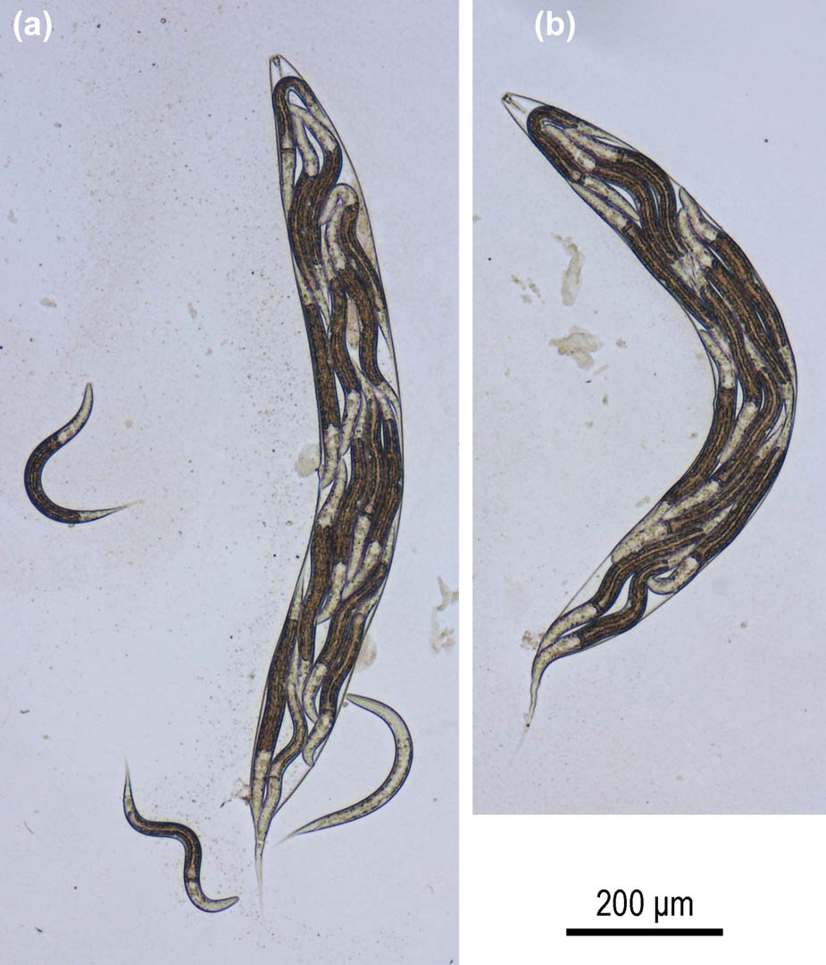
Figure 9
Juveniles of Pristionchus endotocus n. sp. retained in the dead body of their hermaphrodite mothers. (a, b) Two different individuals showing bagging.
Table 1
Morphometric measurements of Pristionchus endotocus n. sp. All measurements are in µm and formatted as “mean ± standard deviation (range).”
| Character | RS6393 | |
|---|---|---|
| Stenostomatous male | Stenostomatous female | |
| n | 10 | 10 |
| L | 779 ± 76.9 (644–906) | 1292 ± 159.5 (1038–1521) |
| L′ | 664 ± 77.9 (541–799) | 1118 ± 145.4 (884–1321) |
| a | 15 ± 1.7 (11.7–16) | 13 ± 0.6 (13–14) |
| b | 5.5 ± 0.5 (4.5–6.2) | 7.8 ± 0.8 (6.5–9.0) |
| c | 6.7 ± 0.8 (5.8–8.5) | 7.5 ± 0.4 (7.0–8.1) |
| c' | 3.3 ± 0.2 (2.8–3.6) | 4.6 ± 0.3 (4.0–4.9) |
| Ant. stoma length (cheilo- + gymnostom) | 6.8 ± 0.4 (6.2–7.4) | 6.6 ± 0.9 (4.9–7.6) |
| Total stoma length | 10 ± 0.7 (8.5–11) | 10 ± 1.0 (8–11) |
| Stoma width | 6.0 ± 0.5 (5.1–6.7) | 7.5 ± 0.4 (6.8–8.0) |
| Ant. pharynx (pro + metacorpus) | 76 ± 1.3 (73–77) | 91 ± 5.3 (84–103) |
| Post. pharynx (isthmus + basal bulb) | 55 ± 3.1 (50–761) | 68 ± 12.1 (58–99) |
| Total pharynx length | 131 ± 3.8 (125–137) | 159 ± 11.4 (144–182) |
| Ant./total pharynx % | 58 ± 1.3 (55–60) | 57 ± 4.5 (46–61) |
| Median bulb diameter | 20 ± 1.3 (18–22) | 26 ± 1.3 (24–28) |
| Terminal bulb diameter | 22 ± 1.5 (20–25) | 29 ± 3.2 (24–35) |
| Ant. end to cardia | 141 ± 4.3 (135–147) | 165 ± 7.1 (155–178) |
| Ant. end to S–E pore | 134 ± 14.1 (116–163) | 161 ± 13.4 (141–181) |
| Ant. end to nerve ring | 98 ± 3.8 (93–104) | 107 ± 4.2 (100–113) |
| Testis length | 543 ± 91.6 (370–652) | — |
| Ant. end to vulva distance | — | 620 ± 83.6 (493–730) |
| Vulva to anus distance | — | 498 ± 70.2 (409–602) |
| T or V | 69 ± 7.9 (51–84) | 48 ± 1.8 (46–53) |
| Maximum body diameter | 54 ± 8.8 (44–68) | 97 ± 15.2 (75–119) |
| Cloacal or anal body diameter | 36 ± 2.2 (31–39) | 38 ± 5.5 (31–49) |
| Tail length | 117 ± 6.9 (107–130) | 172 ± 18.0 (142–204) |
| Spicule curve | 40 ± 3.1 (32–42) | — |
| Spicule chord | 35 ± 2.7 (28–37) | — |
| Gubernaculum length | 18 ± 1.1 (16–19) | — |