
The genus Pristionchus (Kreis, 1932) is emerging as a system for biodiversity research in nematodes and animals in general, largely due to the importance of the species Pristionchus pacificus (Sommer, Carta, Kim, and Sternberg, 1996) as a model organism of nematode evo-devo and phenotypic plasticity research (Sommer, 2015; Kanzaki et al., 2021; Herrmann et al., 2024). Over the years, complete sequencing of the genome, coupled with a wide array of molecular techniques and tools, has been used to study the evolution, development, and biology of P. pacificus (Sommer et al., 1996; Dieterich et al., 2008; Tian et al., 2008; Schlager et al., 2009; Witte et al., 2015; Rödelsperger et al., 2017; Igreja et al., 2022). Alongside these invaluable resources, a robust phylogeny and deep taxon sampling have been indispensable for making inferences in P. pacificus and Pristionchus evolution (Rödelsperger et al., 2024; Yoshida et al., 2023, 2024).
Considerable efforts over the past two decades have been devoted to the collection, description/identification, and cryopreservation of natural diplogastrid and Pristionchus isolates. Notably, these efforts have uncovered more than 50 species of Pristionchus (Herrmann et al., 2024), 38 of which are described from Asia. These findings indicate that while the genus has a nearly worldwide distribution, Asia is the biodiversity hotspot for Pristionchus nematodes (Kanzaki et al., 2021; Herrmann et al., 2024). Nevertheless, despite the large number of species described from the region, Pristionchus sampling efforts in Asia are far from saturation. With Asia as the focus of searches for new Pristionchus species, extensive sampling efforts in 2024 and 2025 in the Philippines yielded the ninth androdioecious species of Pristionchus, which represents at least the eighth transition from gonochorism within the genus (Yoshida et al., 2024). The new species, Pristionchus endotocus n. sp., has notable traits in standard laboratory conditions when compared with other Pristionchus species: consistent matricidal hatching, a high ratio of male offspring, and a strong bias toward the stenostomatous form.
In the present study, this new androdioecious species, P. endotocus n. sp., is described. Morphological, morphometric, and molecular data that distinguish it from the most closely related previously described Pristionchus species are presented. In addition to providing another branch where androdioecy is observed, the new species offers significant potential for comparative studies of life history traits within the Pristionchus system.
1. Materials and methods
1.1. Nematode isolation
July–August 2024 sampling resulted in 22 total nematode isolates frozen from 253 beetle specimens (2 of which were identified as Pristionchus endotocus n. sp.). July 2025 sampling resulted in 18 total nematode isolates frozen from 449 beetle specimens (3 of which were identified as P. endotocus n. sp.). 2024 strains RS6393 and RS6394 were isolated from the same Papuana philippinica specimen. 2025 strains RS6422, RS6423, and RS6424 were isolated from Anomala sp., Amphitrichia sp., and Anomala sp., respectively.
Nematodes were isolated using standard procedures as previously described (Herrmann et al., 2006). Briefly, beetles were dissected on 2% nematode growth medium (NGM) agar plates. Over a time window of 2 weeks, the plates were scanned at room temperature for developing nematodes. Single gravid females were transferred to new plates of NGM seeded with Escherichia coli strain OP50. Established isogenic lines are kept in laboratory cultures and were simultaneously frozen in liquid nitrogen. All isolated strains of P. endotocus n. sp. are available as frozen stocks in the Department of Integrative Evolutionary Biology, Max Planck Institute for Biology Tübingen, Germany, and can be provided as living culture upon request.
1.2. Morphological observation
Adult P. endotocus n. sp. males and hermaphrodites from the designated reference strain RS6393 were used for morphological observations. As all attempts to induce the eurystomatous form using known eurystomatous-form-inducing bacterial diets were unsuccessful in the new species, we attempted to induce this form using starvation (Herrmann et al., 2024). Indeed, starvation resulted in some eurystomatous worms. Type material was prepared by rinsing nematodes in distilled water and killing them at 65°C. The specimens were then fixed in 5% formalin and processed using Seinhorst’s method (Eisenback and Hunt, 1986). The specimens were then mounted on labeled microscope slides with glycerol and embedded in wax. Light microscope observations for drawings and morphometrics were conducted on live material using a compound microscope (Nikon Eclipse Ni) with differential interference contrast optics, a digital camera system (Leica MC170HD), and a drawing tube (Kanzaki, 2013; Ragsdale et al., 2013). Figures presented in this article were formatted using Adobe Photoshop Elements 2020.
1.3. Electron microscopy
For scanning electron microscopy (SEM) and focused ion beam scanning electron microscopy (FIB-SEM) observations, nematodes were collected into phosphate-buffered saline (PBS) buffer and cleaned of bacteria and debris, followed by killing at 65°C. The animals were then fixed in 2.5% glutaraldehyde and 4% formaldehyde in PBS. Fixed specimens were postfixed in 1% osmium tetroxide for 4 h on ice and dehydrated through a graded ethanol series, followed by critical-point drying (CPD300, Leica Microsystems) from carbon dioxide. Specimens were mounted on double-sided carbon tape in desired positions and sputter coated with 4 nm platinum (CCU-010, Safematic). The same sample preparation protocol was used for both SEM and FIB-SEM imaging. SEM images were obtained with a field emission scanning electron microscope (Regulus 8,230, Hitachi High Technology) operated at 3 kV. FIB-SEM images were obtained with a Crossbeam 550 scanning electron microscope (Zeiss) operated at 2 kV. Milling to reveal internal structures was achieved with a gallium ion beam.
1.4. Remarks concerning morphometric measurements
The previous findings have shown that measurements of nematode specimen dimensions can change over time as a result of different culture conditions and many Pristionchus species are morphologically cryptic (Kanzaki et al., 2012; Fonderie et al., 2013; Ragsdale et al., 2015; Herrmann et al., 2016). The new species is hereby described using a multidimensional and integrative approach that combines molecular, biological, morphological, and ecological information.
1.5. Molecular characterization and phylogeny
To integrate Pristionchus endotocus n. sp. strains RS6393 and RS6394 into the most recent version of the Pristionchus species phylogeny (Theam et al., 2025), transcriptomes were generated by growing worms on NGM plates with E. coli OP50 at 20°C and isolating total RNA from two to three mixed-stage culture plates using standard Trizol extraction following the manufacturers’ instructions (Zymo Research, USA). Illumina sequencing libraries were generated and sequenced by the company Novogene, resulting in more than 10 million reads per sample (2 × 150 nt, deposited at the European Nucleotide Archive under the study accession PRJEB20959). Analysis of the raw sequencing reads was performed as described previously (Rödelsperger et al., 2018). In short, raw reads were assembled de novo by the software Trinity (version 2.2.0 with -normalize_reads option) (Grabherr et al., 2011), followed by open reading frame calling and orthology clustering by orthAgogue (Ekseth et al., 2014). Multiple sequence alignments of orthologous groups without duplications were computed with MUSCLE (version 3.8.31) (Edgar, 2004), and a concatenated alignment of more than 140,000 amino acids was taken as input to reconstruct a maximum likelihood tree using the RAxML program (version 8.2.12 with -m PROTGAMMAILG -f a -N 100 options) (Stamatakis, 2014).
1.6. Mating experiments
To test the reproductive isolation of the new species, we performed crosses with the most closely related strains from our collection based on the reconstructed Pristionchus species phylogeny tree. In this case, the closest gonochoristic species are P. quartusdecimus RS5230, P. hangukensis RS6271, and P. purgamentorium RS6138 (Fig. 1). Mating experiments were set up as follows for each species: three virgin females of the gonochoristic species to be tested were transferred to 2% NGM agar mating plates seeded with 50 µl of OP50 along with three males of P. endotocus n. sp. All mating experiments were done in quintuplicate.
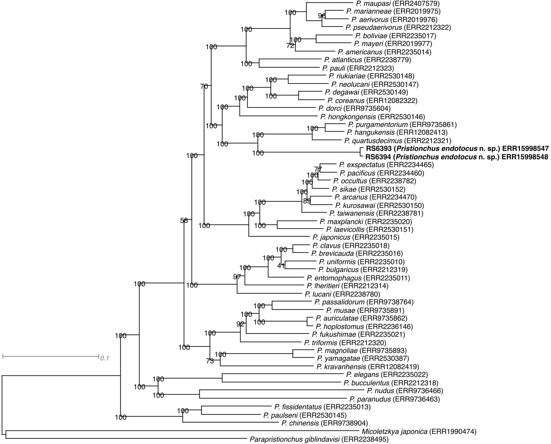
Figure 1
Molecular phylogeny of the genus Pristionchus as inferred from a maximum likelihood tree reconstructed from a concatenated protein alignment, with Micoletzkya japonica and Parapristionchus giblindavisi as outgroups. The identifiers after the species names indicate the Run accessions for the corresponding transcriptomic data at the European Nucleotide Archive.
2. Results
2.1. Molecular profiles and phylogeny
(Fig. 1)
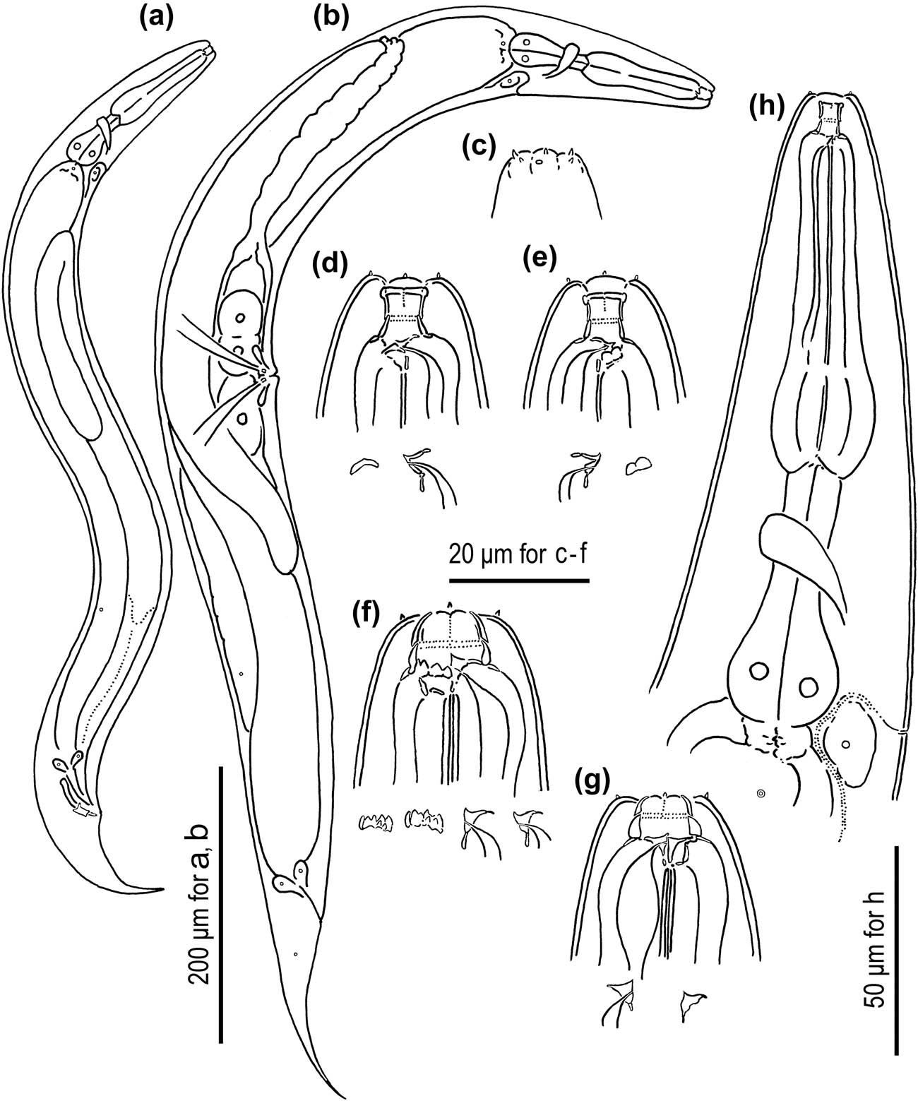
Figure 2
Adult male and hermaphrodite of Pristionchus endotocus n. sp. (a) male; (b) hermaphrodite; (c) lip region of male; (d, e) stomatal region of stenostomatous hermaphrodite where tooth and ridges are drawn separately; (f, g) stomatal region of eurystomatous hermaphrodite where tooth and ridges are drawn separately; (h) anterior part of the body of hermaphrodite. All subfigures are in the right lateral view except for d (left lateral view).
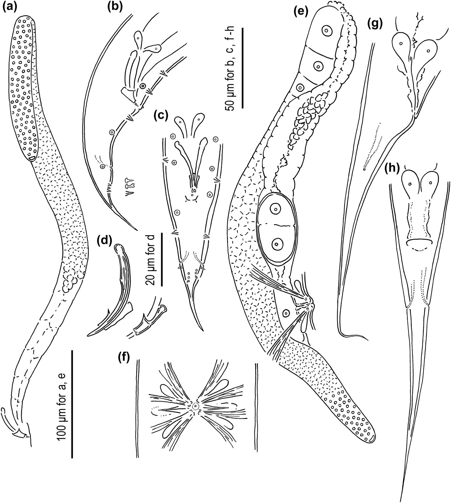
Figure 3
Adult male and hermaphrodite of Pristionchus endotocus n. sp. (a) Whole testis; (b, c) male tail; (d) spicule and gubernaculum; (e) whole part of anterior ovotestis; (f) vulval region of hermaphrodite; (g, h) tail region of hermaphrodite. a, b, e, and g are in the right lateral view; c, f, and h are in the ventral view; and d is in the left lateral view.
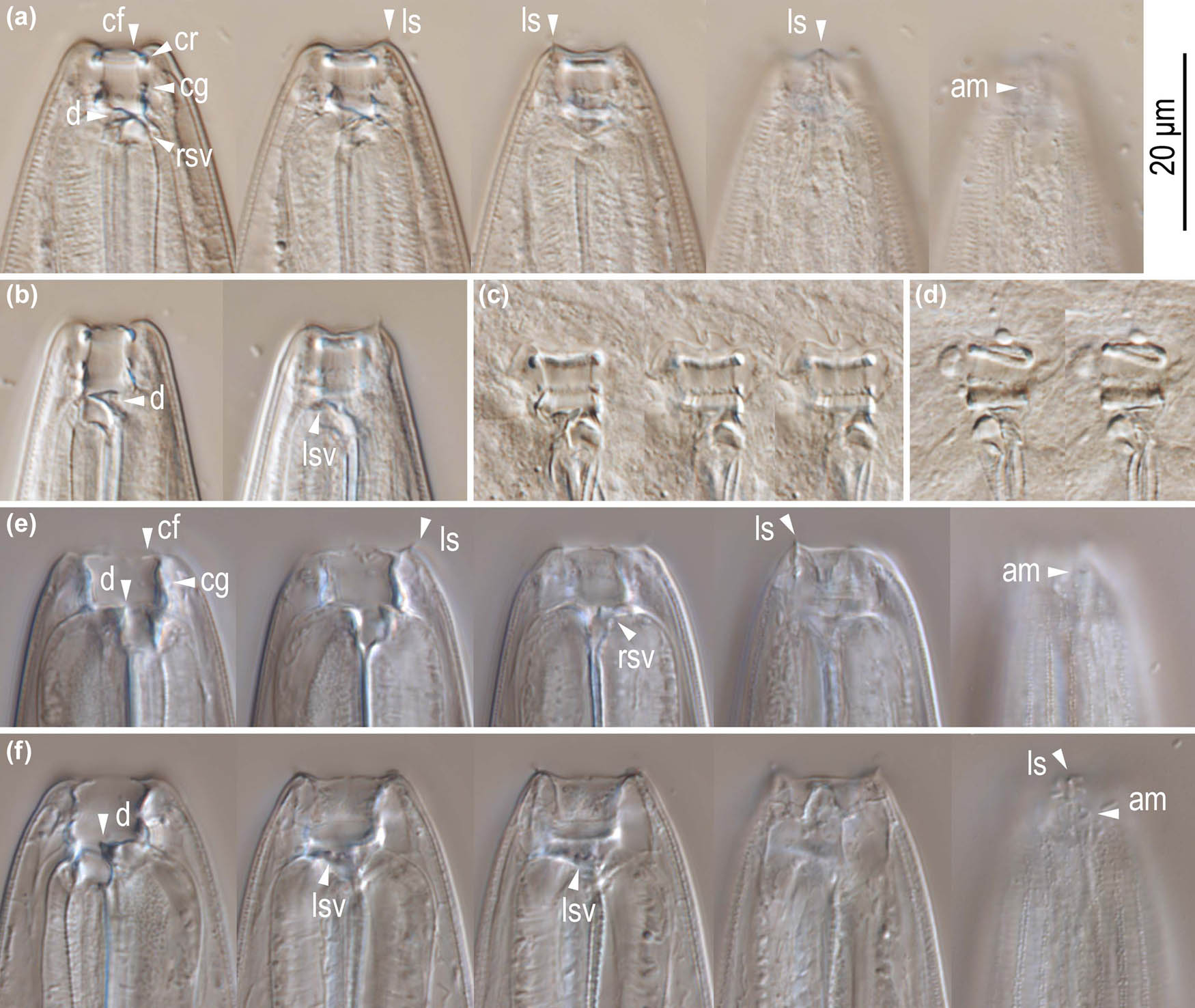
Figure 4
Stomatal morphology of Pristionchus endotocus n. sp. hermaphrodites. (a) Right lateral view of stenostomatous form in five different focal planes; (b) left lateral view of stenostomatous form in two different focal planes; (c) right lateral view of squashed stenostomatous form in three different focal planes; (d) left lateral view of squashed stenostomatous form in two different focal planes; (e) left lateral view of eurystomatous form in five different focal planes; (f) right lateral view of eurystomatous form in five different focal planes. Labels are as follows: am = amphid; cf = cheilostomatal flap; cg = cheilostom-gymnostom margin; cr = cheilostomatal rim; d = dorsal tooth; ls = labial sensilla; lsv = left subventral ridges; rsv = right subventral ridges.
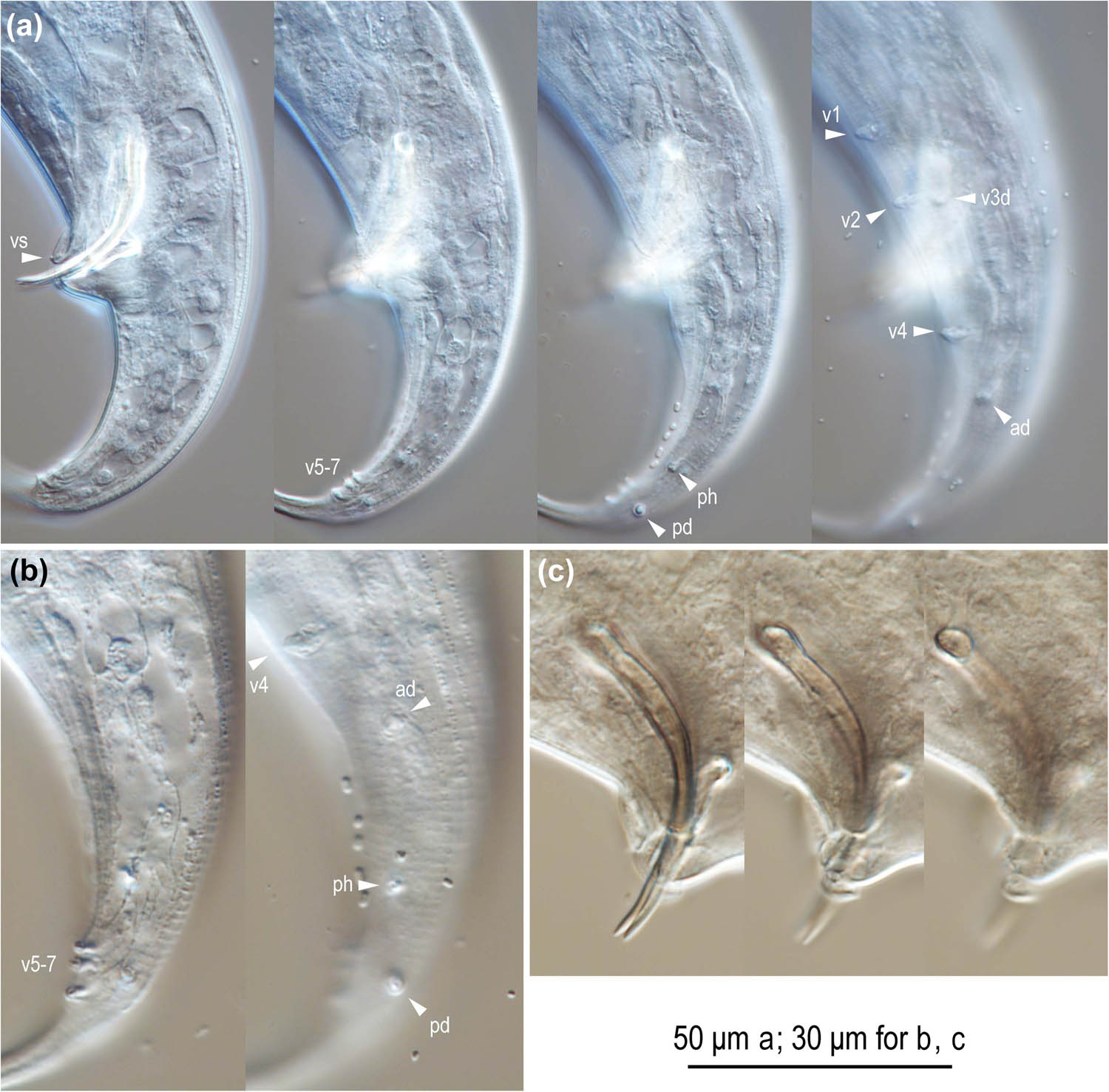
Figure 5
Left lateral view of male tail characters of Pristionchus endotocus n. sp. (a) Whole tail in five different focal planes; (b) posterior part of the tail in two different focal planes; (c) spicule and gubernaculum in three different focal planes. Labels are as follows: ad, pd, and v + number = genital papillae following the terminology in Sudhaus and Fürst von Lieven (2003); ph = phasmid; vs = ventral single papilla.
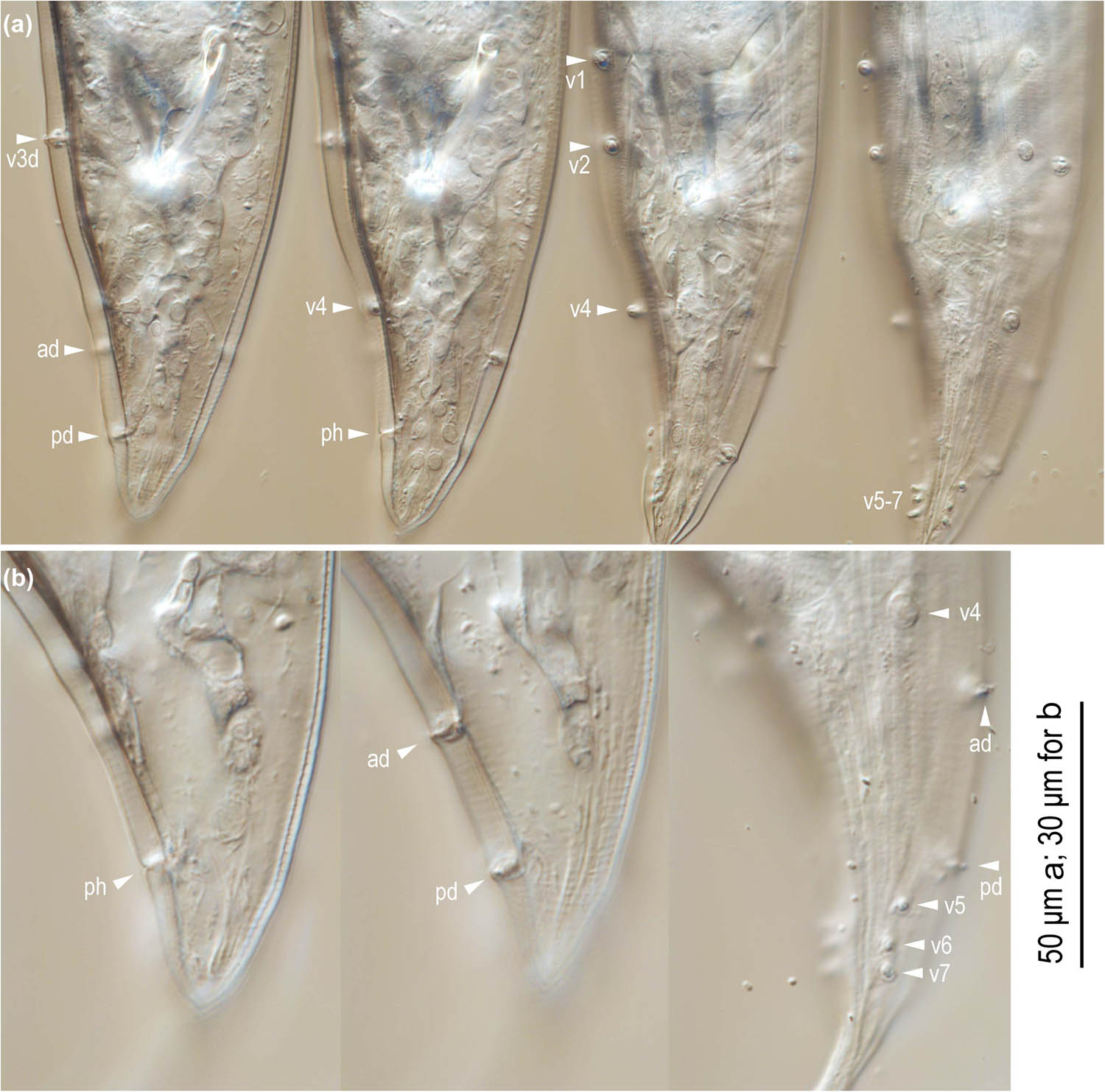
Figure 6
Ventral view of male tail characters of Pristionchus endotocus n. sp. (a) Whole tail in four different focal planes; (b) posterior part of the tail in three different focal planes. Labels are as follows: ad, pd, and v + number = genital papillae following the terminology presentd by Sudhaus and Fürst von Lieven (2003); ph = phasmid; vs = ventral single papilla.
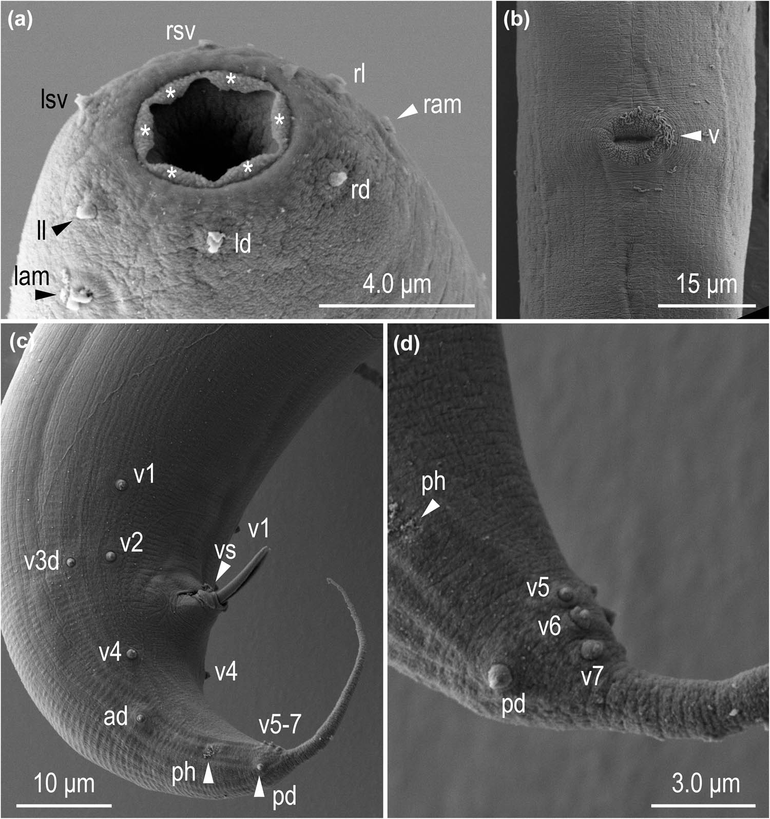
Figure 7
Scanning electron micrographs of Pristionchus endotocus n. sp. (a) Stomatal opening of stenostomatous hermaphrodite; (b) Vulval region of hermaphrodite; (c) Whole tail of male; (d) Tail tip region of male. Labels are as follows: ad, pd, and v + number = genital papillae following the terminology in Sudhaus and Fürst von Lieven (2003); asterisk = cheilostomatal flap-like extension; lam = left amphid; ld = left dorsal labial sensillum; ll = left lateral labial sensillum; lsv = left subventral labial sensillum; ram = right amphid; rd = right dorsal labial sensillum; rl = right lateral labial sensillum; rsv = right subventral labial sensillum; ph = phasmid; v = vulva; vs = ventral single papilla.
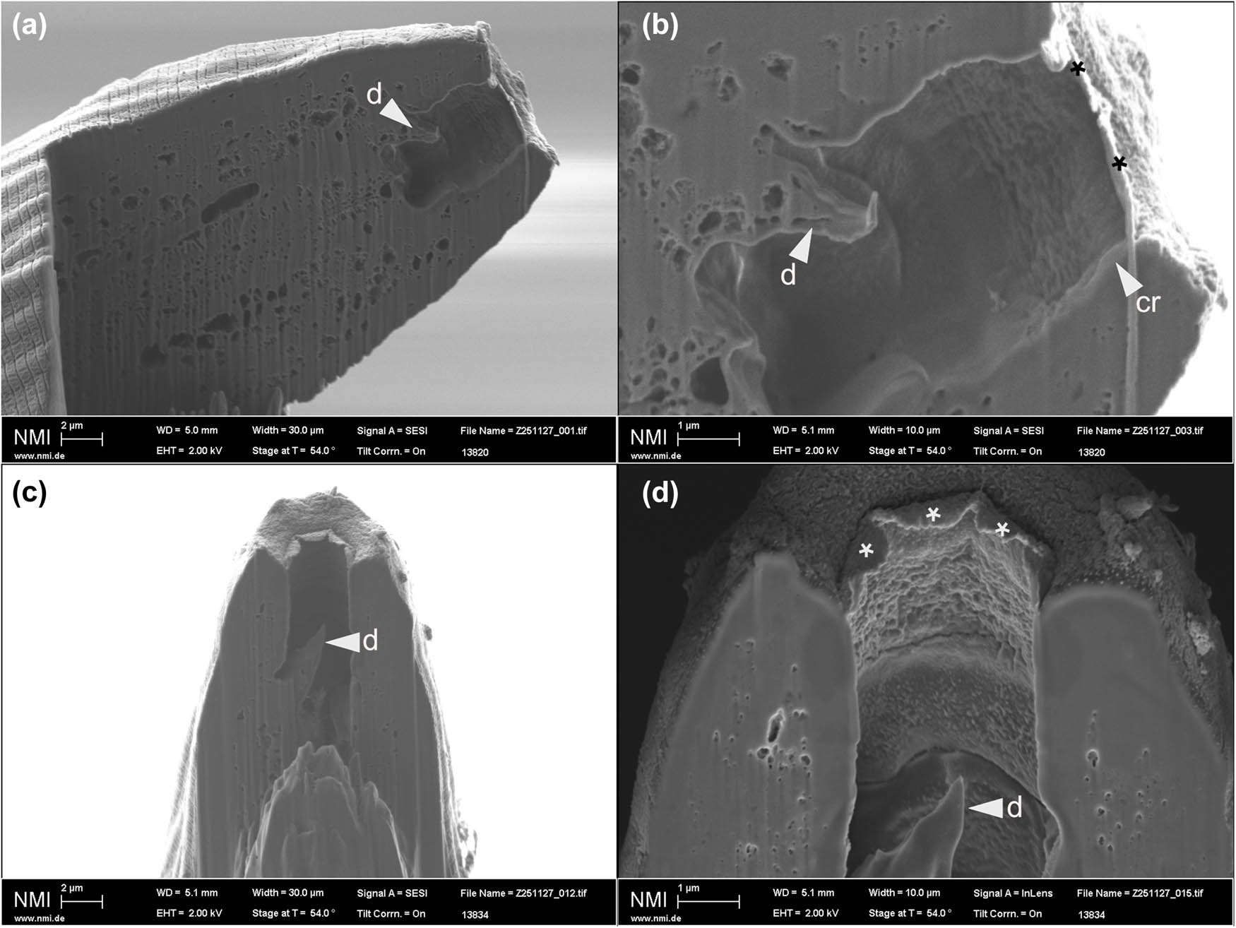
Figure 8
Focused ion beam-scanning electron micrographs of Pristionchus endotocus n. sp. (a, c) Sagittal section of the stomatal opening of stenostomatous hermaphrodites; (b, d) close-up views showing the fusion of cheilostomatal plates. Labels are as follows: asterisk = cheilostomatal flap-like extension; d = dorsal tooth; cr = rim-like thickened anterior end of cheilostom.
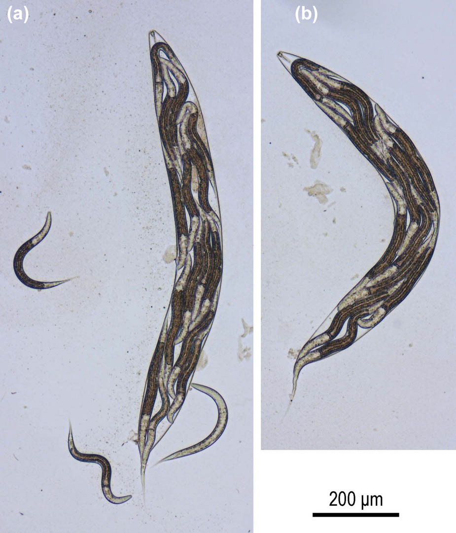
Figure 9
Juveniles of Pristionchus endotocus n. sp. retained in the dead body of their hermaphrodite mothers. (a, b) Two different individuals showing bagging.
Table 1
Morphometric measurements of Pristionchus endotocus n. sp. All measurements are in µm and formatted as “mean ± standard deviation (range).”
| Character | RS6393 | |
|---|---|---|
| Stenostomatous male | Stenostomatous female | |
| n | 10 | 10 |
| L | 779 ± 76.9 (644–906) | 1292 ± 159.5 (1038–1521) |
| L′ | 664 ± 77.9 (541–799) | 1118 ± 145.4 (884–1321) |
| a | 15 ± 1.7 (11.7–16) | 13 ± 0.6 (13–14) |
| b | 5.5 ± 0.5 (4.5–6.2) | 7.8 ± 0.8 (6.5–9.0) |
| c | 6.7 ± 0.8 (5.8–8.5) | 7.5 ± 0.4 (7.0–8.1) |
| c' | 3.3 ± 0.2 (2.8–3.6) | 4.6 ± 0.3 (4.0–4.9) |
| Ant. stoma length (cheilo- + gymnostom) | 6.8 ± 0.4 (6.2–7.4) | 6.6 ± 0.9 (4.9–7.6) |
| Total stoma length | 10 ± 0.7 (8.5–11) | 10 ± 1.0 (8–11) |
| Stoma width | 6.0 ± 0.5 (5.1–6.7) | 7.5 ± 0.4 (6.8–8.0) |
| Ant. pharynx (pro + metacorpus) | 76 ± 1.3 (73–77) | 91 ± 5.3 (84–103) |
| Post. pharynx (isthmus + basal bulb) | 55 ± 3.1 (50–761) | 68 ± 12.1 (58–99) |
| Total pharynx length | 131 ± 3.8 (125–137) | 159 ± 11.4 (144–182) |
| Ant./total pharynx % | 58 ± 1.3 (55–60) | 57 ± 4.5 (46–61) |
| Median bulb diameter | 20 ± 1.3 (18–22) | 26 ± 1.3 (24–28) |
| Terminal bulb diameter | 22 ± 1.5 (20–25) | 29 ± 3.2 (24–35) |
| Ant. end to cardia | 141 ± 4.3 (135–147) | 165 ± 7.1 (155–178) |
| Ant. end to S–E pore | 134 ± 14.1 (116–163) | 161 ± 13.4 (141–181) |
| Ant. end to nerve ring | 98 ± 3.8 (93–104) | 107 ± 4.2 (100–113) |
| Testis length | 543 ± 91.6 (370–652) | — |
| Ant. end to vulva distance | — | 620 ± 83.6 (493–730) |
| Vulva to anus distance | — | 498 ± 70.2 (409–602) |
| T or V | 69 ± 7.9 (51–84) | 48 ± 1.8 (46–53) |
| Maximum body diameter | 54 ± 8.8 (44–68) | 97 ± 15.2 (75–119) |
| Cloacal or anal body diameter | 36 ± 2.2 (31–39) | 38 ± 5.5 (31–49) |
| Tail length | 117 ± 6.9 (107–130) | 172 ± 18.0 (142–204) |
| Spicule curve | 40 ± 3.1 (32–42) | — |
| Spicule chord | 35 ± 2.7 (28–37) | — |
| Gubernaculum length | 18 ± 1.1 (16–19) | — |
2.2. Description
2.2.1. Pristionchus endotocus n. sp. (Figs 2–9, Table 1) Adults
The body is cylindrical and stout. The cuticle is thick, with fine annulation and clear, but shallow longitudinal striations. The lateral field consists of two lines, only weakly distinguishable from the body striation, with the presence of a deirid. The head bears six short and papilliform labial sensillae, without apparent lips; four small, papilliform cephalic papillae are present in males, which is typical for diplogastrid nematodes. Amphidial apertures are located on the lateral sector, slightly dorsally shifted, at the level of the margin of cheilo- and gymnostom. Stomatal dimorphism is seen. Dorsal pharyngeal gland is clearly observed, with penetrating dorsal tooth to the gland opening; the anterior part of the pharynx (= pro- and metacorpus) is 1.5 times long as the posterior part (= isthmus and basal bulb); procorpus is very muscular, stout, occupying half to two-thirds of corresponding body diameter; metacorpus is very muscular, forming well-developed median bulb; isthmus is narrow and not muscular; basal bulb is glandular; pharyngo-intestinal junction is clearly observed, well developed; nerve ring is usually surrounded by the middle region of isthmus; secretory-excretory pore is not conspicuous, which is ventrally located around the level of basal bulb to pharyngo-intestinal junction, i.e., overlapping with basal bulb to approximately 1 body diameter posterior to the basal bulb; excretory duct extends to the anterior part of the body and is reflexed back to the position of pore; deirid is observed laterally, located almost approximately one-third to half body diameter posterior to secretory–excretory pore; hemizonid is not observed; lateral glands, small pores connected to secretory cell, are present and observed on the lateral body surface, with positions inconsistent among individuals, ranging from 5 to 8 for males and 9 to 13 for females.
2.2.2. Stenostomatous form
Cheilostom consists of six per- and interradial plates fused to form a solid, ring-like structure not typical for the genus. The anterior end of each plate thickens to form the rim-like structure, and a very short flap-like expansion is seen on the rim-like part, but the flap is difficult to confirm in the light microscopic observation; gymnostom is short, cuticular ring or short tube like, and the anterior end overlaps cheilostom internally; dorsal gymnostomatal wall is slightly thickened compared to the ventral side; stegostom is separated into three subsections: pro-meso, meta, and telostegostom; pro-meso stegostom forms a weakly cuticularized ring surrounding the anterior edge of pharynx. Metastegostom bears a conspicuous and movable triangular or small claw-shaped dorsal tooth with a strongly sclerotized surface, pointed toward the left subventral ridge with 1 or 2 minute adventitious denticles (bumps) on a plate and pointed toward the right subventral ridge, with one or two blunt distal adventitious denticle(s). Telostegostom forms a weakly sclerotized cup-like cavity connecting the stoma and pharynx.
2.2.3. Eurystomatous form
Cheilostom consists of six per- and interradial plates. Incision between plates is not easily distinguished by light microscopy. The anterior end of each plate extends to form a short flap-like expansion; gymnostom is seen as short, cuticular ring like or short tube like anterior end that overlaps the cheilostom internally; the dorsal gymnostomatal wall is slightly thickened compared to the ventral side; stegostom is separated into three subsections: pro-meso, meta, and telostegostom; pro-meso stegostom forms a weakly cuticularized ring surrounding the anterior edge of the pharynx. Metastegostom bears movable dorsal and right subventral teeth and left subventral ridges. Dorsal tooth is mid-small in size, almost the same size as the dorsal tooth in the stenostomatous form, triangular with slightly anteriorly curved tip; right subventral tooth is almost the same size as the dorsal tooth, triangular with pointed tip; left subventral ridges are separated into three groups, each with two or three small ridges. Telostegostom forms a weakly sclerotized cup-like cavity connecting stoma and pharynx.
2.2.4. Male
Males are ventrally arcuate, strongly ventrally curved at the tail region when killed by heat. Males have a single testis that is ventrally located with the anterior part reflexed to the right side. Spermatogonia are arranged in three to five rows in the reflexed part; then well-developed spermatocytes are arranged as three to four rows in the anterior half of the main branch; and then mature amoeboid spermatocytes are arranged in one to two row(s) in the proximal part of gonad. Vas deferens is formed by relatively large cells, occupying approximately 25% of the whole gonad. Three (two subventral and one dorsal) cloacal gland cells are observed at the distal end of the vas deferens and the intestine. Spicules are paired, separate, smoothly curved in a ventral view, adjacent to each other for the distal fourth of their length, each smoothly tapering to the pointed distal end. Spicules in lateral view smoothly ventrally arcuate, giving them a ca 100° curvature, rounded manubrium sometimes appearing to have a small anterior notch present at the anterior end, lamina/calomus complex (blade) weakly expanded slightly posterior to manubrium (ca one-fifth of the blade length from the anterior), then smoothly tapering to a pointed distal end. Gubernaculum conspicuous, about one-third of the spicule length, broad anteriorly such that the dorsal wall is slightly recurved with dorsal and ventral walls separated at 50°–60° angle at the posterior end. The dorsal side of the gubernaculum possesses a single, membranous, anteriorly directed process; and the lateral pair is more sclerotized, anteriorly and obliquely ventrally directed processes. In the lateral view, the anterior half of gubernaculum with two serial curves is separated by an anteriorly and obliquely ventrally directed process, with the anterior terminal curvature highly concave and almost closed, and with the deep posterior curvature being one-third of the gubernaculum length; the posterior half forms a tube-like process enveloping spicules. Cloacal opening (CO) forms a simple slit. One small, ventral, single genital papilla (vs) is seen on the anterior cloacal lip; nine pairs of genital papillae (v1–v7, ad, pd) and a pair of phasmids (ph) are present, with the arrangement < v1, (v2, v3 d),/, v4, ad, ph, pd, (v5–7) >, as shown in the nomenclature in the study by Sudhaus and Fürst von Lieven (2003), where v1 is located at approximately one cloacal body diameter (CBD) anterior to CO; v2 and v3d pairs are almost at the same level and are approximately one-quarter CBD anterior to CO; v4 is approximately one-quarter CBD posterior to CO and ad is at approximately 1 CBD posterior to CO; the ph is somewhat variable in position: midway between ad and pd to just anterior to pd; pd is slightly anterior to v5–7 triplet, not overlapping with the triplet; v5–7 forms a linearly arranged triplet at the ventral side of the body, close to each other around immediately anterior to the root of tail spike. Anterior five pairs of papillae (v1–4 and ad) are almost equal in size, rather large and conspicuous; v7 and pd papillae are obviously smaller than the anterior five pairs; and v5 and v6 are conspicuous, but smaller than v7 and pd. Anterior two pairs of the ventral triplet papillae (v5 and v6 of v5–v7) are papilliform and borne from socket-like base, v7 simple or typical thorn-like in shape. Tip of v6 papillae is split into two small papilla-like projections. Tail is conical, with a short spike occupying approximately one-third of the whole tail length; the spike smoothly tapered to the bluntly pointed tip. Bursa or bursal flap is absent.
2.2.5. Hermaphrodite
Relaxed or slightly ventrally arcuate when killed by heat. Gonad (ovotestis) is didelphic, amphidelphic; each gonadal system is arranged from the vulva/vagina as uterus, oviduct, and ovary. Anterior and posterior gonads are basically the same in their structure, and only the anterior gonad is described in detail here. Anterior gonad is at the right of the intestine, with the uterus and oviduct extending ventrally and anteriorly on the right of the intestine and with a totally reflexed (= antidromous reflexion) ovary extending dorsally. Oocytes in the ovary are mostly arranged in three to four rows in the distal two-thirds to three-quarters of the ovary and in double or single row in the rest of ovary, the distal tip of the ovary reaching the vulva to the oviduct of the opposite gonad branch depending on the developmental condition. The anterior end of the oviduct (= junction tissue between the ovary and the oviduct) consists of rounded cells. Spermatheca is not clearly distinct, but the anterior part of the oviduct immediately posterior to the junction consists of rounded cells that work as spermatheca. Eggs in single to multiple-cell stage or even further developed at the posterior part of oviduct (= uterus). A mature hermaphrodite often carries more than 15 eggs in each uterus. Receptaculum seminis is not observed, i.e., the organ is not independent, and a part of the oviduct/uterus works as the organ. Vaginal glands are present but obscure. Vagina is perpendicular to the body surface, surrounded by the sclerotized tissue; the vulva is protuberant in the lateral view and pore like in the ventral view. Rectum is approximately one anal body diameter long; the intestine/rectum junction is surrounded by the well-developed sphincter muscle; and three anal glands (two subventral and one dorsal) are present but not obvious. Anus is seen in the form of a dome-shaped slit, posterior anal lip slightly protuberant. Phasmid is approximately 1.7 anal body diameter posterior to the anus. Tail forms elongate conoid with a sharply pointed terminus.
2.2.6. Etymology
Pristionchus endotocus n. sp.
Hermaphrodites of this species obligately show endotokia matricida, or bagging, in standard laboratory conditions.
2.2.7. Diagnosis and relationship
In addition to its generic characters, the new species is characterized by its 1) androdioecious reproductive mode; 2) stenostomatous form with cheilostomatal plate forming a rim-like anterior end; 3) eurystomatous form with mid-small–sized triangular dorsal and right subventral teeth and three left subventral ridges each bearing two to three small ridges; 4) the arrangement of male genital papillae (<v1, (v2, v3d), /, v4, ad, ph, pd, (v5–7) >; 5) and male tail with a short spike that occupies approximately one-third of the whole tail; and 6) elongate conoid hermaphrodite tail with a sharply pointed terminus.
The genus Pristionchus consists of more than 50 species (Susoy et al., 2016; Theam et al., 2025), and although there are several cryptic species complexes (e.g., Kanzaki et al., 2012), they can be typologically distinguished from each other based on the stomatal morphology, structure of the cuticle, arrangement of the genital papillae, and the shape of the tail in males and hermaphrodites/females (Ragsdale et al., 2015; Kanzaki et al., 2021).
Compared with congeners, the new species has three unique characters. First, the cheilostomatal plates of stenostomatous P. endotocus n. sp. worms have a rim-like anterior thickening with a small indistinctive flap, vs the conspicuous flap-like anterior extension of most Pristionchus species (Ragsdale et al., 2015; Kanzaki et al., 2021), or stick-like plates or membrane-like extension of the fig associates (Susoy et al., 2016). Second, the eurystomatous teeth are unique to the new species. The dorsal tooth is rather small (almost the same as the stenostomatal dorsal tooth), and both the dorsal and right subventral teeth are triangular in comparison to all other Pristionchus species, which have more claw-like teeth (e.g., Ragsdale et al., 2015; Susoy et al., 2016; Kanzaki et al., 2021). Third, the male pd genital papillae are located anterior to v5–7 triplet, vs the pd papillae either overlap with or are located posteriorly to the v5–7 triplet in all other species (Ragsdale et al., 2015; Susoy et al., 2016; Kanzaki et al., 2021).
Typologically, P. endotocus n. sp. shares the tube-like stoma with a triangular dorsal tooth in the stenostomatous form, thick and striated cuticle, and the relative position of v2/v3 papillae being at almost the same level (but with v3 being slightly posterior) with P. pacificus, P. arcanus, P. exspectatus, P. mayeri, and P. seladoniae. Nevertheless, the new species can be distinguished from the former four species by the length of the male tail spike, occupying approximately one-third of the whole tail, vs more than half of the whole tail, and the aforementioned unique characters (Sommer et al., 1996; Kanzaki et al., 2012; Kanzaki et al., 2013; Kanzaki et al., 2024). Pristionchus endotocus n. sp. is thus typologically closest to P. seladoniae, but can be distinguished by the aforementioned unique characters, the rim-like anterior end of the cheilostomatal plates in the stenostomatous form, and pd papillae being located anterior to v5–7 triplets in the male tail (Kanzaki et al., 2024).
In addition to these typological characters, the new species has three unique biological characters in standard lab conditions: (1) all mature hermaphrodites retain the last clutch of their eggs in the uterus and the juveniles hatch inside the mother’s body (bagging/endotokia matricida), (2) a strong stenostomatous bias that is uncommon for most androdioecious Pristionchus species (Lightfoot et al., 2021), and (3) a much higher ratio of males in self-offspring compared with other species. These characters have not been reported in the other described Pristionchus species so far.
In mating experiments, P. endotocus n. sp. strains did not produce viable offspring when crossed with phylogenetically close gonochoristic species P. hangukensis, P. quartusdecimus, and P. purgamentorium.
2.2.8. Type habitat and locality
The reference strain of the species is located at Mt. Kitanglad, Mindanao Island, the Philippines, N8°03.406′ E125°00.421′ 1,245 m a.s.l.; and is isolated from Papuana philippinica (Coleoptera: Dynastidae).
2.2.9. Type material
One slide holotype male (RS6393) kept in Max Planck Institute for Biology Tübingen Department for Integrative Evolutionary Biology slide collection, Germany; one paratype slide with one male and one female sent to Mindanao State University, Iligan Institute of Technology, the Philippines. Further paratypes were sent to Staatliches Museum für Naturkunde Karlsruhe, Germany; the museum of Mindanao State University; Iligan Institute of Technology, the Philippines; and the Central Mindanao University, Bukidnon, the Philippines. The new species has been registered in the ZooBank database (zoobank.org) under the identifiers urn:lsid:zoobank.org:pub:2C21E614-A927-4C07-A147-C29B2442235C urn:lsid:zoobank.org:act:978542BA-4122-4CD1-8E9A-EE00B9502E97.
2.2.10. Remarks
Pristionchus endotocus n. sp. has multiple morphological and biological characters that have not been described in congeners so far. These include the cheilostomal ring in the stenostomatous form, a mid-small sized dorsal tooth in the eurystomatous form (Figs 2, 4, 7 and 8), vulval protrusions and the formation of a ‘plug’ in older animals (Figs 3 and 7), retention of eggs over time and their eventual internal hatching (bagging) (Fig. 9). It is worth noting that these characters set P. endotocus n. sp. apart from its closest gonochoristic sister species (P. hangukensis, P. purgamentorium, P. quartusdecimus), except for the stenostomatous bias that is more common in gonochoristic Pristionchus species (Lightfoot et al., 2021).
The changes in the buccal armature of both eurystomatous and stenostomatous animals are not observed in other Pristionchus species and may serve a function. In addition, the stenostomatous bias observed in standard lab conditions, which is unlike most other hermaphrodites of Pristionchus (Lightfoot et al., 2021), may suggest a not-yet-described function of the cheilostomal ring and the smaller-than-usual anterior flaps.
The vulval protrusion in older adults that retain a large number of eggs is well visible, along with a vulval plug of yet unknown structure and function. The plug appears dark red on light microscopy, and is formed by virgin adult hermaphrodites. It is located directly on the vulva with approximately half of it sticking outside in most animals. Current observations suggest that the plug is formed by the hermaphrodites themselves and results in a stark decrease in the number of eggs laid.
The plug itself, while seemingly tied to the apparent bagging phenotype in the new species, does not fully explain it, as not all bagging P. endotocus n. sp. specimens have a visible plug. We report such a structure for any Pristionchus species for the first time in the current work; however, we have observed this plug in other bagging specimens of other species in rare instances (P. pacificus, P. mayeri, P. triformis). This may suggest a yet uncharacterized mechanism tied to egg-laying in Pristionchus.
The retention of eggs in both gonads is not restricted to P. endotocus n. sp., as it is observed in most species of the maupasi clade (Merrill and Ford, 1916). In P. endotocus n. sp., juveniles that hatch from the oldest retained eggs have access to the tissues of the mother, which provides enough energy for them to molt into later stages and exit the cuticle of the mother through the natural orifices (i.e., anus, vulva, mouth), while younger eggs hatch into an empty cuticle and end up as dauer larvae if they are not able to get out. Thus, most hermaphrodites of P. endotocus n. sp. end up producing dauers. Bagging in hermaphroditic species has been shown to be advantageous for the continuation and dispersal of the lineage through dauer larvae, even if it is detrimental for the mother (Vigne et al., 2021). The plug in this case may also help with keeping the progeny inside the cuticle to form dauers.
3. Discussion
The current study describes a new species of Pristionchus that represents another independent transition to androdioecy within the genus (Yoshida et al., 2024). Pristionchus endotocus n. sp. might provide a new reference point to life history trait evolution studies as it exhibits (1) obligate bagging/endotokia matricida, (2) a strong bias to the stenostomatous mouth form, and (3) a high percentage of spontaneous males in all isolated populations in standard laboratory conditions. Future studies on these life history traits may uncover links between the stark morphological differences (i.e., changes in the buccal armature in both mouth forms and protrusion of the vulva with the formation of a ‘plug’) and observed biological characters (i.e., higher ratio of males).
With the current description of Pristionchus endotocus n. sp., androdioecious species comprise close to 20% of the genus Pristionchus (9 out of 54 species). This makes androdioecy more common in Pristionchus when compared to other nematode genera and present in all major subdivisions of the genus.
While most hermaphrodites of Pristionchus spp. are preferentially eurystomatous, P. endotocus n. sp. differs in its apparent stenostomatous bias in standard laboratory conditions. Along with P. endotocus n. sp., P. mayeri and P. boliviae are the only other described androdioecious Pristionchus species that are preferentially stenostomatous in most, if not all, isolated populations (Lightfoot et al., 2021). This finding may suggest that stenostomatous androdioecious Pristionchus species may be more common than initially thought, and androdioecious Pristionchus species may be more diverse in terms of their mouth-form ratios and food preference.
Collection permits
Material was collected under the permits of Prof. Alma B. Mohagan, Biology Department, Central Mindanao University and exported under permit Wildlife Export Certification No. “R10-2025-14” granted by the Department of Environment and Natural Resources (DENR).
Acknowledgements
We would like to thank Alma and Dave P. Mohagan and Eddie P. Mondejar for their generous and indispensable help and support during our fieldwork, Dr. Katharina Hipp and Brigitte Sailer for discussion and help with SEM imaging, Dr. Birgit Schröppel for FIB-SEM imaging, and Heike Haussmann for freezing isolated strains.
Funding information
Authors state no funding involved.
Author contributions
BG, NK, CR, RJS, & MH wrote the manuscript. BG did the measurements, mating experiments, and took EM images. NK did the drawings, took Nomarski images, prepared the figures, and did morphological observations. CW, NHNS, & MH did field sampling. CW & MH did nematode isolation. CR did the bioinformatic analysis and generated the phylogeny tree. NHNS helped with field work and organizing permits.
Conflict of interest statement
Author states no conflict of interest.