
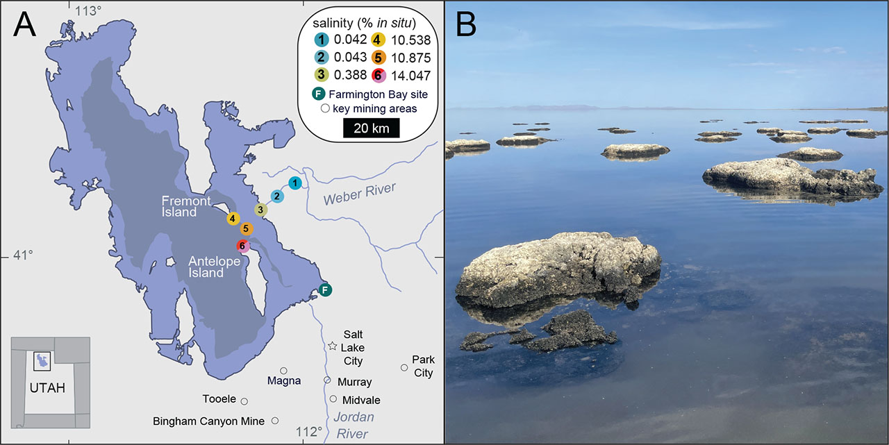
Figure 1:
Sampling sites within the GSL. (A) Locations and average in situ water salinity of six sample sites around (1–3) and within (4–6) the GSL. Lake topographies depict NASA SRTM2 v.2 data from 2007 and UGRC LiDAR data from 2016. Figure adapted from Jung et al. (2024a, 2024b). (B) Representative microbialite mounds at Site 6 from which nematodes were extracted. GSL, Great Salt Lake.
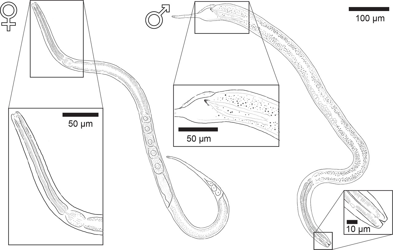
Figure 2:
Diplolaimelloides woaabi sp. nov. Left: holotype female with anterior end inset. Right: holotype male with anterior and posterior end inset, to scale.
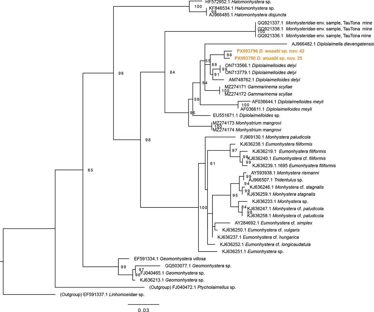
Figure 3:
Maximum-likelihood phylogenetic tree of Monhysteridae nematodes, based on aligned SSU sequences, including two paratype male specimen of D. woaabi sp, nov. indicated in orange. Alignment made with MUSCLE and tree made with IQ-TREE (see section “Materials and Methods”). Branches with less than 50 bootstrap support are not reported.
Table 1:
Measurements (µm) of Diplolaimelloides woaabi sp. nov. (range, mean value in parentheses).
| Holotype female | Paratype female (N = 20) | Holotype male | Paratype male (N = 9) | |
|---|---|---|---|---|
| L | 1,059.4 | 441.0–1,159.4 (712.5) | 1,011.0 | 559.6–1,100.3 (835.5) |
| a | 33.8 | 25.5–34.0 (28.8) | 23.8 | 24.3–37.7 (31.9) |
| b | 6.7 | 4.0–7.4 (5.7) | 5.6 | 5.4–10.7 (6.6) |
| c | 6.5 | 3.6–7.2 (4.8) | 10.2 | 5.4–17.3 (10.0) |
| Stl | 5.2 | 1.5–7.0 (3.5) | 5.8 | 2.9–5.2 (4.0) |
| Stw | 2.9 | 1.2–4.3 (2.2) | 4.7 | 2.3–2.9 (2.6) |
| Bdcs | 11.9 | 4.5–11.0 (7.1) | 13.7 | 6.2–11.2 (7.8) |
| Csla | n/a | 0.6–0.8 (0.7) | n/a | 0.3–0.3 (0.3) |
| Aw | 3.4 | 1.3–4.2 (2.5) | 2.3 | 3.4–3.6 (3.5) |
| Al | 3.5 | 1.3–4.2 (2.6) | 2.3 | 3.4–3.6 (3.5) |
| Daa | 14.4 | 10.8–20.0 (15.9) | 17.8 | 13.8–15.6 (14.7) |
| A% | 23.9 | 10.0–31.4 (16.5) | 13 | 22.0–26.2 (24.1) |
| Bda | 17.6 | 8.8–19.5 (11.8) | 10.9 | 13.6–17.3 (15.5) |
| Ol | 3.8 | 2.1–3.5 (3.1) | 5.3 | 2.3–4.3 (2.9) |
| Ow | 1.9 | 2.1–3.5 (2.5) | 3.6 | 1.4–2.7 (2.1) |
| Dao | 38.7 | 17.6–33.4 (25.5) | 29.3 | 25.4–33.4 (29.0) |
| Bdo | 20.1 | 13.5–19.9 (17.1) | 25.4 | 1.2–18.7 (15.1) |
| Bdph | 26.2 | 15.3–29.8 (20.6) | 35.3 | 19.3–25.4 (23.0) |
| Daph | 156.9 | 102.0–167.6 (126.3) | 181 | 102.9–161.7 (136.0) |
| Mbd | 31.4 | 17.0–35.5 (24.7) | 42.4 | 22.6–31.7 (26.1) |
| Abd | 23.3 | 15.0–28.2 (17.9) | 34.3 | 17.1–28.7 (22.4) |
| Tl | 164.2 | 87.8–210.1 (155.5) | 99.3 | 53.4–122.7 (87.7) |
| Vbd | 30.1 | 19.2–31.3 (23.8) | n/a | n/a |
| Vta | 236.9 | 125.8–285.5 (165.2) | n/a | n/a |
| V% | 62.1 | 49.8–61.8 (54.1) | n/a | n/a |
| B | n/a | n/a | 54.5 | 22.1–52.3 (37.3) |
| S | n/a | n/a | 89.5 | 57.1–77.0 (66.3) |
| Lfilt | 134.4 | 29.5–139.4 (87.2) | 70.4 | 26.6–87.0 (64.2) |
| Ltip | 6.1 | 3.8–12.0 (5.9) | 8.1 | 3.6–6.6 (5.0) |
[i] Abbreviations: Abd, anal/cloacal body diameter; Aw, amphidial fovea width; Al, amphidial fovea length; A%, amphidial diameter (average of length and width) as a percentage of body diameter; Bdcs, body diameter at level of cephalic setae; Bda, body diameter at level of the amphid; Bdph, body diameter at the level of pharyngeal end; Bdo, body diameter at level of ocelli; B, bursa length along the arc; Csl, cephalic setae length; Daa, distance from anterior end to amphid; Dao, distance from anterior end to ocelli; Daph, distance from anterior end to pharyngeal end; L, total length; Lfilt, length of filiform part of the tail; Ltip, length of transparent tail tip; Mbd, maximum body diameter; Ol, ocelli diameter from anterior to posterior; Ow, ocelli diameter from side to side; Pb, pattern of papillae on bursa, number of pairs/on one side only; Ppc, pattern of pre-cloacal papillae; Pcd, pattern of caudal papillae; S, spicule length along the arc; Stl, stoma length; Stw, stoma width; Tl, tail length or distance from anus/cloacal opening to posterior end; Vbd, body diameter at position of vulva; Vta, distance from vulva opening to anus; V%, distance from anterior end to vulva opening in percentage of total length; De Man’s ratios, a, b, and c used in this paper are calculated as standard.
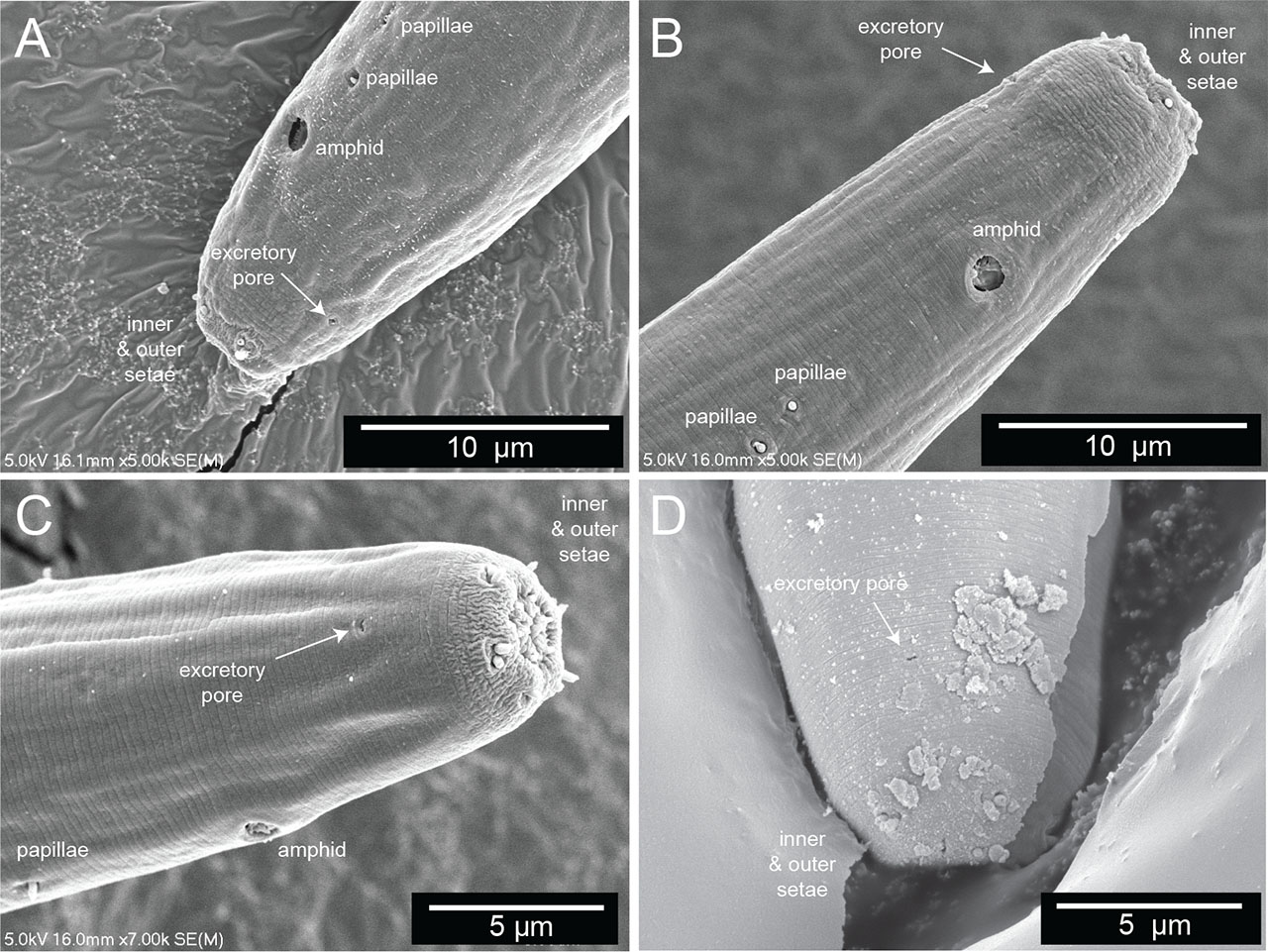
Figure 4:
The secretory–excretory pore. (A–D) Scanning electron microscopy of the anterior end of four different Diplolaimelloides woaabi sp. nov. individuals. The position of the secretory–excretory pore on the ventral side of the nematode is annotated with a white arrow. Other miscellaneous salient features are labeled as landmarks.
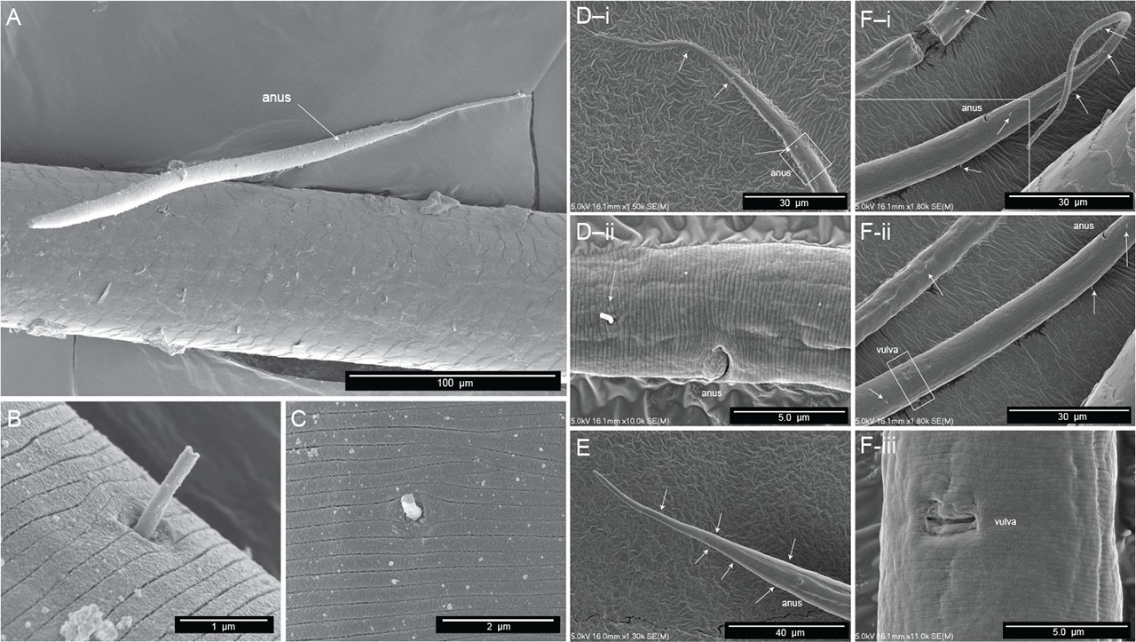
Figure 5:
Scanning electron microscopy of female Diplolaimelloides woaabi sp. nov. shows several features, including the posterior pattern of papillae on (A) the whole body, (B,C) a closeup of integument and single papilla, and (D–F) the posterior ends of nematodes, with each letter depicting a different individual and successively zoomed-in shots of the anus and/or vulva areas with visible papillae labeled with white arrows.
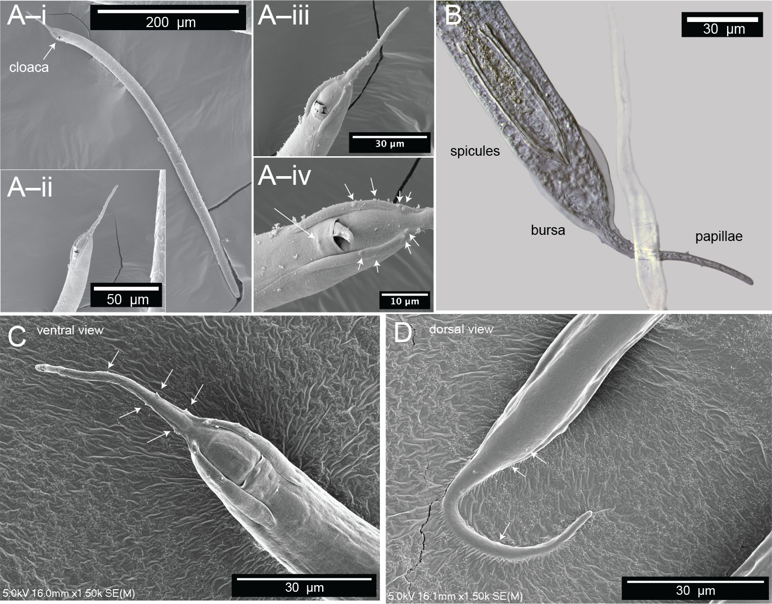
Figure 6:
Microscopy of male Diplolaimelloides woaabi sp. nov. shows the posterior pattern of papillae on the whole body or posterior ends (A–D). Each letter depicts a different individual and successively zoomed-in shots with papillae labeled with white arrows. (B) Image taken with DIC microscopy; all others taken with scanning electron microscopy. DIC, differential interference contrast.
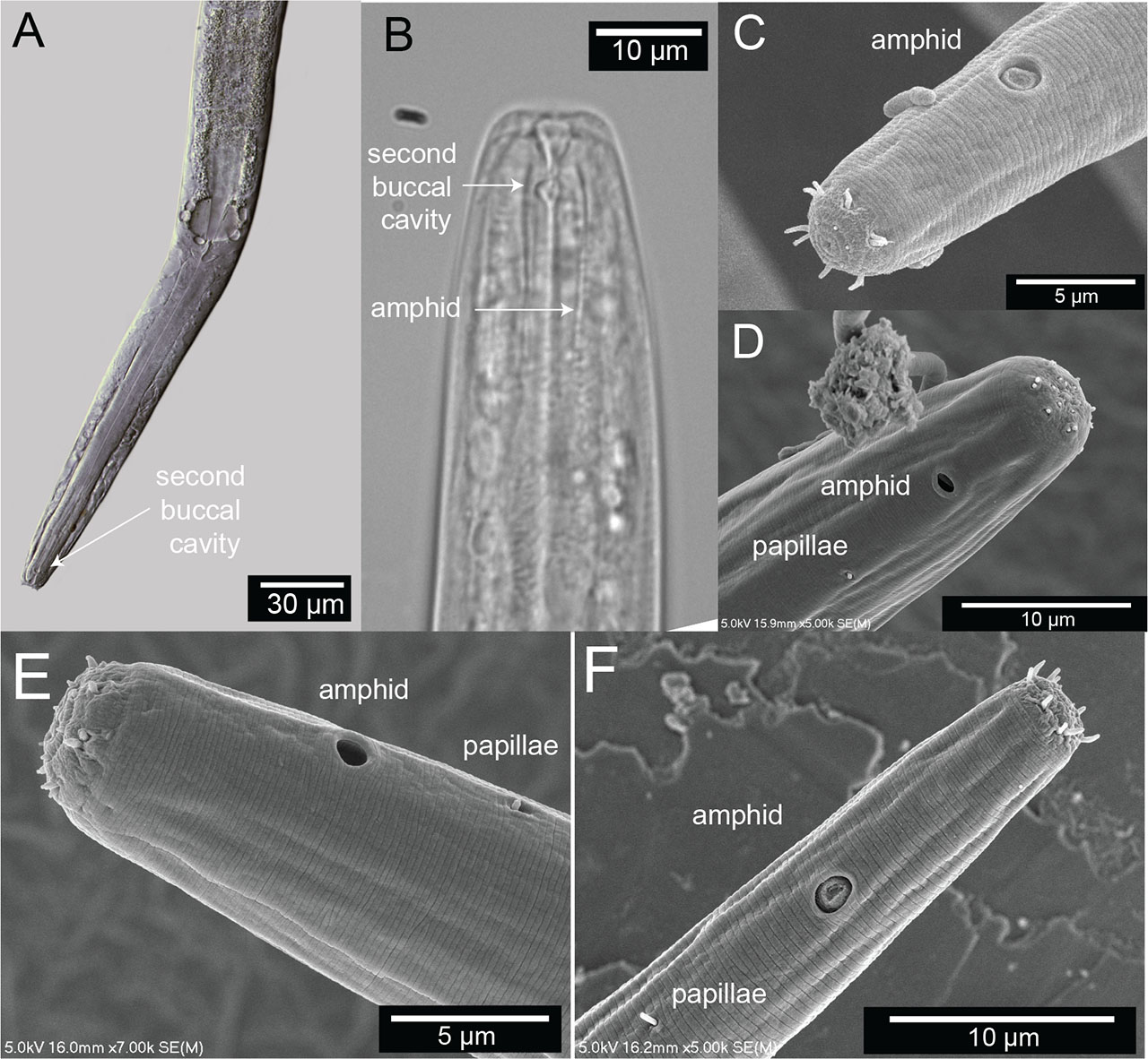
Figure 7:
Terminal head region of Diplolaimelloides woaabi sp. nov. (A) DIC microscopy reveals stoma with a double cavity, or two distinct compartments, the muscular pharynx and the prominent cardia. (B) Section of the anterior end, arrows pointing to the second chamber of the buccal cavity and the position of the amphidal fovea. Scanning electron microscopy shows (C–F) completely fused lip plates that carry a concentration of sense organs in a 6 (inner) + 6 + 4 (outer) pattern, and amphids and somatic papillae visible slightly posterior from the anterior end. DIC, differential interference contrast.
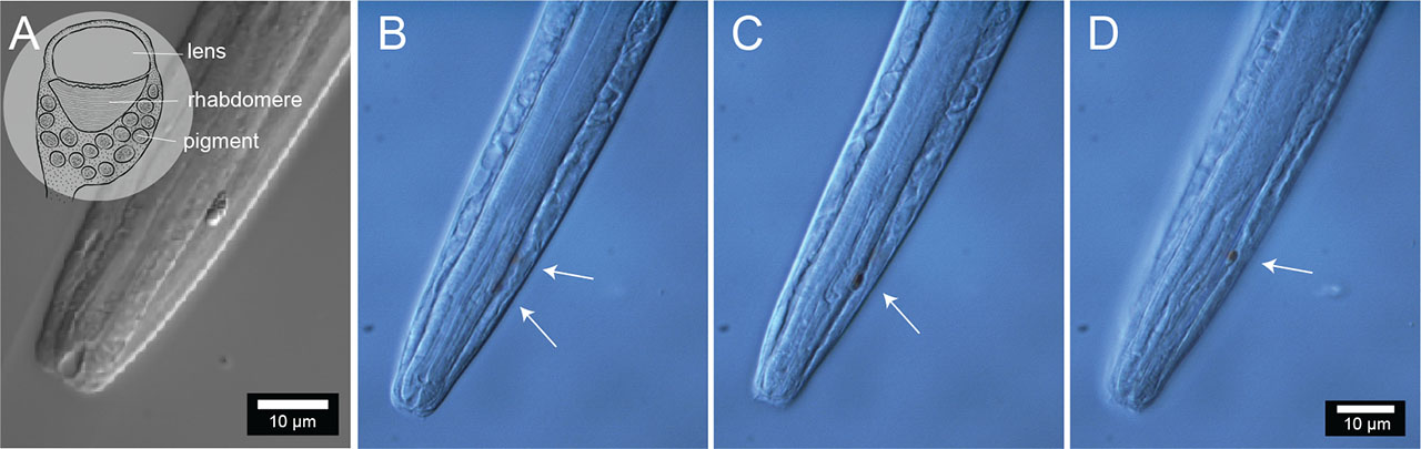
Figure 8:
Photoreceptors of Diplolaimelloides woaabi sp. nov. (A) Anterior body end showing the position of one of the ocelli and detail of the pigment area. Inset shows a schematic reconstruction based on the ultrastructure of an ocellus, adapted from a TEM photograph (Van De Velde and Coomans, 1988). Anterior body end of a different specimen, showing the positions and colored pigment of (B) both, (C) one, and (D) the other ocelli.
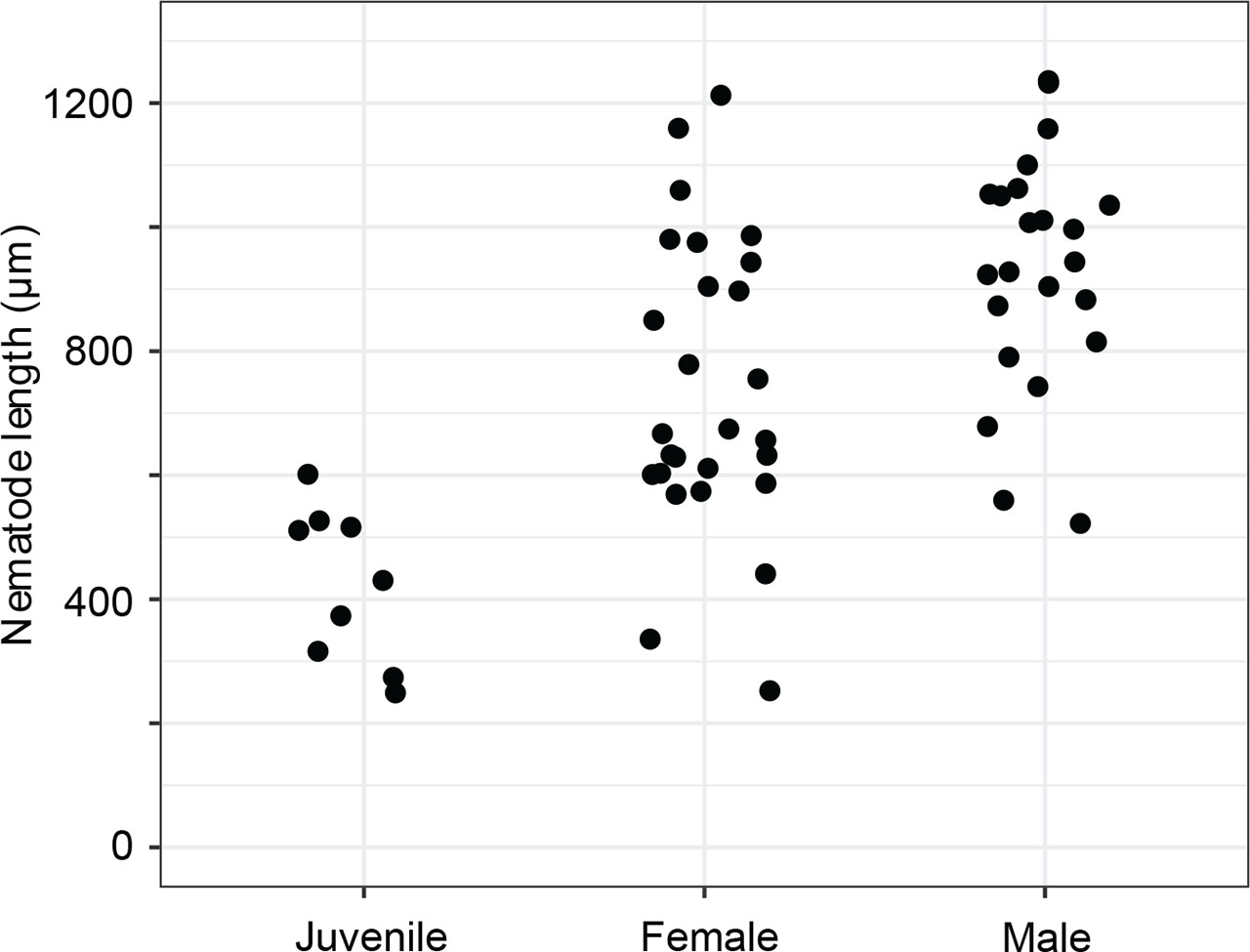
Figure 9:
Diplolaimelloides woaabi sp. nov. total length as a factor of stage and sex.
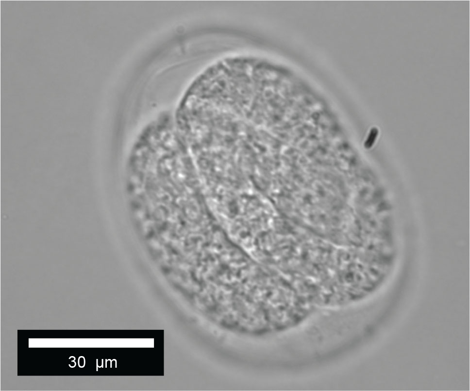
Figure 10:
Diplolaimelloides woaabi sp. nov. egg.
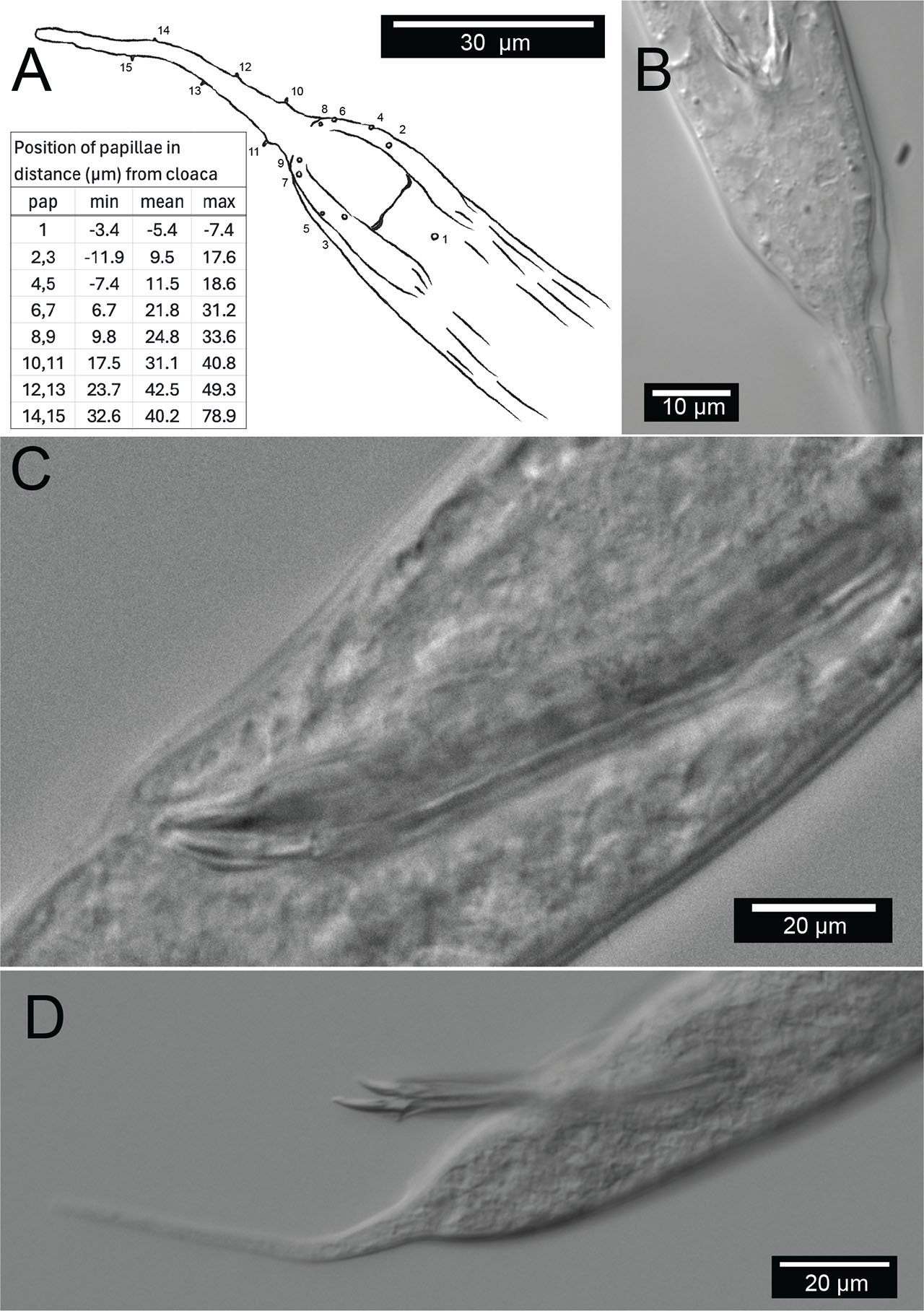
Figure 11:
Male copulatory apparatus, Diplolaimelloides woaabi sp. nov. (A) Diagram of male papillae pattern and (inset) position. Gubernaculum in (B) ventral and (C) lateral view with non-emergent spicule. (D) Lateral view with emergent spicule. Images taken with or traced over DIC microscopy. DIC, differential interference contrast.
Table 2.
Measurements (µm) of Diplolaimelloides woaabi sp. nov. (range) compared with measurements of all other described Diplolaimelloides species.
| D. woaabi | D. altherii | D. brucei | D. conctortus | D. deconincki | D. delyi | D. elegans | D. islandicus | D. meyli | D. oschei | D. palustris | D. rushikondai | D. tehuelchus | D. warwicki | |||||||||||||||
|---|---|---|---|---|---|---|---|---|---|---|---|---|---|---|---|---|---|---|---|---|---|---|---|---|---|---|---|---|
| Ref | Herein | (Meyl, 1954) | (Hopper, 1970) | (Chen et al., 2023) | (Gerlach, 1951) | (Andrássy, 1958, Chen et al., 2023) | (Gagarin and Thanh, 2008) | (De Coninick, 1943) | (Timm, 1961) | (Meyl, 1954) | (Tsalolikhin, 1985) | (Sufyan et al., 2014) | (Pastor de Ward & Lo Russo, 2009) | (Pastor de Ward and Lo Russo, 2009) | ||||||||||||||
| Sex | ♀ | ♂ | ♀ | ♂ | ♀ | ♂ | ♀ | ♂ | ♀ | ♂ | ♀ | ♂ | ♀ | ♂ | ♀ | ♂ | ♀ | ♂ | ♀ | ♂ | ♀ | ♂ | ♀ | ♂ | ♀ | ♂ | ♀ | ♂ |
| L | 441–1159 | 560–1100 | 616–988 | 508–832 | 690–880 | 730–890 | 737–854 | 713–844 | 736 | 860 | 810–1410 | 690–1310 | 809–826 | 835 | 680 | 717–730 | 750–1200 | 820–1250 | 554–850 | 587–717 | 776–851 | 824 | 620–748 | 545–683 | 760–880 | 600–700 | 1000–1010 | 770–1100 |
| a | 25.5–34.0 | 23.8–37.7 | 33.0–40.0 | 37.7–42.2 | 41.9 | 36.9 | 23.0–29.5 | 25.9–32.5 | 21.2 | 24.8 | 26.3–40.2 | 27.3–44.5 | 28.0–30.0 | 35 | 30.2 | 42.7–43.7 | 25.7–37.9 | 31.4–43.5 | 33.0–41.7 | 34.8–37.0 | 27.4–31.0 | 34.3 | 26.0–37.0 | 32.1–42.7 | 33.0–35.0 | 32.9–35.0 | 35.7–40.4 | 30.8–35.5 |
| b | 4.0–7.4 | 5.4–10.7 | 6.5–7.7 | 5.5–6.4 | 7.0 | 6.5 | 6.6–7.1 | 6.1–7.0 | 6.8 | 6.3 | 6.2–8.9 | 5.8–7.9 | 7.0–7.4 | 7.2 | 5.6 | 5.2–5.4 | 5.1–6.7 | 5.0–6.5 | 6.0–7.1 | 6.0–6.8 | 6.6–6.7 | 6.2 | 6.8–8.0 | 6.0–7.6 | 5.7–6.3 | 5.6–6.2 | 7.4–7.8 | 5.7–7.2 |
| c | 3.6–7.2 | 5.4–17.3 | 5.0–6.1 | 5.5 | 9.0 | 12.7 | 3.7–4.1 | 4.4–4.5 | 4.2 | 5.9 | 3.9–7.5 | 5.1–10.3 | 4.2–5.3 | 7.2 | 9.7 | 9.8–10.4 | 9.8–14.4 | 9.4–17.7 | 3.2–4.2 | 3.4–4.5 | 4.5 | 6.7 | 4.0–6.0 | 4.7–8.8 | 4.9–6.0 | 7.6–8.8 | 10.0–10.6 | 14.0–20.8 |
| Stl | 1–7 | 3–6 | n/a | n/a | 5 | 5 | 8–9 | 8–9 | n/a | n/a | 8–9 | 8–9 | n/a | n/a | n/a | n/a | n/a | n/a | n/a | n/a | n/a | n/a | 7–8 | 6–8 | n/a | n/a | n/a | n/a |
| Stw | 1–4 | 2–5 | n/a | n/a | n/a | n/a | 3 | 2–3 | n/a | n/a | 3 | 3–4 | n/a | n/a | n/a | n/a | n/a | n/a | n/a | n/a | n/a | n/a | n/a | n/a | n/a | n/a | n/a | n/a |
| Bdcs | 4–12 | 6–14 | n/a | n/a | 6–7 | 6–7 | 8 | 8–9 | n/a | n/a | n/a | n/a | 10–11 | 11 | n/a | n/a | n/a | n/a | n/a | n/a | n/a | n/a | 8–10 | 6–7 | 7–9 | 6–9 | 7–8 | 7–9 |
| Csla | 0.6–0.8 | 0.3–0.3 | n/a | n/a | Absent | Absent | n/a | n/a | n/a | n/a | 1–2 | 1–2 | 1 | 1 | n/a | n/a | n/a | n/a | n/a | n/a | n/a | n/a | n/a | n/a | 0.4–0.5 | 0.4–0.5 | 1.4–1.5 | 1.5 |
| Aw | 1.3–4.2 | 2.3–3.6 | n/a | n/a | 2 | 2 | 3–4 | 3 | n/a | n/a | 3 | 3–4 | n/a | n/a | n/a | n/a | n/a | n/a | n/a | n/a | n/a | n/a | 3–5 | 3–5 | 2.8–4.0 | 2.8–3.0 | 4 | 3.5–4.0 |
| Al | 1.3–4.2 | 2.3–3.6 | n/a | n/a | 2 | 2 | 3–4 | 3 | n/a | n/a | n/a | n/a | n/a | n/a | n/a | n/a | n/a | n/a | n/a | n/a | n/a | n/a | 3–5 | 3–5 | 3.0–4.0 | 3.5–4.0 | 3.5–4 | 3.5–4.0 |
| DaA | 10.8–20.0 | 13.8–17.8 | n/a | n/a | 3–8 | 3–8 | 9–10 | 9 | n/a | n/a | 10–14 | 10–13 | 17–18 | 18 | n/a | n/a | n/a | n/a | n/a | n/a | n/a | n/a | 7.8–11.7 | 7.8–11.7 | 10.0–12.0 | 10.0–11.0 | 8–9 | 8–9 |
| A% | 10.0–31.4 | 13.0–26.2 | n/a | n/a | 10.7 | 1–8 | 23.0–30.6 | 23.6–29.8 | n/a | n/a | n/a | n/a | n/a | n/a | n/a | n/a | n/a | n/a | n/a | n/a | n/a | n/a | n/a | n/a | 21.5–30.8 | 23.1–27.3 | 30.8–36.4 | 30.8–33 |
| BdA | 8.8–19.5 | 10.9–17.3 | n/a | n/a | n/a | n/a | 11–12 | 11–12 | n/a | n/a | n/a | n/a | n/a | n/a | n/a | n/a | n/a | n/a | n/a | n/a | n/a | n/a | n/a | n/a | 10–12 | 10–11 | 11–13 | 11–13 |
| Ol | 2.1–3.8 | 2.3–5.3 | n/a | n/a | n/a | n/a | n/a | n/a | n/a | n/a | n/a | n/a | n/a | n/a | n/a | n/a | n/a | n/a | n/a | n/a | n/a | n/a | absent | absent | 2 | 1.5–2 | 3 | 3 |
| Ow | 1.9–3.5 | 1.4–3.6 | n/a | n/a | n/a | n/a | n/a | n/a | n/a | n/a | n/a | n/a | n/a | n/a | n/a | n/a | n/a | n/a | n/a | n/a | n/a | n/a | absent | absent | 2 | 1.5–2 | 3 | 3 |
| Dao | 17.6–38.7 | 25.4–33.4 | n/a | n/a | 27–37 | 27–37 | 35–44 | 40–49 | n/a | n/a | 43–49 | 49–59 | 42–45 | 45 | n/a | n/a | n/a | n/a | n/a | n/a | n/a | n/a | absent | absent | 20–36 | 21–26 | 30–45 | 30–38 |
| Bdo | 13.5–20.1 | 1.2–25.4 | n/a | n/a | n/a | n/a | 17–20 | 17–18 | n/a | n/a | n/a | n/a | n/a | n/a | n/a | n/a | n/a | n/a | n/a | n/a | n/a | n/a | absent | absent | 14–18 | 13–15 | 17 | 14–20 |
| Bdph | 15.3–29.8 | 19.3–35.3 | n/a | n/a | n/a | n/a | 21–24 | 21–24 | n/a | n/a | n/a | n/a | n/a | n/a | n/a | n/a | n/a | n/a | n/a | n/a | n/a | n/a | n/a | n/a | 19–23 | 19–20 | 130–136 | 122–152 |
| Daph | 102–168 | 103–181 | n/a | n/a | 97–130 | 97–130 | 108–123 | 113–118 | n/a | n/a | n/a | n/a | n/a | n/a | n/a | n/a | n/a | n/a | n/a | n/a | n/a | n/a | 85–115 | 80–95 | 120–122 | 109–126 | 130–136 | 122–152 |
| Mbd | 17–35 | 23–42 | 22 | 17 | 19 | 22 | 26–33 | 25–32 | 35 | 35 | 40 | 34 | 28 | 24 | 23 | 17 | 31 | 28 | 19 | 18 | 28 | 24 | 20–25 | 15–17 | 20–26 | 20–21 | 27 | 28 |
| Abd | 15–28 | 17–34 | n/a | n/a | 15–17 | 15–17 | 14–16 | 15–17 | n/a | n/a | 23–24 | 22–26 | n/a | n/a | n/a | n/a | n/a | n/a | n/a | n/a | n/a | n/a | 15–16 | 12–18 | 15–21 | 16–19 | 18–24 | 20–23 |
| Tl | 88–210 | 53–123 | n/a | n/a | 65–98 | 34–60 | 188–209 | 164–186 | n/a | n/a | 233–268 | 152–190 | 154–196 | 116 | n/a | n/a | n/a | n/a | n/a | n/a | n/a | n/a | 110–175 | 85–110 | 138–147 | 77–87 | 95–100 | 53–54 |
| Vbd | 19–31 | n/a | n/a | n/a | n/a | n/a | 24–28 | n/a | n/a | n/a | n/a | n/a | n/a | n/a | n/a | n/a | n/a | n/a | n/a | n/a | n/a | n/a | 20–22 | n/a | n/a | n/a | n/a | n/a |
| VtA | 126–285 | n/a | n/a | n/a | n/a | n/a | n/a | n/a | n/a | n/a | n/a | n/a | 168–175 | n/a | n/a | n/a | n/a | n/a | n/a | n/a | n/a | n/a | 50–165 | n/a | n/a | n/a | n/a | n/a |
| V% | 50–62 | n/a | 58–65 | n/a | 64–65 | n/a | 52–54 | n/a | 54 | n/a | 53–64 | n/a | 56–59 | n/a | 65 | n/a | 62–68 | n/a | 49–54 | n/a | 55–56 | n/a | 55–62 | n/a | 57–61 | n/a | 69–74 | n/a |
| B | n/a | 22–55 | n/a | n/a | n/a | n/a | n/a | n/a | n/a | n/a | n/a | n/a | n/a | n/a | n/a | n/a | n/a | n/a | n/a | n/a | n/a | n/a | n/a | n/a | n/a | 38.4 | n/a | 50 |
| Pb | n/a | 2 + 2 | n/a | 1 + 2 + 2 | n/a | 2 + 2 | n/a | 2 + 2 | n/a | 1 + 2 | n/a | 2 + 2 | n/a | 1 + 2 + 1 + 1 | n/a | 1 + 2 | n/a | 1 + 2 | n/a | 2 + 2 | n/a | 1 + 1 + 1 + 1 + 1 | n/a | 2 + 2 | n/a | 2 + 2 | n/a | 1 + 2 |
| Ppc | ≥1 | 1 | n/a | 0 | n/a | n/a | n/a | n/a | n/a | 1 | n/a | 0 | n/a | n/a | n/a | n/a | n/a | n/a | n/a | n/a | n/a | n/a | n/a | n/a | n/a | 2 | n/a | n/a |
| Pcd | ≥3 | ≥3 | n/a | 2 | n/a | 2 | 4 + 2 | 2 | n/a | n/a | n/a | 2 | n/a | 0 | ||||||||||||||
| S | n/a | 57–90 | n/a | 62–70 | n/a | 31–38 | n/a | 45–50 | n/a | 45–50 | n/a | 48–78 | n/a | 46 | n/a | 27 | n/a | 35–41 | n/a | 27–33 | n/a | 45 | n/a | 46 | n/a | 60–67 | n/a | 33–39 |
| Lfilt | 30–139 | 27–87 | n/a | n/a | n/a | n/a | n/a | n/a | n/a | n/a | n/a | n/a | n/a | n/a | n/a | n/a | n/a | n/a | n/a | n/a | n/a | n/a | n/a | n/a | n/a | 51–64 | 20–26 | 18–26 |
| Ltip | 4–12 | 4–8 | n/a | n/a | n/a | n/a | n/a | n/a | n/a | n/a | n/a | n/a | n/a | n/a | n/a | n/a | n/a | n/a | n/a | n/a | n/a | n/a | n/a | n/a | n/a | 6–8 | 4–5 | 4–6 |
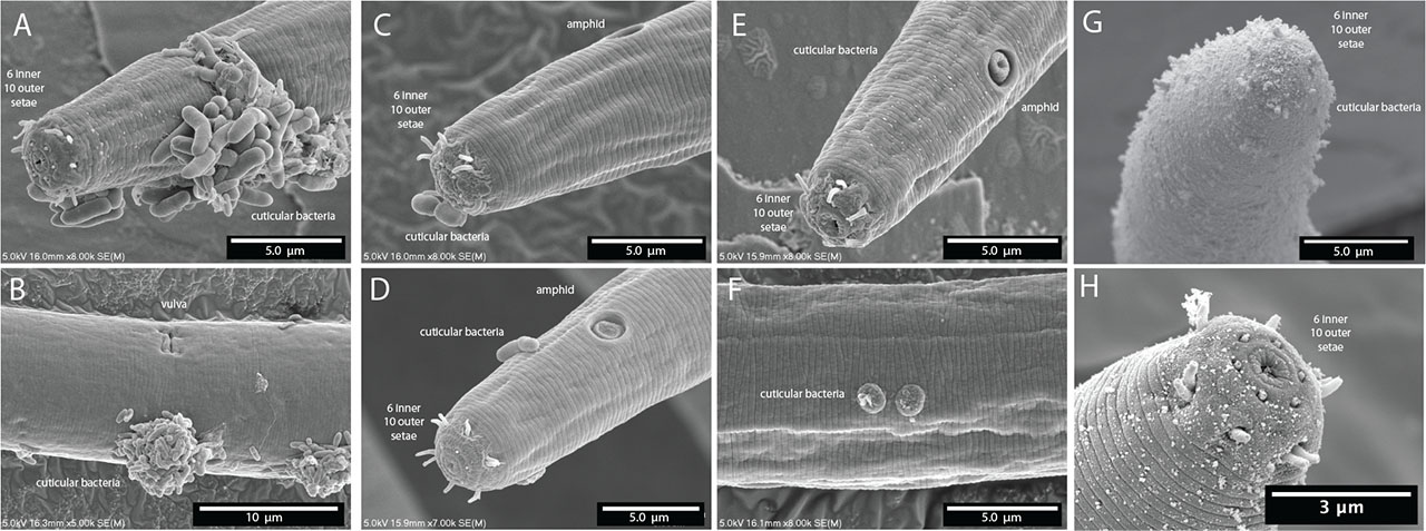
Figure 12:
Scanning electron microscopy of body ornamentation or adherence of other organisms and/or substances on Diplolaimelloides woaabi sp. nov. that are (A–D) rod-like, (E,F) wart-like, or (G,H) fuzz-like.
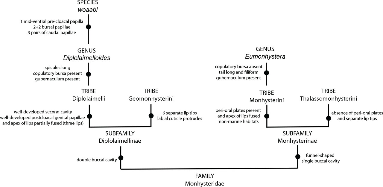
Figure 13:
Differential diagnosis to identify Diplolaimelloides woaabi sp. nov.
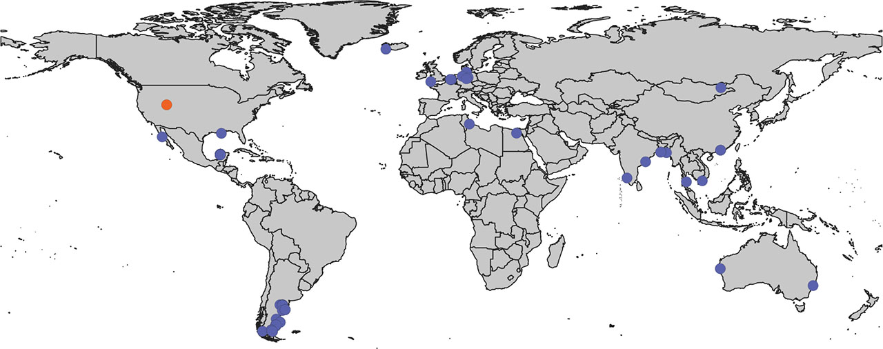
Figure 14:
Global map of Diplolaimelloides. GIS map of documented Diplolaimelloides recovered from the wild (n = 63), representing 14 species. Where possible, exact GPS coordinates are used directly from source material including species-description manuscripts, images of notebooks, and the GBIF webserver. In a few cases, approximate coordinates are estimated from geographical description. D. woaabi sp. nov. depicted in orange. GBIF, Global Biodiversity Information Facility; GIS, Geographic Information Systems.