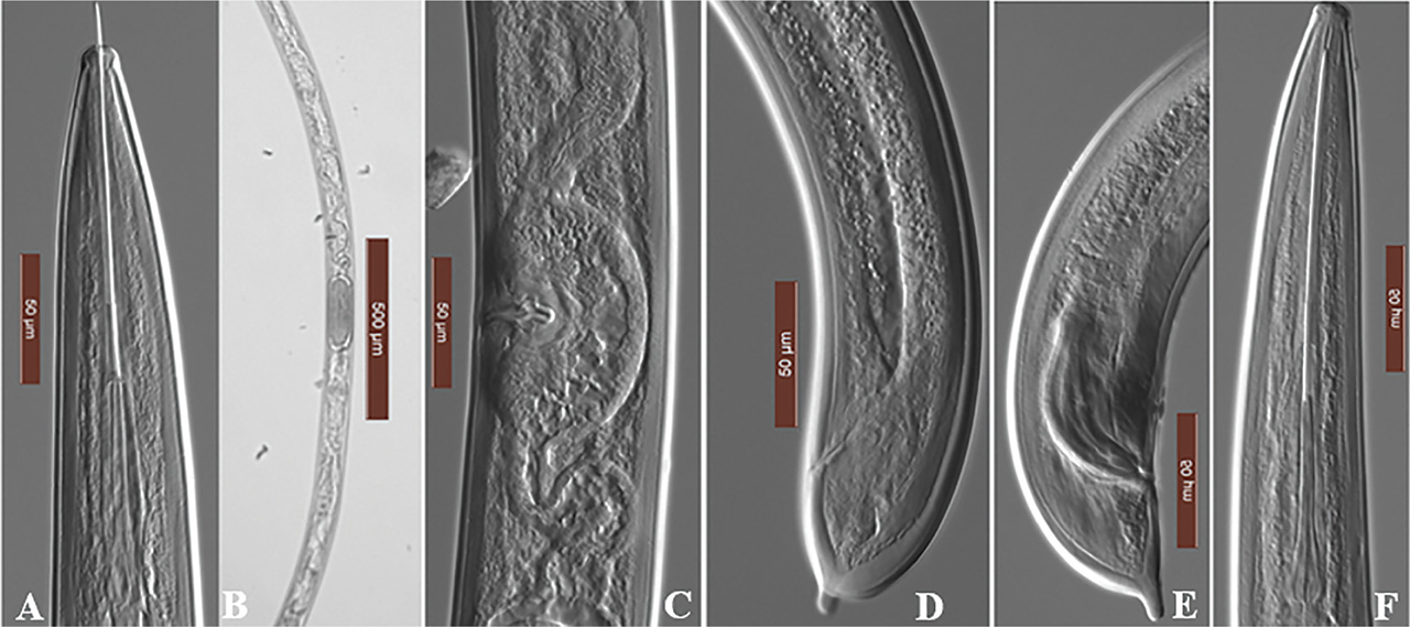
Figure 1:
Micrographs of Xiphinema diversicaudatum: A–C: female anterior region; D: female posterior region; E: male posterior region, F: male anterior region.

© 2024 Jerry Akanwari, Qing Yu, Tahera Sultana, published by Society of Nematologists, Inc.
This work is licensed under the Creative Commons Attribution 4.0 License.