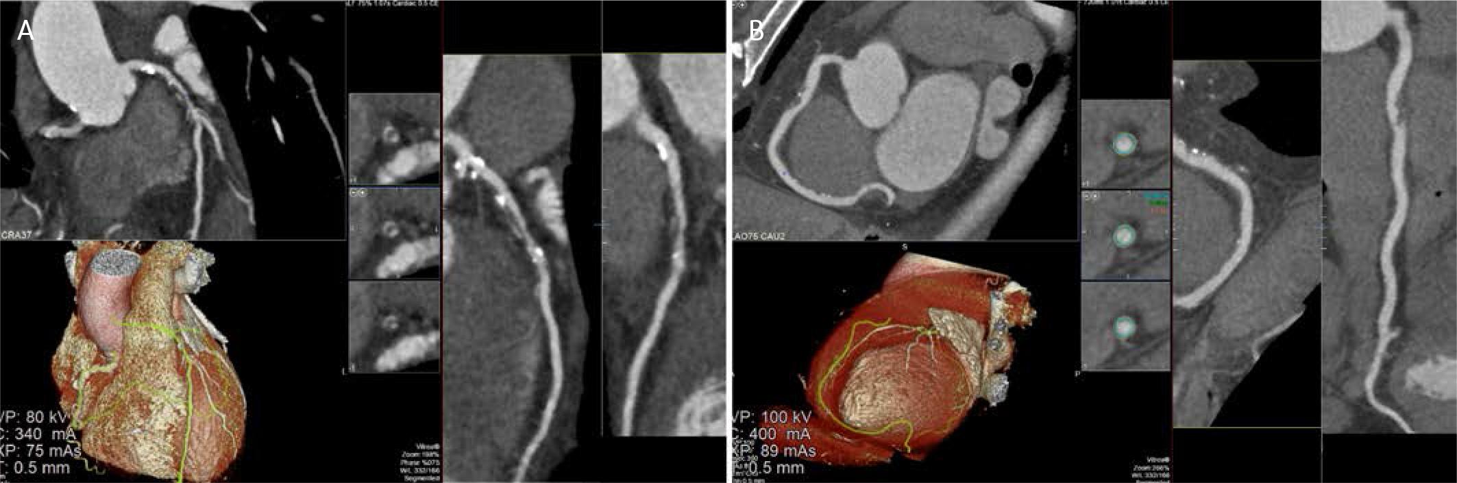
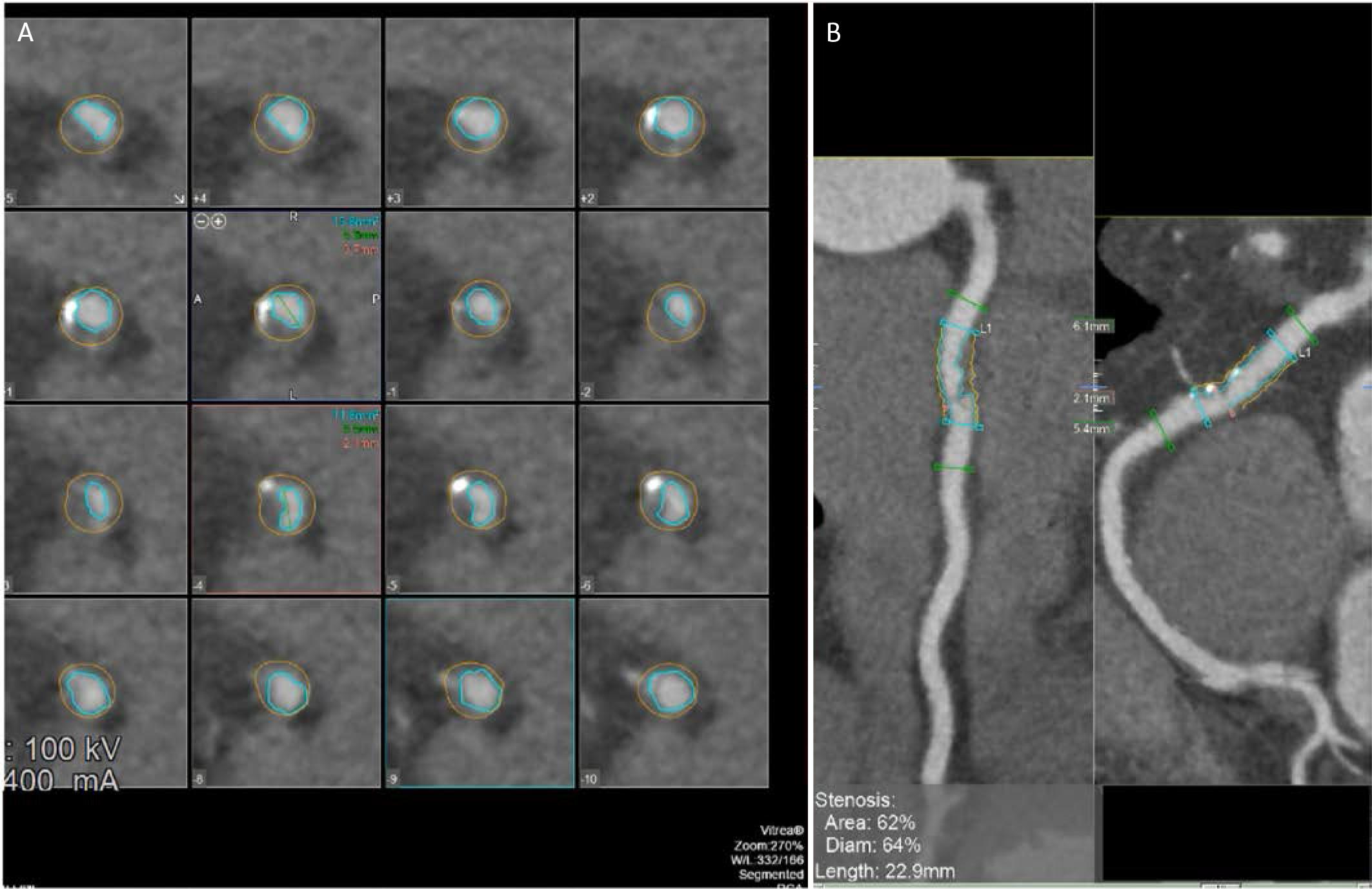
Have a personal or library account? Click to login
Coronary Computed Tomography Angiography in Non-ST Elevation Acute Coronary Syndromes: Present Evidence and Potential Advances Cover

Coronary Computed Tomography Angiography in Non-ST Elevation Acute Coronary Syndromes: Present Evidence and Potential Advances

Open Access
|Mar 2026

Figures & Tables

FIGURE 1.

CCTA demonstrating high-risk atherosclerotic plaque morphology in the left anterior descending and right coronary arteries. A. Multiplanar reconstruction of the left anterior descending artery demonstrates an eccentric, predominantly non calcified, low attenuation plaque in the proximal–mid segment, associated with positive vessel remodeling and superimposed spotty calcifications. Volume rendered 3D reconstruction confirms the extent and distribution of the lesion along the LAD. B. CCTA images illustrating high-risk plaque characteristics. Axial cross-sectional images (upper right) demonstrate the typical features of vulnerable plaque, including the napkin-ring sign, spotty calcification, and low-attenuation plaque. The 3D volume rendered reconstruction (lower left) depicts the spatial relationship of the high-risk plaque within the coronary anatomy.

FIGURE 2.

Automated coronary plaque quantification and stenosis assessment using CCTA post-processing software. A. Serial crosssectional images along the lesion, with automated luminal (inner contour) and vessel (outer contour) segmentation enabling precise assessment of plaque burden and vessel remodeling. B. Multiplanar reconstruction views of the affected coronary segment with software-derived measurements indicating a 62% area stenosis and 64% diameter stenosis over a lesion length of 22.9 mm, consistent with hemodynamically significant obstructive CAD as defined by contemporary CCTA criteria.

Key randomized trials of CCTA in suspected NSTE-ACS

TrialPopulationPrimary outcomeKey findings
RAPID-CTCA15Suspected/provisional ACS; intermediate risk (n = 1,748)1-year death or type 1/4b MINo difference in MACE; reduced ICA use; modest reduction in subsequent testing, no major LOS difference
VERDICT8Confirmed NSTE-ACS (n = 1,023)Diagnostic performance vs. ICAHigh NPV for ruling out ≥50% stenosis; accurately characterized CAD distribution
BEACON22ED suspected ACS, low-intermediate riskMACE at follow-upNo difference in MACE; shorter LOS and lower costs in low-risk patients
PROTECCT21Intermediate hs-cTn ‘observe zone’12-month MACENo change in MACE; trend to fewer downstream tests
DOI: https://doi.org/10.2478/jce-2026-0005 | Journal eISSN: 2457-5518 | Journal ISSN: 2457-550X
Language: English
Page range: 1 - 11
Submitted on: Jan 13, 2026
|
Accepted on: Mar 3, 2026
|
Published on: Mar 27, 2026
In partnership with: Paradigm Publishing Services
Publication frequency: 4 issues per year

© 2026 Dan-Alexandru Cozac, Maria Teresa Savo, Gabriele Cordoni, Martina Palmisano, Ali Al-Enazi, Talal Al Otaibi, Raffaella Motta, Valeria Pergola, published by Asociatia Transilvana de Terapie Transvasculara si Transplant KARDIOMED
This work is licensed under the Creative Commons Attribution-NonCommercial-NoDerivatives 3.0 License.