
Atrial fibrillation (AF) is associated with a significant burden on healthcare systems and poses considerable risks to affected individuals. The condition is a major risk factor for stroke, patients with AF having a fivefold increased risk compared to those without AF. Strokes associated with AF tend to be more severe and debilitating, leading to higher rates of mortality and long-term disability. Beyond stroke, AF is linked to an increased risk of heart failure, myocardial infarction, and cognitive decline. Given the substantial clinical implications, early diagnosis and effective management of AF are critical. Traditional diagnostic methods, such as electrocardiogram (ECG) and Holter monitoring, provide valuable information but have limitations, particularly in detecting paroxysmal AF.1 This has led to growing interest in the potential of artificial intelligence (AI) to enhance the detection, prediction, and management of AF, leveraging advanced algorithms to improve patient outcomes and optimize healthcare resources. Using the advanced computational capabilities provided by modern technology and AI, we explore the current landscape of machine learning (ML) methods in predicting and screening for AF, as well as the implications and future direction of this rapidly progressing field.
Several methods for AF risk estimation have been developed over time, most of them based on traditional clinical risk factors. The Framingham Heart Study2 followed 4,764 participants for up to 10 years and developed a risk prediction model to estimate the probability of AF occurrence based on factors such as age, sex, body mass index (BMI), systolic blood pressure, treatment for hypertension, PR interval, presence of a significant murmur, and history of heart failure. Similarly, the Atherosclerosis Risk in Communities (ARIC) study investigated the causes and outcomes of atherosclerosis and its clinical manifestations, including AF, in more than 15,000 participants. Data from ARIC were used to develop and validate risk prediction models for various cardiovascular diseases, aiding in the identification of high-risk individuals and guiding preventive strategies.3 Subsequently, the Cohorts for Heart and Aging Research in Genomic Epidemiology – Atrial Fibrillation (CHARGE-AF) consortium combined data from several large cohort studies, including the Framingham Heart Study, ARIC, the Cardiovascular Health Study (CHS) and the Rotterdam Study. The CHARGE-AF4 study investigates genetic and epidemiological factors contributing to cardiovascular disease and aging-related conditions, including AF. Its risk prediction model has been validated across different cohorts, demonstrating robust performance in predicting the risk of developing AF. The CHARGE-AF model incorporates age, sex, BMI, blood pressure, diabetes mellitus, smoking status, previous cardiovascular disease, alcohol consumption, ethnicity, genetic factors, and electrocardiographic parameters. Recent studies report that the CHARGE-AF score has good discriminatory ability for incident AF and represents a promising prediction tool for this arrhythmia. In addition, new screening tools, such as smartphone applications and smartwatches, are rapidly evolving. Therefore, the broader application of the CHARGE-AF score in clinical practice, together with emerging health technologies, may improve AF prediction and facilitate more effective stroke prevention, especially in high-risk patients.5
Recently, there has been a surge of interest in the use of ML techniques to predict and screen for AF. Even before the integration of AI into clinical medicine, research had already identified numerous clinical risk factors that could predict the development of AF. These risk factors include prior medical diagnoses, laboratory data (cardiac and inflammatory biomarkers), imaging data (cardiac CT, MRI, echocardiography), as well as electrophysiological data. Advances in cardiac monitoring technologies have significantly expanded our knowledge of the true clinical burden of AF. These data are easily accessible in electronic health records and can be automatically processed by AI algorithms.
Imagistic features play a crucial role in predicting AF by providing valuable insights into the electroanatomic alterations associated with this arrhythmia. Studies have shown that combining image-derived radiomics phenotypes with ECG features enhances the discrimination of prevalent AF, particularly in women, compared with ECG alone.12 Additionally, radiomics analysis applied to preprocedural cardiac CT scans in patients with AF has demonstrated superior predictive capability for persistent AF compared with left atrial diameter alone.13
AI, ML, and deep learning (DL) are interrelated fields in computer science, each with distinct characteristics but significant overlap. AI is the broadest concept, encompassing any technique that enables computers to mimic human intelligence, including problem-solving, reasoning, learning, planning, natural language understanding, and perception.14 ML is a subset of AI that involves training algorithms to learn from and make predictions of decisions based on data. Instead of being explicitly programmed to perform a task, ML systems improve their performance as they are exposed to more data. Clearly, a high volume of heterogenous data is needed to enhance learning.15 DL is a subset of ML that focuses on deep neuronal networks with many layers, inspired by the structure of the human brain. These networks are particularly effective at capturing complex patterns in large datasets.16
Recent studies using AI models for predicting AF
| Year | Study | Journal | AI method | Dataset | Key findings |
|---|---|---|---|---|---|
| 2024 | Lin et al.6 | Med | DL, HBBIs | 23,763 24-h Holter ECG recordings | HBBI-AI effectively predicted AF risk using only HBBI information through evaluating autonomic imbalance. |
| 2023 | Hygrell et al.7 | EP Europace | CNN | 478 963 single-lead ECGs | A single-lead ECG machine learning can identify individuals at risk of undetected paroxysmal AF. |
| 2023 | Hill et al.8 | European Heart Journal | ML | Medical records | The AF risk-prediction algorithm was effective in identifying participants at high risk of undiagnosed AF. |
| 2023 | Chen et al.9 | Helyion | DL | 443,053 CCTA images | Automatically filling defects assessment of LAA on CT images detecting clinical or subclinical AF |
| 2021 | Grout et al.10 | BMC Med Inform Decis Mak | ML | Electronic health data | Using only pre-existing electronic health records, this streamlined model for predicting the risk of undiagnosed atrial fibrillation within a 2-year period achieved a C-statistic of 0.81. |
| 2020 | Baek et al.11 | European Heart Journal | RNN | 2,585 ECGs from hospital patients | AI identified AF with an AUC of 0.79, recall of 82%, and overall accuracy of 72.8%; useful in identifying AF in patients with unexplained strokes. |
CNN, convolutional neural network; HBBI, heart beat-to-beat interval; RNN, recurrent neural network
AF and enlargement, together with abnormal movement patterns of the left atrium caused by low-amplitude electrical activity, can lead to subtle ECG changes. These changes are often missed by the human eye but may be detectable by an AI model, thereby improving earlier diagnostic methods.17 Recent studies have highlighted the potential of AI models to estimate AF risk with reasonable accuracy by using ECG waveform data to extract predictive information beyond traditional clinical risk factors. DL approaches have been successfully applied to automatically diagnose AF from ECG signals, combining ML techniques with neural networks to achieve high accuracy in AF detection.18 The first study to use a convolutional neural network for ECG analysis was conducted by Attia et al. at the Mayo Clinic ECG Laboratory.19 This landmark study identified subtle patterns in normal sinus rhythm ECGs that are often imperceptible to the human eye. The authors trained the ML model using a dataset of over 450,000 ECGs from 126,526 patients and discovered that the model could accurately distinguish patients with a history of AF using a single 12-lead ECG. The STROKESTOP I, STROKESTOP II, and SAFER studies from Sweeden recorded a total of 248,964 intermittent ECGs from 6,658 participants. These studies used DL techniques, such as convolutional neural networks, suitable for signal procession and image recognition. An AI-based algorithm was applied to predict paroxysmal AF from a sinus rhythm ECG, achieving moderate accuracy in an age-homogeneous cohort (area under the curve (AUC) of 0.62). The SAFER study is ongoing, with preliminary findings expected to provide insights into the feasibility and impact of AF screening.20–22
Different imaging techniques, such as echocardiography, CT, and cardiac MRI, provide functional and anatomical information about the heart. Information processing using AI, particularly advanced ML and DL algorithms, can analyze these high-resolution images to predict the onset of AF.23
It is well known that atrial structural abnormalities, including fibrosis and enlargement, are key factors in AF perpetuation.24 Previous studies have shown that echocardiographycally measured left atrial volume, diastolic dysfunction, strain, and ventricular wall thickness are associated with the development of new-onset AF.25,26 Assessment of left atrial fibrosis using MRI, particularly through the detection of late gadolinium enhancement, has also been associated with new-onset AF. Siebermair et al. examined the predictive value of left atrial fibrosis in 182 patients without AF and without apparent heart disease. Their results showed that the development of AF may be preceded by fibrotic alterations exceeding 6%, yielding an AUC of 0.67. This predictive performance improved to an AUC of 0.80 when a history of hypertension and left ventricular ejection fraction were included.27
CT imaging, by analyzing plaque characteristics as well as left atrial and pulmonary vein morphology, is also indicative of AF development. Fat attenuation index (FAI), a promising AI-based CT biomarker, predicts AF by quantifying perivascular and epicardial adipose tissue (EAT) inflammation, which is linked to AF perpetuation.28,29 Recent studies indicate higher levels of perivascular inflammation in the region of the left coronary circulation, suggesting a potential predictive value for future risk assessment.30
EAT, a metabolically active form of visceral fat, may reflect metabolically unhealthy obesity and metabolic syndrome. Increased EAT volume has been associated with several cardiovascular diseases, including coronary artery disease and AF.31,32 The Oxford Risk Factors and Noninvasive imaging (ORFAN) study,33 an international multicenter prospective cohort study, has collected 3,720 coronary CT angiography scans and clinical data since 2005. Including approximately 75,000 patients in the UK and 2,5000 internationally, the investigators validated a deep learning network for automated quantification of EAT volume. Their findings demonstrated that EAT volume is an independent predictor of postoperative arrhythmogenesis, particularly an increased burden of AF (Figure 1).
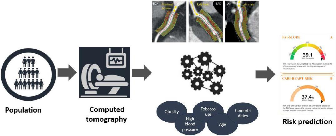
ORFAN study diagram.
Sieweke et al. developed the EAHsy-AF Risk Score based on echocardiographic parameters, including septal PATDI and the ratio of indexed left atrial volume and mitral annulus velocity during atrial contraction (LAVI/a’) in a validation cohort of 290 patients. In addition to age and hypertension, these parameters were identified as independent predictors of subclinical AF.34 The Multi-Ethnic Study of Atherosclerosis (MESA) study used a DL model for automatic cardiac chamber volumetry on non-contrast CT scans obtained during coronary artery calcium (CAC) scoring. The study included 6,814 patients from six centers without known cardiovascular disease who underwent ECG-gated non-contrast CT for CAC assessment. The investigators compared the predictive value of Al–enabled left atrial volumetry with the CHARGE-AF score and NT–proBNP values. Their results demonstrated good predictive performance of left atrial volumetry for AF, suggesting its potential usefulness in selecting participants for AF prevention clinical trials.35
However, future research is needed to establish the role of ML in prognosis and in the detection of non-imaging diagnoses such as AF. Large population-based studies may be impractical because of the costs associated with screening asymptomatic individuals and the substantial selection bias toward patients undergoing cardiac imaging. Nevertheless, ML applied to cardiac imaging in AF is likely to have an important role in periprocedural prognosis and management. With well-designed studies, it may also contribute to improved AF prediction.