
Figure 1
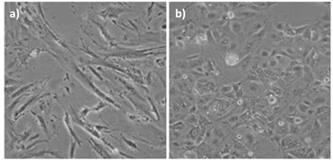
Microscopic appearence of the cells a) CoN normal colon epithelial cells, b) Caco-2 colon cancer cells.
Table 1
Oligonucleotide sequences and PCR conditions
| Primer pairs | Sequence | Product size | Annealing T (°C) | Cycle |
|---|---|---|---|---|
| NFE2L2 Sense | 5’GCGACGGAAAGAGTATGAGC3’ | 181 bp | 60 | 25 |
| NFE2L2 Antisense | 5’GTTGGCAGATCCACTGGTTT3’ | |||
| β-actin Sense | 5’TGAGCGCGGCTACAGCTT3’ | 120 bp | 56 | 35 |
| β-actin Antisense | 5’TCCTTAATGTCACGCACGATTT3’ |
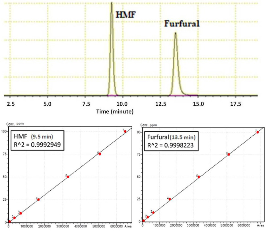
Figure 2
Schematic representation of a) HPLC chromatogram of HMF and furfural and b) Calibration curves of HMF and furfural standards.
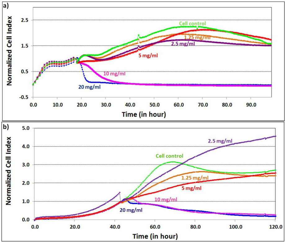
Figure 3
Real-time cell proliferation analysis: Effects of XOS hydrolysate supplementation on proliferation ofa) CoN cormal colon cells, b) Caco-2 colon cancer cells.
Table 2
Duplication time (td) of cell lines after XOS hydrolysate supplementation
| Treatment | Duplication time (h) | |
|---|---|---|
| XOS hydrolysate | CoN (normal colon cells) | Caco-2 (colon cancer cells) |
| Control | 30.78 ± 0.50 | 53.65 ± 4.56 |
| 1.25 mg mL-1 | 28.75± 0.67 | 37.70 ± 1.35 |
| 2.50 mg mL-1 | 25.97± 0.32 | 26.97 ± 0.54 |
| 5.00 mg mL-1 | 27.05± 0.21 | 47.19 ± 0.78 |
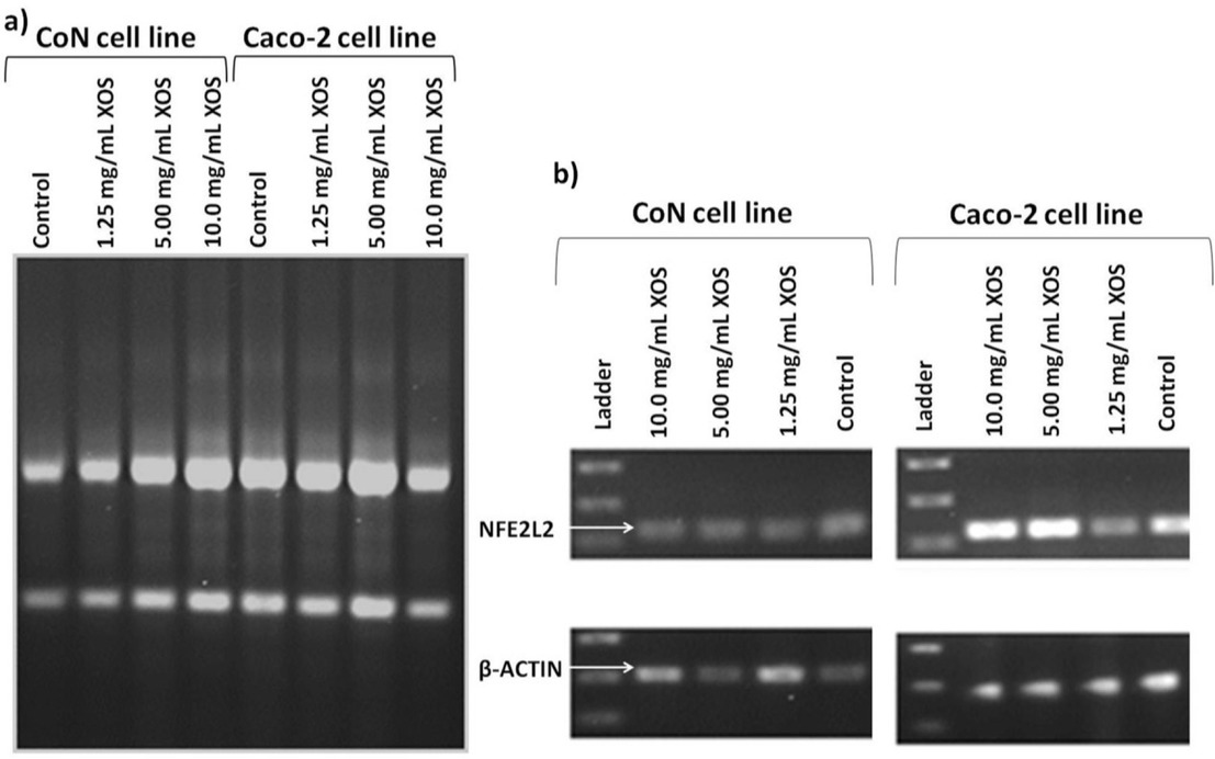
Figure 4
a) CoN and Caco-2 total RNA samples; ribosomal RNA subunits (28S, 18S) on 1% agarose gel.b) RT-PCR products for control and treated cells (NFE2L2 and β-actin genes).
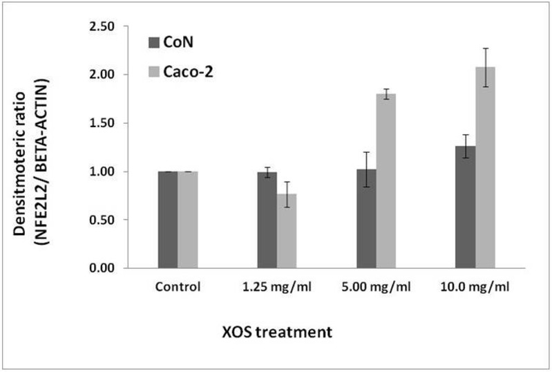
Figure 5
Schematic representation of densitometric analysis of normalized NFE2L2 gene expression levels in normal (CoN) and colon cancer cells (Caco-2).
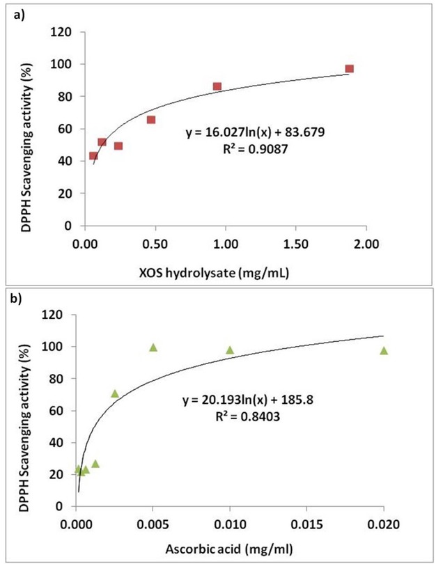
Figure 6
Graphical representations of DPPH scavenging activities of a) XOS hydrolysate and b) Ascorbic acid (L-ASA).