
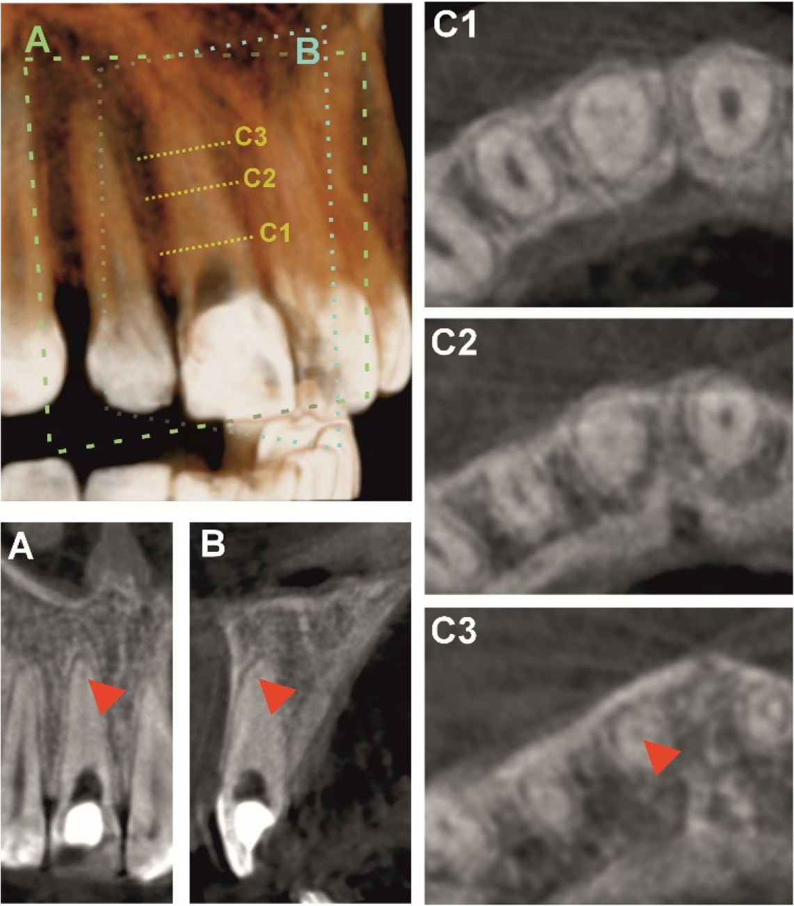
Figure 1.
A three-dimensional CBCT reconstruction illustrating the orientation of the imaging planes used for sectional analysis. The line labeled A represents the coronal plane, while B denotes the sagittal plane selected for detailed evaluation. The dotted markers C1, C2, and C3 indicate the axial slice levels corresponding to the coronal, middle, and apical thirds of the root canal, respectively.
The corresponding coronal (A) and sagittal (B) slices are shown in the lower left portion of the figure. The red arrowheads in these views indicate the location where the previously obstructed root canal becomes patent, marking the point of regained canal lumen continuity. The axial slices (C1–C3) displayed on the right depict cross-sectional morphology in the coronal, middle, and apical thirds of the canal, with the arrowhead in C3 highlighting the same transition point identified in the other planes.
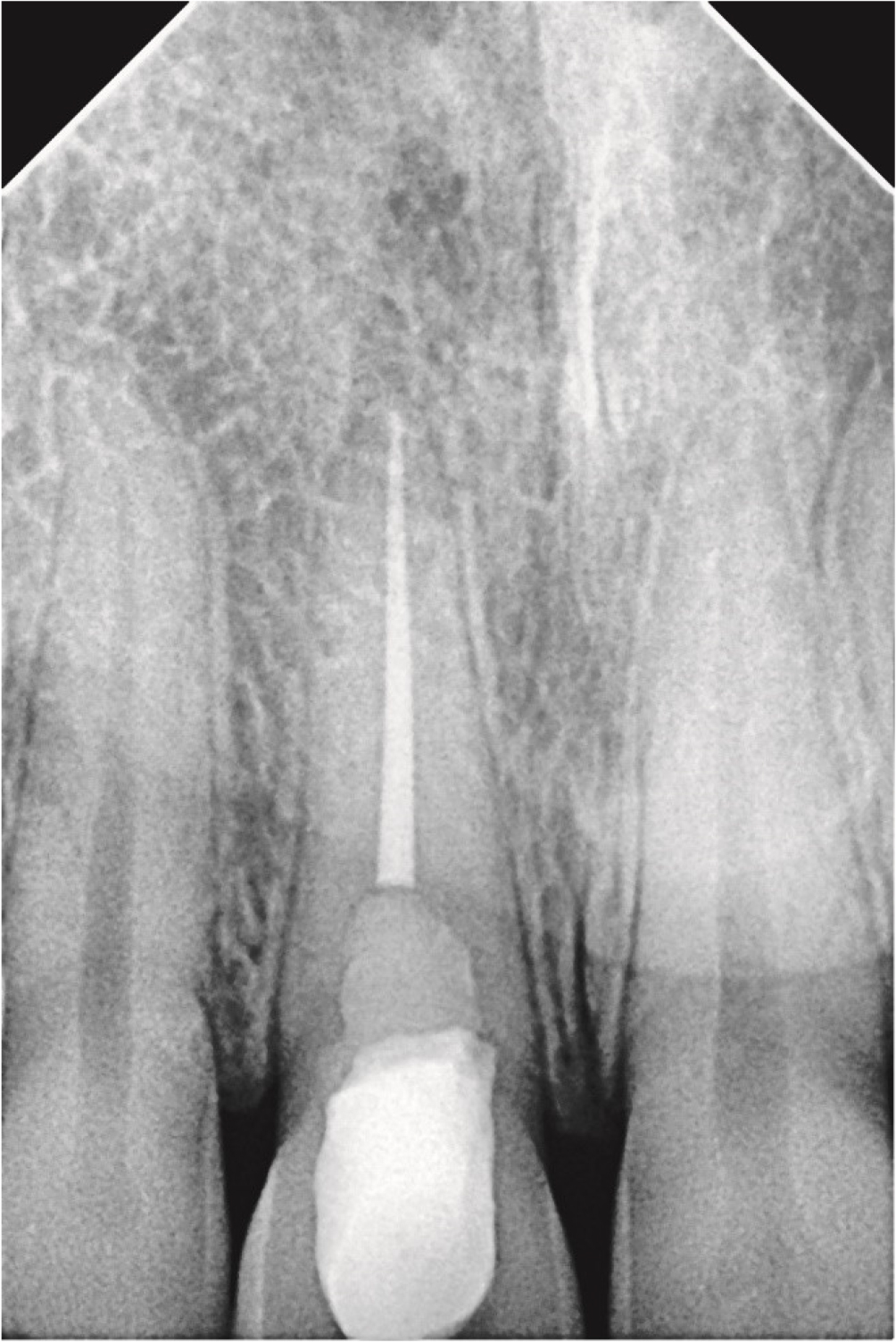
Figure 2-
Retroalveolar radiograph after definitive root canal obturation