
INTRODUCTION
Pulp canal obliteration (PCO) represents progressive deposition of hard tissue within the pulp chamber and root canal (1). PCO is referred to as calcific metamorphosis or pulp calcification (2). The formation of calcified tissue within the pulp space reflects a complex biological reaction of odontoblasts and subodontoblastic cells (3). As a consequence of calcification, the canal lumen becomes partially or completely obliterated by mineralized material resembling tertiary dentin (4). PCO may occur as part of physiological aging or as a response to external irritation, such as caries, restorative procedures, orthodontic forces, or dental trauma (5). Among these, trauma is one of the most recognized predisposing factors, since the disruption of pulpal blood supply following luxation or concussion injuries often triggers accelerated dentin deposition during the reparative phase. The degree of obliteration depends on the severity of the initial trauma and the pulp’s reparative capacity, and may progress gradually over months or years (6).
When endodontic therapy becomes necessary, the absence of a visible canal greatly complicates the procedure. Locating the canal orifice and establishing a glide path are particularly challenging, often requiring the application of modern endodontic therapeutic approaches (7). Excessive removal of coronal dentin during the search for the canal can compromise tooth integrity and increase the risk of perforation or instrument fracture (5). Even when the canal is successfully negotiated, adequate cleaning and shaping may be compromised due to irregular canal geometry and reduced accessibility (8). In trauma-related cases, these challenges are even more pronounced. Traumatized teeth often exhibit not only calcific changes but also crown discoloration, altered dentin morphology, and apical resorption, further complicating diagnosis and treatment (2, 9). Because obliteration develops gradually, it is often diagnosed incidentally at an advanced stage, when the canal is already completely calcified and clinical symptoms such as pulpitis or a periapical lesion have appeared (10).
Diagnosis requires a combination of clinical evaluation and high-resolution imaging, as sensibility tests often yield false-negative results, and conventional radiographs may fail to depict the early stages of calcification (6). Two-dimensional radiography has limited diagnostic accuracy in detecting obliteration, especially in anterior teeth, where overlapping structures obscure subtle changes within the root canal system (4). Advanced imaging modalities, such as conebeam computed tomography (CBCT) provide three-dimensional visualization that enables detection of residual canal space, assessment of the extent and orientation of calcification, and identification of periapical pathology (11). Furthermore, the combination of CBCT and 3D printing has emerged as a state-of-the-art option for canal localization in severely calcified teeth, although its use is currently restricted to specialized clinical and research settings (12). Nevertheless, in everyday practice, diagnosis of pulp canal obliteration still relies primarily on conventional radiography and clinical assessment, with CBCT reserved for complex or unclear cases (13).
The aim of this case report is to evaluate the role of CBCT in the diagnosis and treatment planning of teeth with root canal obliteration, particularly those resulting from trauma, and to emphasize the diagnostic and therapeutic challenges this condition poses in clinical endodontic practice.
CASE REPORT
A 17-year-old female patient, with no reported systemic medical conditions, presented to the Department of Dentistry, Faculty of Medical Sciences, complaining of sensitivity in the region of the maxillary right central incisor. Her dental history revealed a traumatic injury to the same tooth five years prior, for which no treatment had been undertaken. The patient reported occasional dull pain and sensitivity to thermal stimuli. Clinical examination revealed discoloration of the crown of the affected tooth. Pulp vitality testing yielded a negative response, while percussion and palpation tests were unremarkable. Conventional radiography did not reveal any periapical pathology, and the root canal could not be visualized. Based on the patient’s medical history, clinical examination, and additional diagnostic findings, a diagnosis of pulp necrosis was established for the maxillary right central incisor, indicating the need for endodontic treatment. The patient gave written informed consent before the treatment.
Endodontic treatment was performed in two sessions. At the initial appointment, rubber dam (Cerkamed, Stalowa Wola, Poland) isolation was achieved, and an access cavity was prepared using a small round diamond bur under highspeed handpiece. Upon inspection, canal obliteration was noted and the canal orifice could not be located. Therefore, the procedure was postponed to a subsequent visit. For further evaluation, CBCT scan was obtained. The scanning was obtained following the ALARA (as low as reasonably achievable) principle using Orthophos XG 3D device (Sirona Dental Systems GmbH, Bensheim, Germany). The device was set for three-dimensional recording using VOLL HD (85 kV/6 mA, exposure time – 14.3 s) or VOLL HD2 settings (85 kV/10 mA, exposure time – 5.0 s) with a voxel size of 100 μm. CBCT imaging revealed extensive root canal obliteration, with the canal only discernible in the apical third (Figure 1). Additionally, a periapical radiolucency was observed after CBCT analysis. Based on these findings, the initial diagnosis of pulp necrosis was revised to chronic apical periodontitis affecting the maxillary right central incisor.
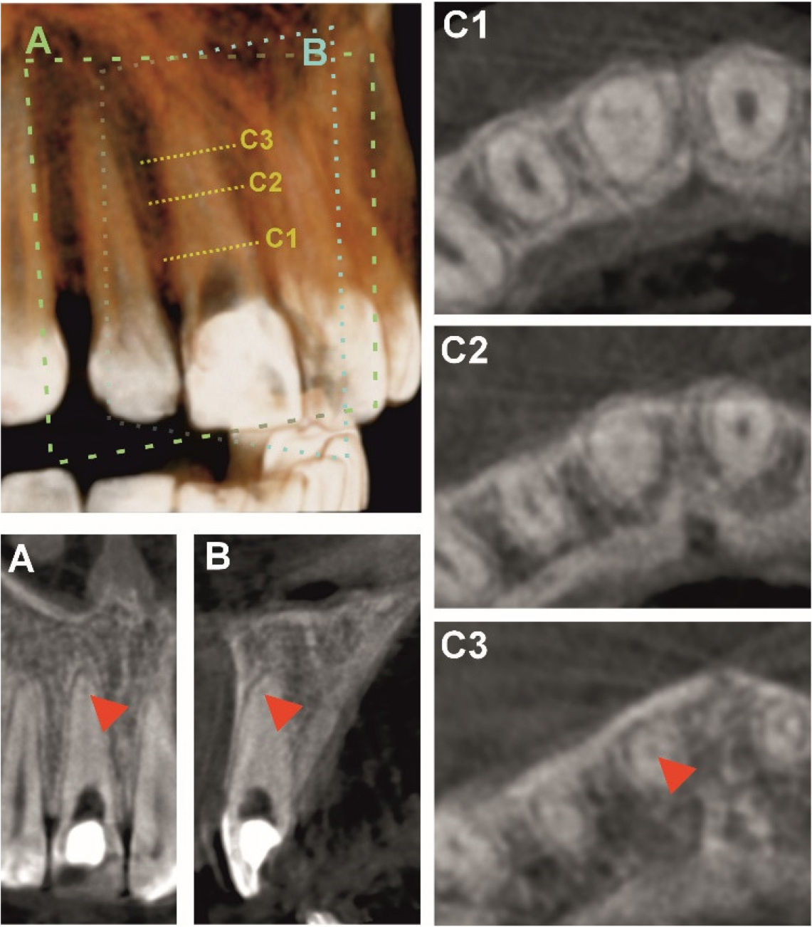
Figure 1.
A three-dimensional CBCT reconstruction illustrating the orientation of the imaging planes used for sectional analysis. The line labeled A represents the coronal plane, while B denotes the sagittal plane selected for detailed evaluation. The dotted markers C1, C2, and C3 indicate the axial slice levels corresponding to the coronal, middle, and apical thirds of the root canal, respectively.
The corresponding coronal (A) and sagittal (B) slices are shown in the lower left portion of the figure. The red arrowheads in these views indicate the location where the previously obstructed root canal becomes patent, marking the point of regained canal lumen continuity. The axial slices (C1–C3) displayed on the right depict cross-sectional morphology in the coronal, middle, and apical thirds of the canal, with the arrowhead in C3 highlighting the same transition point identified in the other planes.
At the second appointment, an absolutely dry operative field was established, and magnification loupes (Illuco loupes 5,5x, Gunpo City, South Korea), were used to aid canal localization. With copious irrigation using 17% EDTA (MD-Cleanser EDTA Solution, Meta Biomed, Chugbuk, South Korea), 2% sodium hypochlorite (Chloraxid 2%, Cerkamed, Stalowa Wola, Poland), and saline, combined with ultrasonic activation, the canal orifice was successfully located. Initial canal negotiation was performed with a #6 K-file, followed by progressive instrumentation using #8, #10, and #15 hand K-files (Dentac, Istanbul, Turkey). Working length was determined using an electronic apex locator (Woodpex III, Woodpecker, China), measuring 22 mm. Mechanical preparation was completed using the RC Blue rotary system (Dentsply Sirona, Charlotte, North Carolina, USA) in reciprocal motion. Chemomechanical preparation was achieved with a single 25/08 instrument using the crowndown technique and abundant ultrasonic-activated irrigation. Continuous irrigation with 2% sodium hypochlorite was maintained throughout instrumentation to prevent debris accumulation. Final irrigation was performed in the following sequence: 5.25% sodium hypochlorite, distilled water, 17% EDTA followed by distilled water, and a final rinse with 5.25% sodium hypochlorite and distilled water. This protocol ensured effective removal of the smear layer and chemical disinfection of the canal system.
After completion of chemomechanical preparation, canal was dried with sterile paper points (Spident Co., Ltd., Incheon, South Korea) and obturated with cold lateral compaction technique and using Vio Seal (Spident Co., Ltd., Incheon, South Korea) sealer. The quality of the root canal filling was evaluated radiographically (Figure 2). The tooth was then temporarily restored, and postoperative instructions were provided. A subsequent visit was scheduled for placement of the definitive restoration.
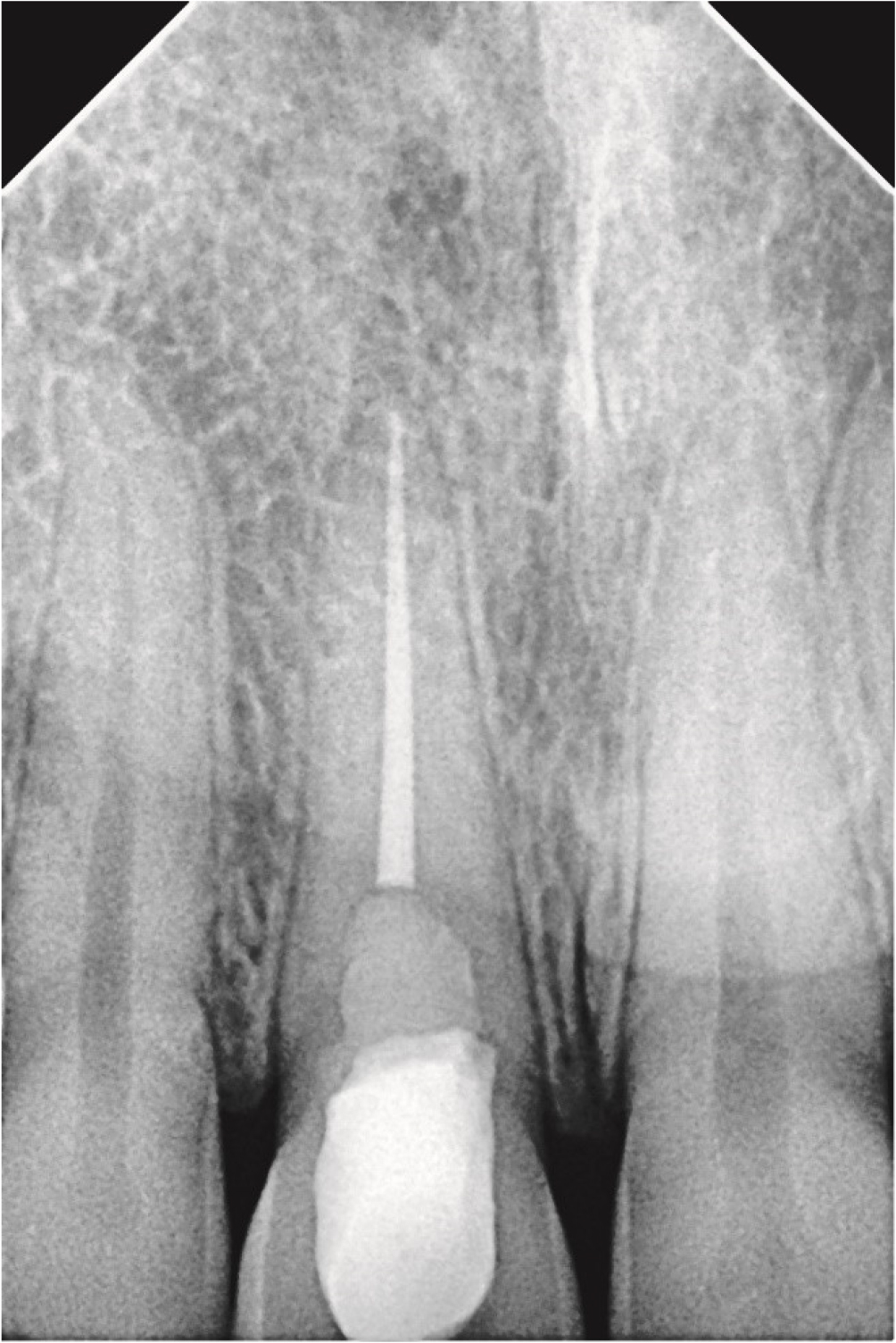
Figure 2-
Retroalveolar radiograph after definitive root canal obturation
DISCUSSION
PCO represents a pathological process characterized by the gradual deposition of mineralized tissue within the pulp chamber and root canal system, frequently resulting in the radiographic loss of canal visibility (6). It may present as either total or partial obliteration. In cases of total calcification, the entire pulp canal system is radiographically indistinguishable, whereas partial obliteration is characterized by a reduced or partially calcified pulp chamber, with root canals still discernible but significantly narrowed (13).
The prevalence PCO has been widely documented in the endodontic literature, although results vary among studies. The incidental prevalence of PCO based on radiographic records has been estimated at 4.7% (14). However, the incidence increases substantially following dental trauma, ranging from 4% to 24% (6), which is consistent with our case where PCO developed after a traumatic injury to the maxillary right central incisor in a young patient. In teeth with immature root formation and who have experienced trauma, prevalence can be 56.9% (14). The prevalence of calcification is also found to be higher in anterior teeth, especially in central and lateral incisors (15). Obliteration occurs more frequently in individuals over 40 years of age, identifying age as a significant factor influencing the occurrence of PCO. In contrast, our case involved a 17-year-old patient, highlighting that PCO can also occur in younger individuals following trauma. Alamoudi et al., in their case-control study, reported that the prevalence of pulp calcifications was higher in females (62%) compared to males (38%) (3). Although the exact etiology of pulpal calcification remains unclear (13), dental trauma is most commonly associated. Age has also been identified as a contributing factor (14). In addition, pulp canal obliteration may occur as a physiological response to chronic irritation, such as caries, deep restorations, trauma from occlusion, physiologic change, and orthodontic movement (7). Research have shown that the use of calcium hydroxide and hydrogen peroxide, can also promote calcification. Dentine dysplasia and long-term use of glucocorticoids have been correlated with calcification of the dental pulp (16).
While epidemiological studies have highlighted the prevalence and contributing factors of pulp canal obliteration, a thorough understanding of its clinical presentation and associated diagnostic challenges is crucial for effective patient management. Recognizing the subtle signs and limitations of conventional diagnostic methods allows clinicians to identify PCO early and implement appropriate treatment strategies. Diagnosing PCO is often challenging due to its complex clinical presentation and the limitations of conventional testing methods, making it essential to use an advanced diagnostic technique to accurately assess pulp status and canal morphology. PCO is most often discovered incidentally during routine radiographic examinations, as it typically progresses without evident clinical symptoms. Nevertheless, as the process advances, crown discoloration and pulp necrosis with periapical lesions and associated symptoms, may develop which represent the primary reasons patients seek dental treatment (10). This was evident in our case, where the patient presented with crown discoloration and sensitivity five years after trauma. Conventional pulp sensibility tests such as electric pulp testing and thermal tests (cold or heat) often yield unreliable or false-negative results. This occurs because these methods rely on neural stimulation, which may be reduced or absent in calcified canals, even when the pulp remains vital. Non-invasive diagnostic techniques such as pulse oximetry and laser Doppler flowmetry evaluate pulpal blood flow and have demonstrated higher accuracy in assessing pulp vitality. Consequently, clinicians should interpret negative sensibility test results in teeth with pulp canal obliteration cautiously and incorporate adjunctive vitality assessment methods when necessary (17).
According to the American Association of Endodontists, PCO is considered to be an endodontic case with a high degree of difficulty. Endodontic treatment is indicated only in the presence of acute symptoms and apical periodontitis, or when PCO complicates the treatment and adversely affects prognosis (18). In such cases, careful preoperative planning is essential. The most frequently reported difficulties include the extensive removal of tooth structure required during conventional access cavity preparation and the difficulty in locating the canal orifice (10). The access cavity preparation stage is a critical determinant of success in managing PCO, as it directly influences the clinician’s ability to identify and negotiate the obliterated canal while minimizing procedural complications (10). The deposition of tertiary dentin in the pulp chamber results in an irregular and constricted canal anatomy, complicating access to the residual pulp tissue, particularly in the apical third of the root. These difficulties increase the risk of iatrogenic complications, such as perforations, ledge formation, loss of peri-cervical dentin and instrument fracture (4,19). Careful use of hand files, chelating agents such as 17% EDTA), and sodium hypochlorite irrigation has been shown to facilitate negotiation even in heavily calcified canals. In contrast, traditional methods, including the use of rigid stainless-steel hand instruments (K-files, C+ files), often prove inadequate in managing the complex and constricted anatomy of calcified canals. (4) Additionally, step-back preparation minimizes the risk of procedural errors, while rotary nickel–titanium (NiTi) systems have proven valuable for efficient mechanical shaping of the canal space (4). These challenges can be further mitigated by modern clinical aids such as CBCT, dental operating microscopy, and the use of ultrasonic tips to enhance precision and safety during treatment (10). In our case, canal negotiation was achieved using magnification loupes without the use of a dental operating microscope, which would have been the optimal method.
Preoperative radiographic evaluation is essential in cases of PCO to assess crown orientation, root curvature, and the degree of canal calcification. Conventional two-dimensional (2D) radiographs, while widely used, have inherent limitations due to superimposition of anatomical structures, geometric distortion, and restricted visualization of the three-dimensional anatomy of root canals (1). Canals that appear patent or calcified on 2D images may not accurately reflect clinical negotiability, and complete radiographic obliteration does not always indicate the absence of a canal space (13). By contrast, CBCT provides a three-dimensional evaluation of root canal anatomy, allowing precise assessment of canal morphology, orientation, extent of calcification, and residual canal space (6). As in our case, CBCT was used to confirm canal location and assess apical third negotiability, justifying its use given the complex obliteration. CBCT facilitates optimal access planning by estimating calcified tissue thickness and predicting the depth and path of the canal (13). However, CBCT is not intended to replace conventional radiography but to complement it by providing additional diagnostic information (20). Its use requires specialized training and a thorough understanding of dental anatomy, as high-density structures can generate artifacts that may affect interpretation. CBCT also involves higher costs, limited availability, and exposure to ionizing radiation, so each scan must be justified by its potential diagnostic benefit. Image quality can be influenced by factors such as field of view, voxel size, number of projections, tube voltage, and the presence of materials within the canal (21). Clinical studies have demonstrated that the combination of CBCT, dental operating microscopy, and ultrasonic instrumentation improves canal negotiation, reduces procedural errors, and increases success rates in the treatment of PCO (1).
The present findings further highlight the clinical relevance of CBCT in the management of pulp canal obliteration, particularly in cases where conventional imaging assessment is limited. In accordance with the recommendations of the European Society of Endodontology, CBCT should be selectively utilized in complex endodontic situations, enhancing diagnostic precision and improving clinical outcomes (22). The present case supports existing evidence that CBCT can significantly reduce iatrogenic complications, improve canal localization, and increase the overall predictability of endodontic treatment in challenging PCO cases (23).
CONCLUSION
This case demonstrates that CBCT plays a crucial role in the diagnosis and management of teeth affected by pulp canal obliteration following trauma. Its ability to visualize root canal anatomy in three dimensions provides valuable information for determining the extent of calcification and planning a safe and conservative access pathway. The combination of CBCT imaging, the use of magnification, ultrasonic activation, and careful instrumentation enables successful negotiation and treatment of calcified canals while minimizing procedural complications. Early and accurate diagnosis supported by advanced imaging techniques can significantly improve the prognosis and long-term outcomes of endodontic treatment in such complex cases. This case highlights the importance of integrating advanced imaging and meticulous endodontic techniques in managing complex calcified teeth.
ACKNOWLEDGMENTS
This research was funded by the Ministry of Education, Science and Technological Development of the Republic of Serbia, agreement No. 451-03-47/2023-01/200111, and the Faculty of Medical Sciences, University of Kragujevac, Serbia, project number JP 07-25.