
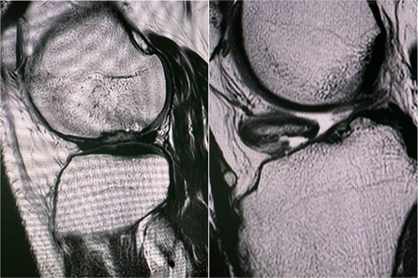
Figure 1.
Preoperative MRI showing an osteochodral lesion of the lateral femoral condyle and intraarticular loose body
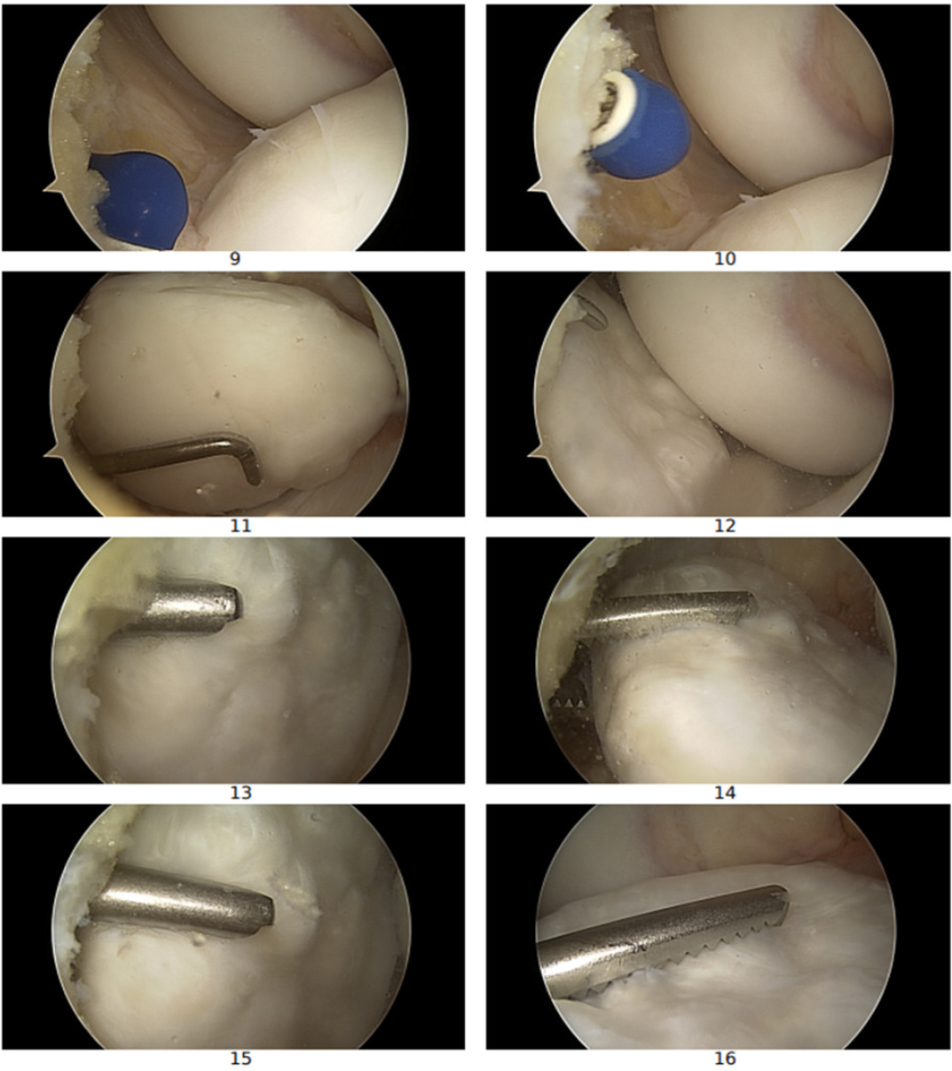
Figure 2.
Arthroscopic view of intaarticular loose body
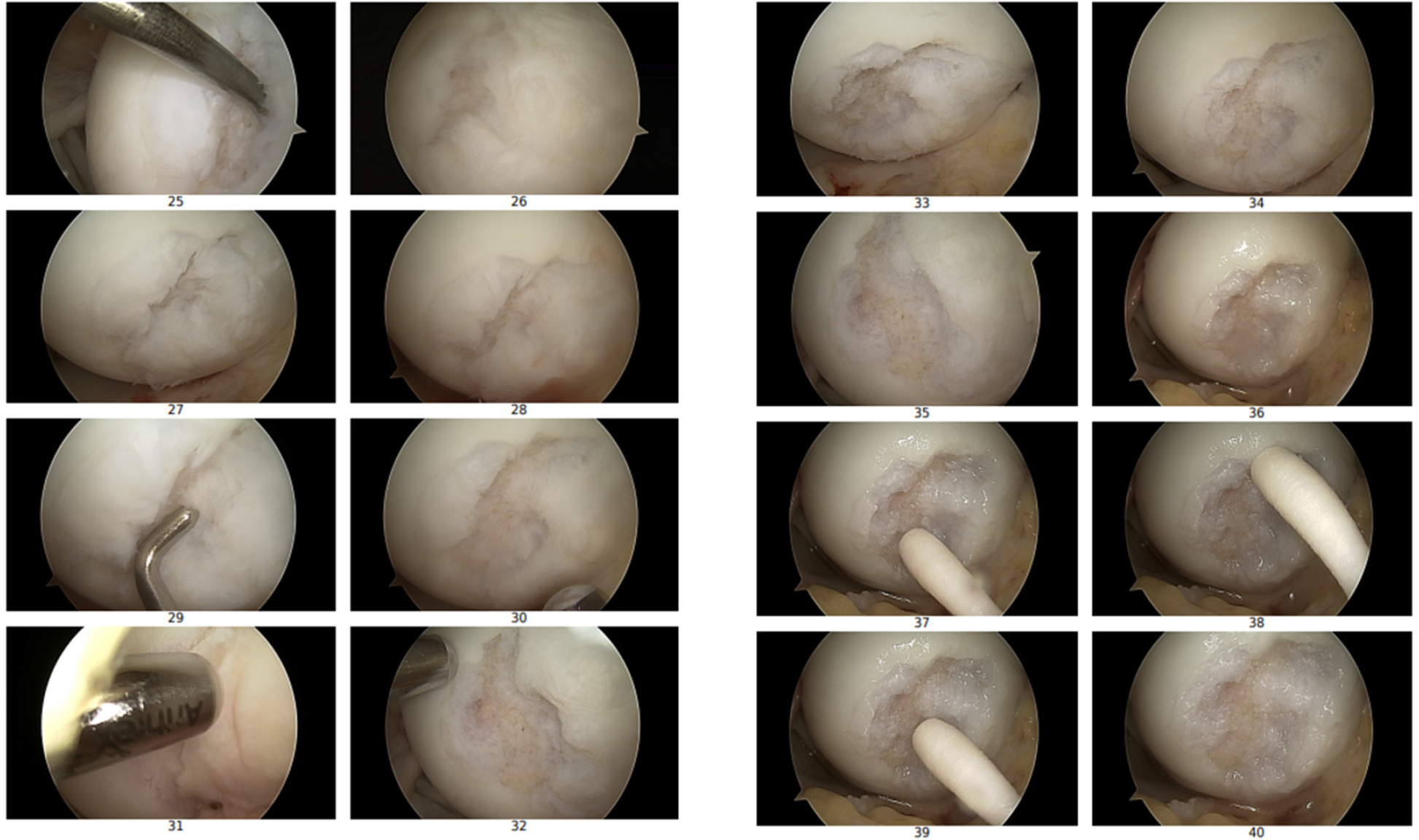
Figure 3.
Arthroscopic view of an osteochondral lesion of the lateral femoral condyle
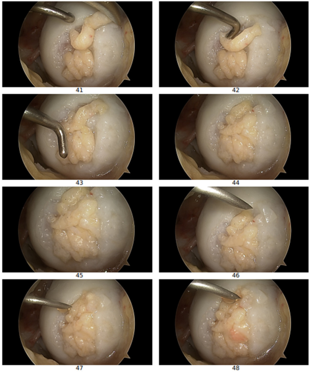
Figure 4.
Arthroscopic images showing the osteochondral lesion after filling with the minced cartilage-PRP composite during the AutoCart procedure with uniform graft distribution
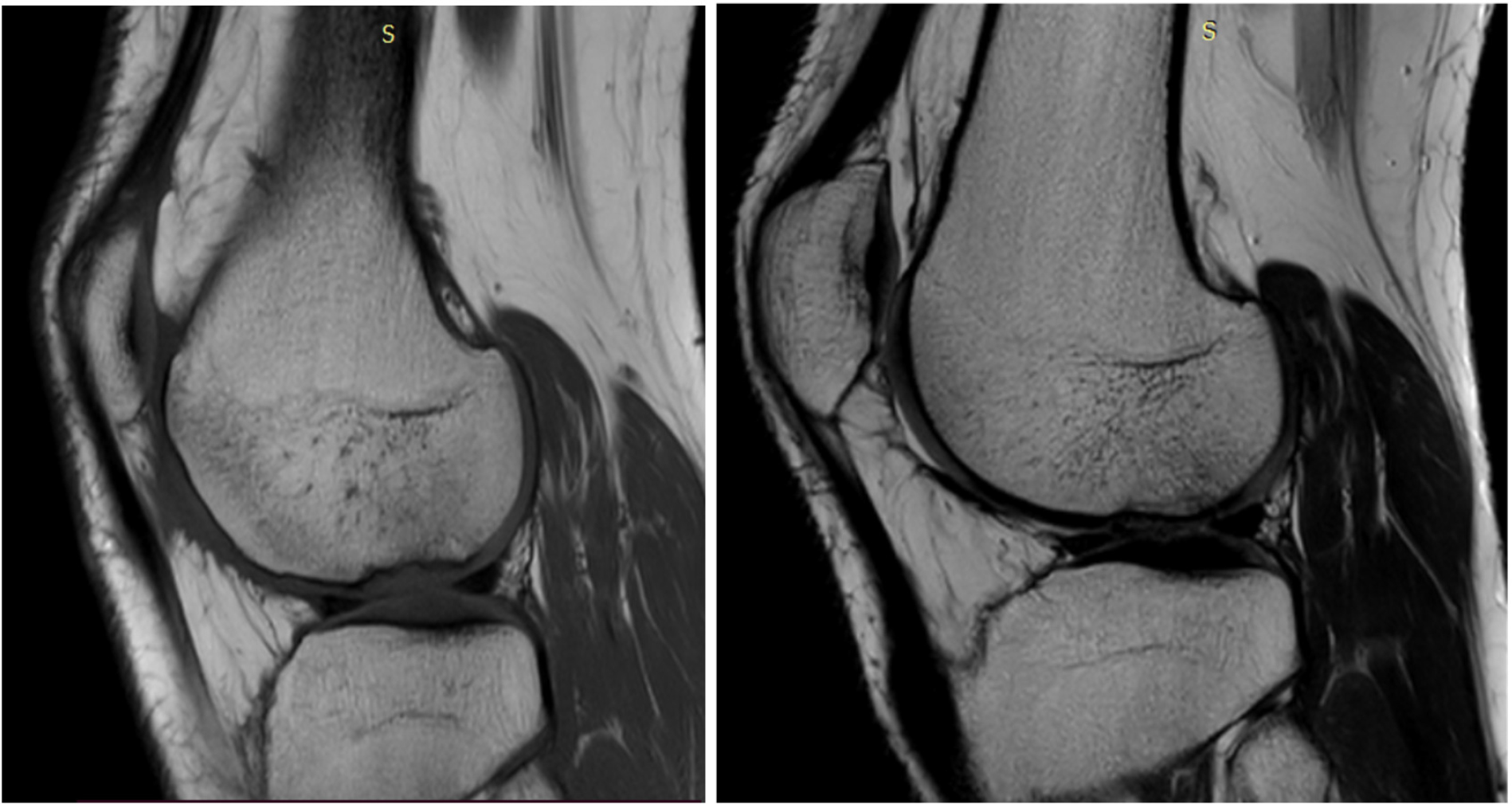
Figure 5.
MRI of the left Knee:12 and 24 months postoperatively