
INTRODUCTION
The true incidence of osteochondral lesions in humans remains uncertain, as a substantial proportion of cases remain asymptomatic.(1,2) Articular cartilage damage leading to chondral lesions may result from metabolic, genetic, vascular, or traumatic factors. These defects may develop after a single episode of excessive joint loading or as a consequence of repetitive microtrauma over time. Depending on the extent of cartilage involvement, lesions may range from small superficial defects to osteochondral injuries affecting the full thickness of the cartilage and the underlying subchondral bone.(3) Although the exact prevalence of cartilage lesions is difficult to determine, several studies have reported that articular cartilage defects are identified in approximately 60–66% of knees undergoing arthroscopic evaluation.(4) The management of focal chondral injuries continues to represent a significant challenge for orthopedic surgeons. The primary goals of treatment are pain reduction, restoration of joint function, and facilitation of a return to normal daily activities and an active lifestyle.Therapeutic strategies include conservative management, palliative procedures, and reparative or restorative surgical techniques aimed at promoting articular cartilage regeneration. Commonly utilized surgical procedures include microfracture, autologous chondrocyte implantation (ACI), osteochondral autologous transfer (OATS), and minced cartilage implantation (MCI). The concept of minced cartilage implantation was first introduced in 1983.(5)In recent years, increasing attention has been directed toward single-stage cartilage repair techniques that avoid the need for in vitro cell expansion while preserving viable chondrocytes capable of producing extracellular matrix and supporting cartilage regeneration.(6,7) Contemporary clinical studies have demonstrated encouraging short- and mid-term outcomes following arthroscopic autologous minced cartilage implantation for focal cartilage defects of the knee.(8)Furthermore, biologically augmented approaches combining minced cartilage fragments with platelet-rich plasma or fibrin-based matrices have been proposed to enhance graft stability and create a more favorable biological environment for cartilage repair. (9) These developments have contributed to the emergence of standardized arthroscopic techniques that allow simultaneous harvesting and implantation of autologous cartilage fragments within a single surgical procedure.The aim of this case report is to present the clinical and radiological outcome of a single-stage arthroscopic treatment of a focal cartilage lesion of the lateral femoral condyle in a young male patient using a biologically augmented minced cartilage technique.
CASE REPORT
A 23-year-old male patient presented to the orthopedic clinic with complaints of pain localized to the left knee. According to his medical history, the pain had been present for approximately one year, with progressive worsening over the preceding several months. The patient denied any history of trauma. On physical examination, the range of motion was within normal limits, although pain occurred at maximum flexion. Clinical tests, including McMurray, Lachman and Ballottement were negative. Radiological evaluation with plain radiographs and MRI revealed an osteochondral lesion located at the lateral femoral condyle and loose body. Based on these findings, surgical management was indicated and the patient was scheduled for knee arthroscopy with an AutoCart procedure.
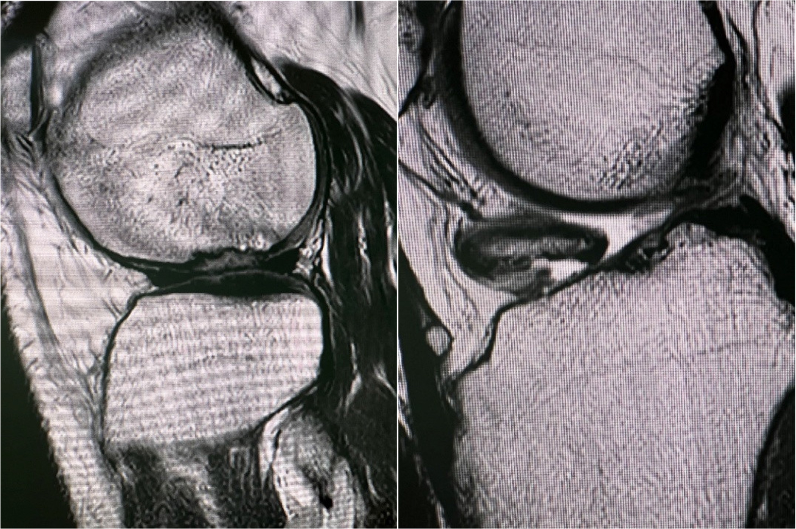
Figure 1.
Preoperative MRI showing an osteochodral lesion of the lateral femoral condyle and intraarticular loose body
Under spinal anesthesia, arthroscopy of the left knee was performed using two standard portals. An osteochondral lesion was identified on the lateral femoral condyle. In addition, a large intra-articular loose body was visualized and subsequently removed. A 5-mm shaver blade was then used to harvest autologous chondral fragments from the healthy cartilage margins surrounding the lesion. A sterile tissue-collection device connected to suction was used to allow simultaneous harvesting and collection of viable cartilage fragments for subsequent implantation. Once harvesting was completed, the tissue collector was opened and the filter chamber containing the cartilage fragments was retrieved. A total of 15 mL of venous blood was aspirated using a doublesyringe system for platelet-rich plasma (PRP) preparation and centrifuged at 1500 rpm for 5 minutes. The resulting plasma was transferred into a smaller syringe. Preparation of autologous thrombin was subsequently performed using a PRP-based activation system according to the manufacturer’s protocol. The plasma was sequentially processed and allowed to incubate in a horizontal position to facilitate thrombin generation, after which it was aspirated into a sterile syringe for intraoperative use.Using a connector system, the harvested chondral fragments were mixed with PRP in a 3:1 ratio. Through several forward-and-back transfers between syringes, a homogeneous paste-like composite was created and loaded into a 1-mL syringe equipped with an application cannula. After evacuation of intra-articular fluid, the osteochondral defect was carefully dried and the arthroscopic trocar was withdrawn to create a dry working environment. The prepared chondral-PRP composite was then applied directly onto the lesion site. Autologous thrombin was subsequently applied to promote graft stabilization and biological fixation within the defect. Care was taken to ensure uniform distribution and stable adherence of the graft material to the surrounding cartilage margins.
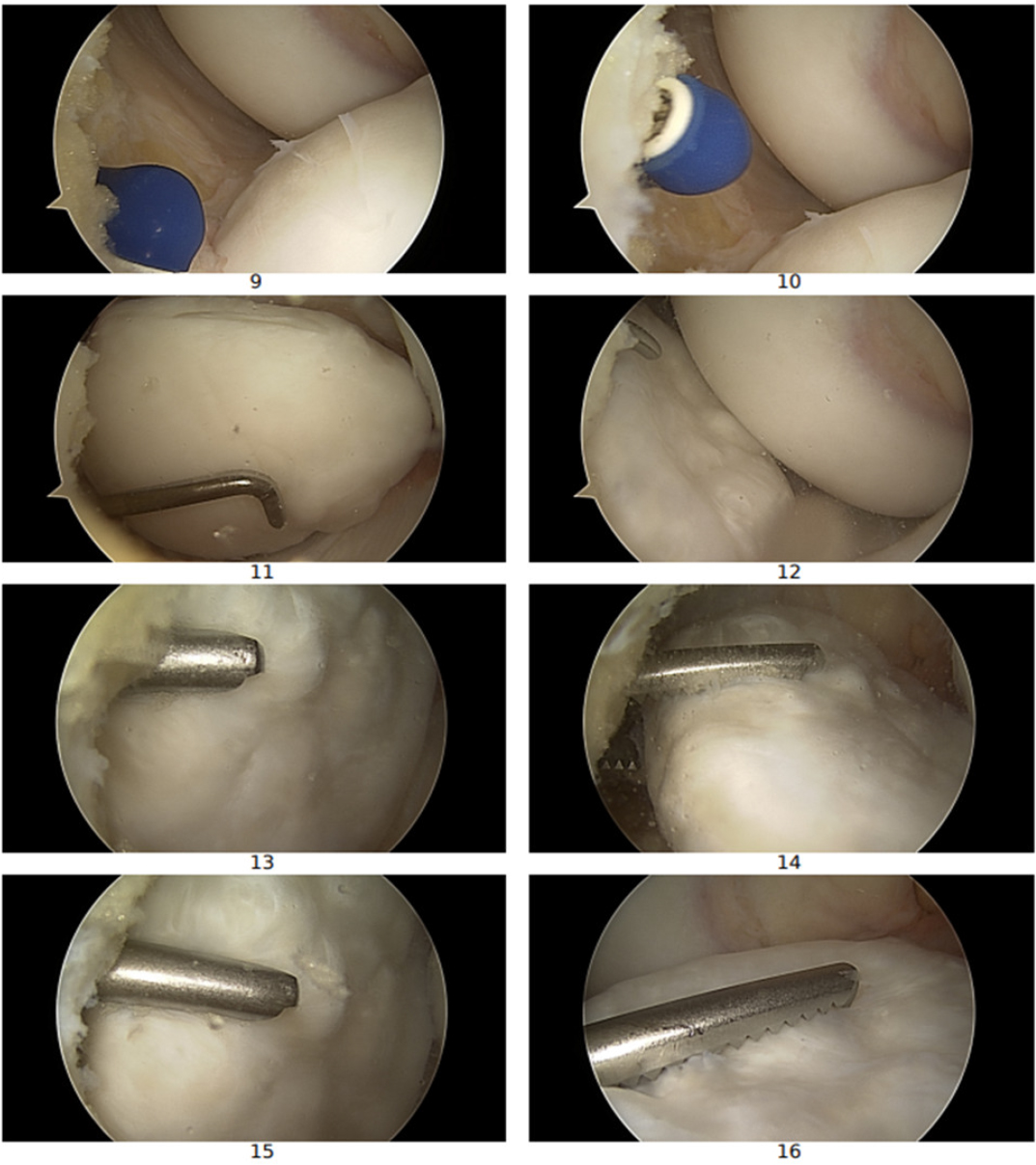
Figure 2.
Arthroscopic view of intaarticular loose body
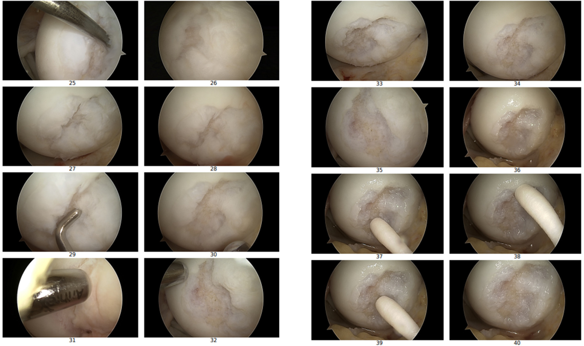
Figure 3.
Arthroscopic view of an osteochondral lesion of the lateral femoral condyle
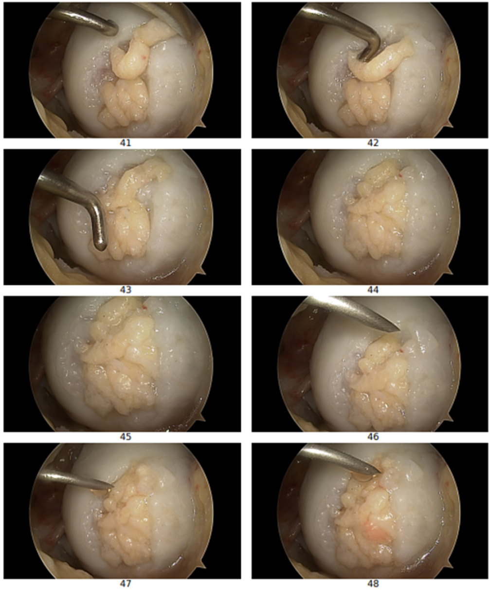
Figure 4.
Arthroscopic images showing the osteochondral lesion after filling with the minced cartilage-PRP composite during the AutoCart procedure with uniform graft distribution
In the immediate postoperative period, early mobilization and structured physical rehabilitation were initiated. Thromboprophylaxis was administered with rivaroxaban 10 mg once daily for two weeks. The patient was instructed to ambulate with two axillary crutches while maintaining a nonweight-bearing regimen during the initial rehabilitation phase.At 24-month follow-up, clinical examination demonstrated full range of motion without joint effusion, instability or mechanical symptoms. Follow-up evaluations were performed at 6, 12, and 24 months postoperatively. Preoperatively, the Visual Analog Scale (VAS) pain score was 6, the International Knee Documentation Committee (IKDC) subjective score was 45, and the Tegner activity level was 3. At 12-month follow-up, substantial clinical improvement was observed, with VAS decreasing to 0, IKDC increasing to 82, and Tegner activity level improving to 5. At 24-month follow-up, the outcome remained stable, with VAS remaining 0, IKDC further improving to 90 and Tegner activity level maintained at 5, indicating sustained functional recovery and complete resolution of pain.
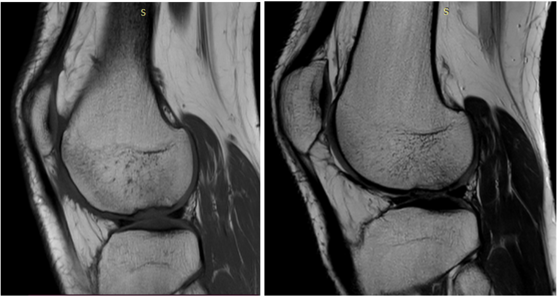
Figure 5.
MRI of the left Knee:12 and 24 months postoperatively
DISCUSSION
The present case demonstrated favorable clinical and radiological improvement following a fully arthroscopic autologous minced cartilage implantation using the AutoCart technique for the treatment of a focal osteochondral lesion of the knee. The encouraging outcome observed in this patient supports the growing interest in single-stage biological cartilage repair strategies that aim to restore joint surface integrity while preserving the biological potential of autologous chondrocytes. Articular cartilage lesions may arise from traumatic injury, degenerative processes, dysplasia, or osteochondritis dissecans. In physically active individuals, repetitive mechanical loading may compromise subchondral bone circulation, which may predispose the joint to progressive cartilage degeneration. If left untreated, focal cartilage defects may eventually lead to the development of degenerative osteoarthritis. Therefore, surgical intervention is frequently required to restore the articular surface and stimulate regeneration of cartilage tissue resembling native hyaline cartilage.(9) Over the past two decades, several cartilage repair strategies have been developed, including microfracture, osteochondral autograft transplantation, autologous chondrocyte implantation (ACI), and more recently, minced cartilage implantation techniques. One of the early clinical experiences with autologous cartilage repair was reported by Stone et al., who demonstrated significant clinical improvement in patients with Outerbridge grade IV cartilage lesions treated using autologous cartilage repair techniques.(10)Subsequent studies have explored biologically augmented approaches combining cartilage fragments with autologous bone or biological scaffolds. Christensen et al. described the use of autologous dual-tissue transplantation (ADTT), which combines autologous cartilage and bone fragments for the treatment of osteochondral lesions. Their results demonstrated significant improvement in clinical outcomes and restoration of subchondral bone structure at one-year follow-up. (11) More recently, single-stage minced cartilage implantation has gained increasing attention as a biologically attractive alternative to traditional cartilage repair techniques. Massen et al. reported significant improvements in clinical scores and patient-reported outcome measures in patients treated with autologous minced cartilage implantation at two-year follow-up. Importantly, minced cartilage fragments preserve viable chondrocytes capable of producing extracellular matrix and contributing to hyaline-like cartilage repair tissue.(12) Similarly, Runer et al. demonstrated favorable mid-term outcomes in patients treated with minced cartilage implantation, with high graft durability observed at 60 months of follow-up and a very low revision rate. (13) Focal chondral lesions of the knee represent a significant clinical challenge due to the limited intrinsic healing capacity of articular cartilage and the potential progression toward early osteoarthritis if left untreated (14). Blanke et al. further reported significant improvements in functional knee scores and satisfactory defect filling on MRI in patients treated with all-autologous minced cartilage procedures (15). Comparable improvements in patient-reported outcomes were also observed in the study by Schneider et al., confirming the clinical effectiveness of this biological cartilage repair technique (7). Regenerative cartilage repair strategies have evolved considerably in recent years, with various biological procedures demonstrating encouraging clinical and functional outcomes (16,17,18,19). These approaches aim to restore the articular surface and improve joint function, particularly in young and active patients with symptomatic cartilage defects. Pohl et al. reported encouraging clinical and radiological outcomes two years after autologous shaver-based minced cartilage implantation for cartilage lesions of the knee. Their findings demonstrated significant improvement in clinical scores and MRI parameters, highlighting the regenerative potential of minced cartilage techniques and their effectiveness as a single-stage cartilage repair strategy.(20) In addition, Başal et al. evaluated autologous minced cartilage implantation combined with an arthroscopic platelet-rich fibrin membrane for the treatment of large chondral defects of the knee. Their prospective comparative study demonstrated significant clinical improvement and satisfactory defect filling on follow-up imaging, suggesting that biological augmentation may further enhance cartilage regeneration and improve treatment outcomes.(21) Compared with conventional autologous chondrocyte implantation (ACI), the AutoCart technique offers several potential advantages. It represents a single-stage procedure that avoids the need for in vitro cell expansion and a second surgical intervention. This reduces treatment costs, regulatory complexity, and overall rehabilitation time. Furthermore, the procedure can be performed arthroscopically, allowing for a minimally invasive approach while preserving the biological properties of autologous cartilage fragments. Despite these encouraging findings, several limitations must be acknowledged. First, the present report describes a single clinical case, which limits the generalizability of the results. Second, although clinical improvement and favorable radiological findings were observed, standardized patient-reported outcome scores such as IKDC, KOOS, or Lysholm were not prospectively recorded. Third, longer follow-up is necessary to determine the durability of the repair tissue and to evaluate the long-term clinical outcomes of the AutoCart technique. Nevertheless, the present case contributes to the growing body of evidence supporting single-stage biological cartilage repair strategies. The favorable clinical course and radiological improvement observed in this patient suggest that arthroscopic autologous minced cartilage implantation using the AutoCart technique may represent a promising treatment option for focal osteochondral lesions of the knee. However, larger prospective studies with standardized outcome measures and longer follow-up are required to confirm the clinical effectiveness and long-term durability of this technique.
CONCLUSION
This case demonstrates the feasibility of the AutoCart procedure as a fully arthroscopic, minimally invasive single-stage technique for the treatment of focal osteochondral lesions of the knee. By utilizing autologous cartilage fragments combined with biological augmentation, the technique may create a favorable biological environment that supports cartilage repair and restoration of the articular surface.
In the present patient, encouraging clinical and radiological improvement was observed during short-term follow-up. However, as this report describes a single clinical case, the findings should be interpreted with caution. Further prospective studies involving larger patient cohorts, standardized clinical outcome measures, and longer follow-up periods are necessary to better define the role and long-term effectiveness of the AutoCart technique in the treatment of osteochondral defects of the knee.