
Introduction
Melanoma is a malignant tumor originating from neuroectodermal melanocytic cells. These are melanin-producing cells found primarily in the skin, but also in the mucous membranes of the respiratory, gastrointestinal, and genitourinary tracts, the uvea of the eye, and the meninges. The most common location for mucosal melanoma is the head and neck region (45–55%), with 25–40% of cases occurring in the oral cavity [1–2].
The most important cause of an increased risk of developing cutaneous melanoma is considered to be ultraviolet radiation (sunlight, especially sunburns in childhood and at a young age, and tanning beds), constant mechanical or chemical irritation, low pigment content in the skin, and genetic predispositions [3]. In contrast, the etiopathogenesis of mucosal melanoma development remains unclear; the impact of UV radiation on its development has not been proven [4]. Distinct molecular features, including a lower incidence of BRAF oncogene mutations, but a higher incidence of KIT oncogene mutations in mucosal melanoma, suggest divergent genetic etiologies between cutaneous melanoma and mucosal melanoma [1–2].
Cutaneous melanoma, as opposed to mucosal melanoma, is characterized by a high mutational burden of BRAF and NRAS, which forms the basis for the efficacy of targeted therapy. There is also a larger number of clinical trials confirming the effectiveness of immunotherapy in the treatment of cutaneous melanoma compared to mucosal melanoma. For this reason, surgical treatment still remains the mainstay of treatment for mucosal melanoma. The disease is detected at a later stage, is characterized by high malignancy, and has a worse prognosis, regardless of the stage of advancement [1]. Among mucosal melanomas, those of the head and neck region have the best survival rates [5]. Recurrence rates are high, and five-year survival remains low. Evidence for the efficacy of systemic treatment is limited to small retrospective cohorts and subgroup analyses, demonstrating lower response and survival rates compared to cutaneous melanoma [4, 6].
Epidemiology
According to the 2022 World Health Organization report, in the Polish population, the highest number of deaths caused by cutaneous melanoma occurs in individuals over 75 years of age [7].
Among all melanomas, mucosal melanomas account for less than 2% of cases, and their incidence remains at a constant level, in contrast to cutaneous melanomas, the number of which is steadily increasing [2]. An analysis of epidemiological indicators from the years 2000–2022 reveals a pronounced, long-term upward trend in the incidence of cutaneous melanoma in Poland, characterized by a more than twofold increase in the registered frequency of this malignancy within the studied population. Furthermore, the transient decline in the incidence curve observed in 2020, followed by a subsequent peak in 2021, may be interpreted as a probable reflection of the healthcare debt phenomenon, stemming from delays in oncological diagnostics during the COVID-19 pandemic. [Fig.1]

Fig 1.
Methodology
The aim of this review is to present current literature data on diagnostic and therapeutic issues in cutaneous melanoma and mucosal melanoma.
In order to gather relevant data, a comprehensive search of the scientific literature was conducted using Polish – and English-language electronic databases. After analysis, 37 sources were selected. The analyzed sources primarily included meta-analyses, randomized controlled trials (RCTs), and epidemiological data from the World Health Organization (WHO). Scientific databases, including PubMed and Google Scholar, were systematically searched using a combination of the following keywords: “cutaneous melanoma”, “mucosal melanoma”, “BRAF/MEK inhibitors”, “immunotherapy”, “Mohs micrographic surgery”, and “wide local excision”.
Etiopathogenesis
The presence of mutations in the RAS/RAF/MEK/ERK MAP kinase (MAPK) pathway is observed in 75% of cutaneous melanoma cases. The dominant mechanism leading to the hyperactivity of the RAS/RAF/MEK pathway in cutaneous melanoma is a mutation of the gene encoding the BRAF kinase, with somatic mutations in the BRAF V600 gene being observed in 50–70% of cutaneous melanomas arising in areas not exposed to chronic sunlight [10].
In their study, Nassar et al. indicate that mucosal melanoma carries distinct genetic alterations and oncogenic factors compared to cutaneous melanoma, and therefore should be treated accordingly. The cause of oral melanoma and the risk factors contributing to the malignant transformation of cells remain unclear, and the associations between human papilloma-virus (HPV), human immunodeficiency virus (HIV), and oral mucosal melanoma (OMM) have not been confirmed [11]. Common factors (BRAF and NRAS) occurring in cutaneous melanoma have a lower mutation rate in mucosal melanoma. In contrast, SF3B1 and KIT show a higher mutation rate in mucosal melanoma compared to cutaneous melanoma [12].
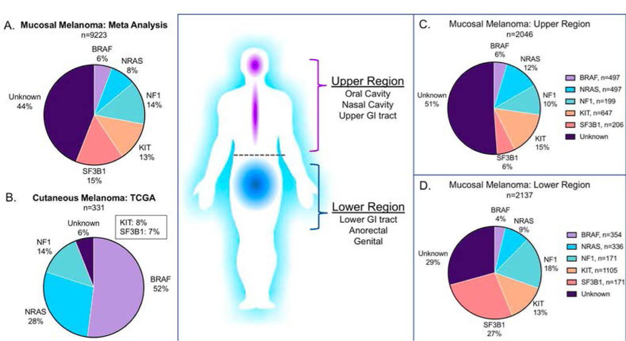
Fig 2.
Comparison of mutation frequencies in cutaneous and mucosal melanoma [12].
In their study, Buchbinder et al. demonstrated that the most common mutations in mucosal melanoma occur in the SF3B1 (27%), KIT (18%), and NF1 (17%) genes, which is a pattern distinct from cutaneous melanomas [13].
In a study by Broit et al., mutations in mucosal melanoma were evaluated depending on the site of occurrence. The analysis revealed that KIT, NF1, BRAF, NRAS, SF3B1, and SPRED1 were significantly mutated genes. ATRX and SF3B1 mutations occurred more frequently in melanomas of the lower body, and CTNNB1 in the upper body [14]. The highest incidence of BRAF and NRAS oncogene mutations applies to the mucous membranes of the head and neck region [2, 12, 14, 15] [Fig. 3].
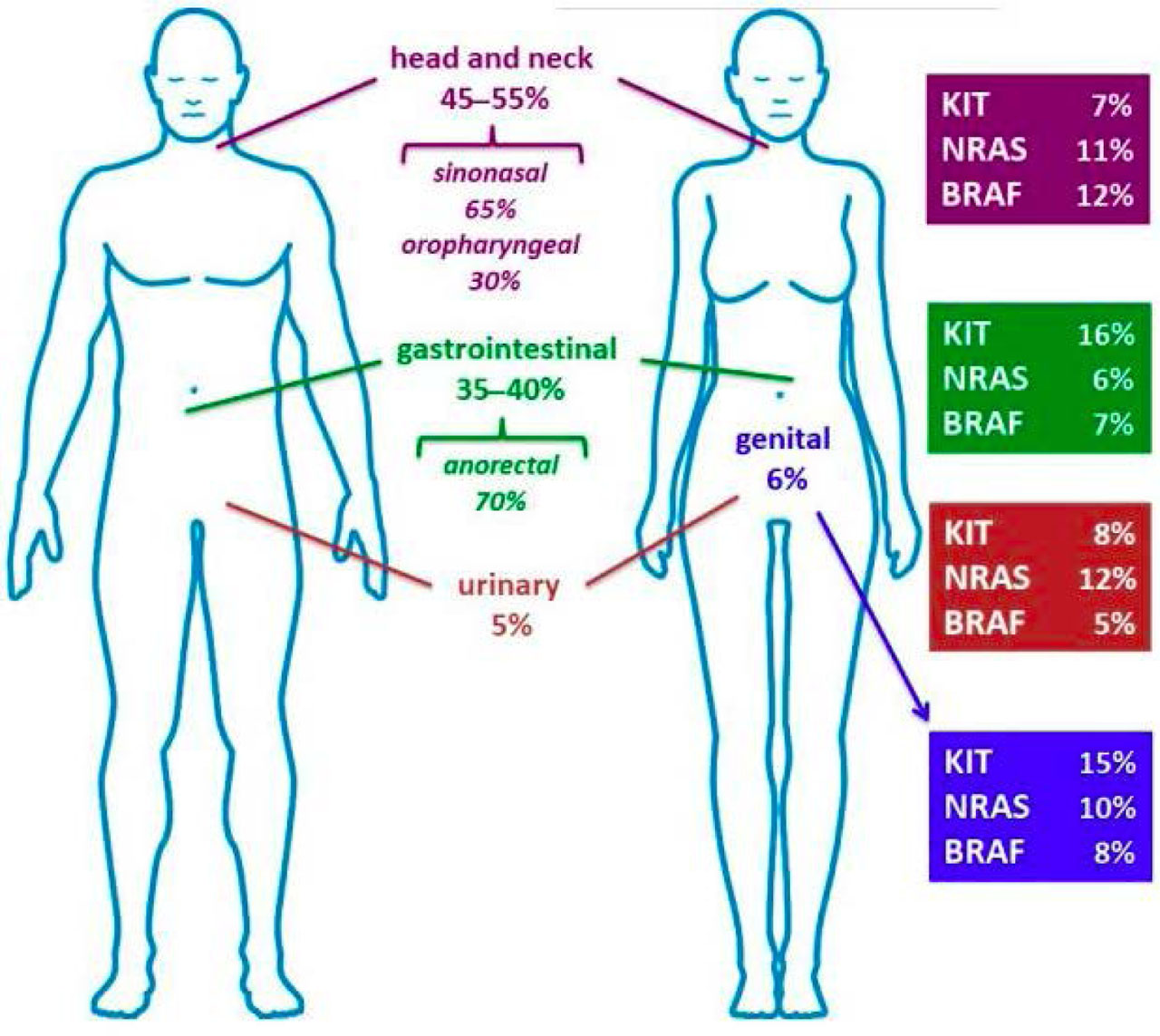
Fig. 3
Distribution of mucosal melanomas and mutation frequency depending on location [2].
Clinical presentation
Mucosal melanomas are detected at a more advanced stage due to an anatomical location that is often overlooked during examination compared to cutaneous melanoma. Mucosal melanomas of the head and neck region most commonly occur in the nasal cavity, where they often remain asymptomatic for a long time. Most nasal and sinus lesions are polyps ranging in color from dark brown and red to white. At a more advanced stage, mucosal melanomas may present with symptoms such as epistaxis (nosebleeds) and/or nasal obstruction, facial pressure, and exophthalmos, diplopia (double vision), or neurological symptoms. These melanomas are most often detected before metastasis to the lymph nodes occurs [16]. In contrast, up to 50% of patients with oral mucosal melanomas and 65% with laryngeal melanomas present with lymph node metastases at the time of diagnosis.
Primary forms of laryngeal mucosal melanoma belong to casuistic entities. Anatomically, the neoplastic process is most frequently located within the epiglottis and subsequently involves the glottic structures. Due to its non-specific symptomatology including dysphonia, hemoptysis, dysphagia, and progressive airway obstruction, this lesion remains virtually indistinguishable from squamous cell carcinoma and other malignant epithelial tumors of the larynx on physical examination. Approximately 20% of cases of mucosal melanoma of the head and neck are located within the oral cavity, showing a clear predilection for involving the hard palate and the maxillary alveolar mucosa. Although the initial stage of tumor development is usually asymptomatic, in the clinically overt phase, symptoms such as pain, tissue swelling, and bleeding predominate. Morphologically, the lesions take the form of macular or nodular eruptions with pigmentation ranging from brown to black. Adjacent satellite lesions may occur in the immediate vicinity of the primary focus. An important piece of information for increasing the detectability of mucosal melanomas is the fact that approximately 30–40% of mucosal melanomas are colorless (amelanotic), whereas in the skin they occur in less than 10% of cases [3, 16].
Therapeutic strategies. Treatment of cutaneous melanoma.
The majority of patients with cutaneous melanoma are diagnosed at an early stage and treated with surgical methods. Despite this, there is a group of patients with stage II–IV disease, where immunotherapy and molecularly targeted drugs may be indicated. Patients treated with immunotherapy respond to treatment more slowly and at a lower rate, but achieve long-term control, whereas those reciving targeted treatment achieve a faster and higher rate of response to treatment, but with a shorter durability of response. Furthermore, according to the latest guidelines, neoadjuvant treatment is utilized, which consists of immunotherapy (e.g., ipilimumab + nivolumab, pembrolizumab) and BRAF and MEK inhibitors (dabrafenib/trametinib) [10, 17].
Surgical oncology in the treatment of cutaneous melanoma
In recent years, significant changes have occurred in the treatment of melanoma, better adapted to the biology of the disease, including immunotherapy and targeted therapy; the basis of treatment and staging of local disease remains surgical resection through wide local excision of the primary lesion [18].
Current guidelines of the National Comprehensive Cancer Network (NCCN) recommend wide excision with a 1 cm margin for melanomas with a thickness of 1.0 mm or less, wide excision with a 1–2 cm margin for melanomas with a thickness of 1–2 mm, and a 2 cm margin for melanomas with a depth greater than 2 mm. The excised tissue should include the skin and subcutaneous tissue down to the level of the fascia, which contains all the local lymphatic tissue. Certain anatomical sites, including but not limited to the fingers, ears, face, and plantar surface of the foot, may not be as easily amenable to rigid surgical criteria without significant concerns regarding poor cosmetic and functional outcomes, and therefore remain poorly investigated [18].
Particular attention should be paid to the MelMarT trial, which brought the topic of excision margins for cutaneous melanoma into discussion. The study randomized 400 patients from 17 centers in 5 countries. The melanomas were located on the trunk (56.9%), extremities (35.6%), and head and neck (7.4%). The study evaluated, among others, the need for reconstruction, perioperative adverse events at wide excision sites, and the patients’ quality of life. It turned out that 34.9% of patients required reconstruction with a skin graft or local flap in the 2 cm group compared to 13.6% in the 1 cm group. In the 2 cm group, a significantly increased need for reconstruction was found across all locations, especially the extremities and the head and neck. Furthermore, a significant increase in wound necrosis was noted in the 2 cm arm compared to the 1 cm arm (3.6% vs. 0.5%). No differences in quality of life were observed between the two groups after a 12-month follow-up [19].
In a study by Vecchiato et al., data from the Italian melanoma registry were analyzed, focusing on complication rates (such as wound infections, suture dehiscence, or the formation of lymphoceles/seromas) following basic procedures performed as part of melanoma surgery: wide excision (WE), sentinel lymph node biopsy (SLNB), and lymphadenectomy (LFND). The researchers arrived at the following results: regarding wound infections, the rate was 1.1% for WE, 1.3% for SLNB, and 4.1% for LFND; regarding postoperative wound dehiscence, the rate was 2.0% for WE, 0.9% for SLNB, and 2.8% for LFND; regarding seroma, the rate was 4.2% for SLNB, while reaching as high as 15.1% for LFND [20].
In their work, Jacobsen et al. investigated the possibilities of Mohs micrographic surgery, particularly in the region of head and neck tumors, because the anatomical and functional complexity of these areas leads to suboptimal surgical treatment, resulting in unacceptably high rates of local recurrence and persistently positive margins with traditional wide local excision. The researchers concluded that patients undergoing Mohs micrographic surgery may have better survival than patients undergoing wide local excision [21].
In another study, Etzkorn et al. evaluated the efficacy of Mohs micrographic surgery (MMS) in the treatment of atypical intraepidermal melanocytic proliferation (AIMP); it was found that MMS with MART-1 immunostaining provides excellent local control of AIMP in a specialized setting and allows for the definitive removal of subclinical spread prior to reconstruction [22].
Williams et al., in a systematic review encompassing over 41,000 patients, indicate a lack of sufficient scientific evidence allowing for an unambiguous comparison of the efficacy of Mohs micrographic surgery with traditional wide local excision in the treatment of invasive melanoma. The authors emphasize that to draw reliable conclusions, it is necessary to conduct precise, randomized clinical trials [23].
Immunotherapy in the treatment of cutaneous melanoma
Immunotherapy in the treatment of melanoma is a highly dynamic field. In recent years, tremendous progress has been made in this matter to the benefit of patients. Drugs that deserve special distinction include nivolumab, ipilimumab, relatlimab, pembrolizumab, atezolizumab, and talimogene laherparepvec, as well as combinations of these agents.
Nivolumab and ipilimumab
Ipilimumab is a monoclonal antibody directed against CTLA-4. Nivolumab, on the other hand, is a fully humanized, monoclonal IgG4 immunoglobulin directed against PD-1. By binding to PD-1, it prevents the interaction of PD-1 with both PD-L1 and PD-L2 [10].
The combination of nivolumab and ipilimumab is one of the most effective first-line options for selected patients with unresectable or metastatic melanoma, particularly when long-term disease control is the treatment goal [10].
A phase III trial by Atkins et al. aimed to determine the optimal treatment sequence for patients with metastatic melanoma harboring a BRAF V600 mutation. The patients were divided into two groups. In one of them, dual immunotherapy (nivolumab with ipilimumab) was administered first, with a switch to targeted therapy (dabrafenib with trametinib) in the event of disease progression. In the second group, the inverse sequence was used (starting with targeted therapy and then switching to immunotherapy). The results clearly favored initiating treatment with immunotherapy, which translated into a significantly higher 2-year overall survival rate (71.8% compared to 51.5%), a longer median duration of response, and better progression-free survival. Due to such a clear clinical advantage for patients in the first group, the study was prematurely terminated by the monitoring committee, and the safety profiles of both regimens remained similar and consistent with predictions. This study clearly demonstrated that the standard of care should be dual immunotherapy, reserving targeted therapy as a second-line treatment in case of disease progression [24].
The registration trial for the combination of nivolumab and ipilimumab was the phase III CheckMate 067 trial. The aim of the study was to evaluate the efficacy of nivolumab with ipilimumab or nivolumab alone compared to ipilimumab alone in previously untreated patients diagnosed with metastatic, locally unresectable cutaneous melanoma. In 2024, the final 10-year results of the study were published. Over a follow-up period of 10 years, the median OS was 71.9 months for the group receiving nivolumab with ipilimumab, 36.9 months for the group treated with nivolumab, and 19.9 months for the group receiving ipilimumab monotherapy, respectively [25].
Pembrolizumab
Pembrolizumab is another agent with strong clinical evidence confirming its therapeutic efficacy. It is a humanized IgG4 monoclonal antibody exhibiting specificity for binding to the PD-1 receptor, thereby blocking the interaction between the PD-1 receptor and its ligands – PD-L1 and PD-L2 [10].
In a study by Luke et al., it was demonstrated that adjuvant therapy with pembrolizumab significantly improves recurrence-free survival (RFS) and distant metastasis-free survival (DMFS) in patients with resected stage IIB or IIC melanoma. The estimated distant metastasis-free survival at 36 months was 84.4% for pembrolizumab and 74.7% for the placebo. Furthermore, the estimated recurrence-free survival at 36 months was 76.2% for pembrolizumab and 63.4% for the placebo. These results confirmed the rationale for using pembrolizumab as an adjuvant therapy in patients following the resection of stage IIB or IIC melanoma [26].
Relatlimab
Relatlimab is an IgG4 class monoclonal antibody directed against the LAG-3 receptor. The phase III RELATIVITY-047 trial showed that the combination of nivolumab with relatlimab provides a significant improvement in progression-free survival (PFS); the median PFS was 10.2 months for the combination of nivolumab with relatlimab compared to 4.6 months for nivolumab monotherapy [27].
Targeted therapy in the treatment of cutaneous melanoma
The introduction of drugs targeted at the RAS/RAF/MAPK pathway, namely BRAF and MEK inhibitors, has led to a significant prolongation of overall survival (OS) in patients treated palliatively, with a median exceeding 2 years. Currently, three combinations of BRAF/MEK inhibitors are available on the market. These are: dabrafenib and trametinib, vemurafenib and cobimetinib, and encorafenib and binimetinib [10].
Dabrafenib and trametinib
The analysis of data derived from the COMBI-v trial clearly indicates that the use of combination therapy (dabrafenib with trametinib) in patients with advanced melanoma with a BRAF mutation brings significant benefits in terms of health-related quality of life (HRQoL) compared to vemurafenib monotherapy. In addition to the previously documented prolongation of survival time, patients undergoing dual therapy reported a significantly higher level of overall functioning and a more effective reduction of burdensome symptoms, with particular emphasis on pain complaints. Importantly, this advantage was maintained at every stage of therapy, including the moment of disease progression occurrence. The above results prove that a comprehensive assessment of clinical benefits, encompassing both hard endpoints and the patient’s perspective regarding treatment tolerance and symptom relief, fully justifies the use of a combined blockade of the BRAF and MEK pathways as the optimal standard of care in this patient population [28].
Vemurafenib and cobimetinib
In a pooled analysis of the BRIM-2, BRIM-3, BRIM-7, and coBRIM clinical trials, a clear advantage of combination therapy over monotherapy was demonstrated in patients with disseminated melanoma with a BRAF V600 mutation. The use of cobimetinib in combination with vemurafenib significantly improved the depth of response to treatment in the form of tumor mass reduction, compared to monotherapy with vemurafenib alone. Importantly, the higher efficacy of combination therapy and the associated long-term survival benefits were observed independently of other baseline prognostic factors [29].
Encorafenib and binimetinib
The long-term efficacy of combination therapy with BRAF and MEK inhibitors in patients with advanced melanoma with a BRAF V600 mutation was confirmed in the 5-year update of the results of the phase III COLUMBUS trial. The analysis showed that the use of encorafenib in combination with binimetinib brings significant clinical benefits compared to vemurafenib monotherapy. This combination therapy, compared to vemurafenib, significantly improved the 5-year PFS (23% vs 10%) and OS (35% vs 21%) rates, with particularly good results in patients with normal LDH levels. This combination also provided a longer median duration of response (18.6 vs 12.3 months) and higher disease control (92.2% vs 81.2%) [30].
Furthermore, the re-administration of BRAF and MEK inhibitors after immunotherapy failure constitutes a safe and clinically justified treatment option for advanced melanoma. Despite lower efficacy relative to the first line therapy, this treatment is characterized by less toxicity and brings measurable benefits, especially in patients in a good general condition, with normal LDH concentrations, and without brain metastases [31].
Tab. 1
Summary of targeted therapies in the treatment of cutaneous melanoma [10].
| Mutation | Targeted therapy |
|---|---|
| PD-1/PD-L1/anty-CLTA-4/LAG-3 | Niwolumab, pembrolizumab, atezolizumab, ipilimumab, relatlimab, T-VEC |
| BRAF V600 | BRAF+MEK inhibitors (vemurafenib + cobimetinib, dabrafenib + trametinib, encorafenib + binimetinib) |
| c-KIT | Tyrosine kinase inhibitors (imatinib, nilotinib) |
Therapeutic strategies. Treatment of mucosal melanoma
Surgical treatment and radiotherapy
The treatment of mucosal melanoma continues to be based primarily on surgical resection, despite the emergence of clinical trials utilizing novel methods with the use of targeted therapy and immunotherapy [32]. In the diagnosis and treatment planning of melanoma, a sentinel lymph node biopsy is performed; however, modern oncology strives to minimize the invasiveness of procedures when they do not translate into a prolongation of the patient’s survival. Therefore, regardless of the result of the sentinel lymph node biopsy, the routine performance of complete lymph node dissection, i.e., complete neck dissection, is currently not recommended in patients with oral melanoma, because it is associated with an increased risk of complications, is mutilating for the patient, and does not increase overall survival. The most important prognostic factor is obtaining clear microscopic margins. In locations such as the oral cavity or paranasal sinuses, where anatomical constraints limit surgical clerance, narrower margins are permissible than on the skin, provided that the treatment is supplemented with radiotherapy to achieve local control or adjuvant therapy to achieve locoregional control [1, 16] [Tab. 2]. Spencer states that combining surgical and radiation therapy, despite improving local efficacy, does not increase the overall survival of patients with mucosal melanoma [1]. In the majority of patients, distant metastases ultimately occur, despite the performance of a radical surgical procedure [3]; in approximately half of the patients, this occurs within the first year, regardless of the primary treatment [16].
Tab. 2
Evolution of management strategies in the event of detecting metastases in the sentinel lymph node in patients with melanoma [16].
| Comparative criterion | Historical paradigm (Radical) | Current guidelines (Selective paradigm) |
|---|---|---|
| Therapeutic strategy | Completion lymph node dissection (CLND) – routine performance of complete lymph node dissection. | Active oncological surveillance – using serial ultrasonographic diagnostics. |
| Indications | Obligatory after confirming the presence of tumor cells in the sentinel lymph node. | Selective; observation preferred in patients without clinical and radiological signs of progression in the regional lymphatic basin. |
| Clinical rationale | Striving for maximal regional control and the elimination of subclinical neoplastic foci. | Results of multicenter studies demonstrating a lack of benefit in overall survival following CLND. |
| Impact on overall survival | Lack of statistically significant improvement in the overall survival rate. | Equivalent overall survival rates compared to the group subjected to CLND. |
| Regional control | Marginally higher rate of locoregional control. | Acceptable regional control; in case of progression on ultrasound examination – salvage lymphadenectomy is performed. |
| Complications and quality of life | High rate of complications: lymphedemas, neurological dysfunctions, neuropathies, limited mobility (e.g., of the shoulder girdle). | Significant reduction in postoperative complications; significant improvement in the patient’s quality of life. |
In a single-arm phase II study conducted at the Sun Yat-sen University Cancer Center, 33 patients with histologically confirmed mucosal melanoma of the head and neck region underwent surgical treatment followed by intensity-modulated radiation therapy (IMRT) with an equivalent dose of 2 Gy per fraction, amounting to 65–70 Gy for CTV1 (high-risk areas, including the tumor bed) and 50–55 Gy for CTV2 (low-risk areas). The study demonstrated that postoperative radiotherapy effectively reduces the risk of local recurrence and is characterized by an acceptable toxicity profile; however, it has no impact on overall survival and disease-free survival due to the high rate of distant metastases. The authors emphasize that it is essential to combine surgical and radiological treatment with systemic therapy [33].
Immunotherapy in mucosal melanoma
The standard of systemic treatment is dual immune checkpoint blockade (anti-PD-1 and anti-CTLA-4). In an analysis of data derived from three studies utilizing pembrolizumab, the response rate to treatment in a group of 84 patients with advanced mucosal melanoma was 19%. Better responses were noted in patients receiving pembrolizumab in the first line of treatment compared to patients previously treated with ipilimumab (22% vs. 15%). The median progression-free survival time and overall survival time were 2.8 months and 11.3 months, respectively [34]. Similar treatment results were observed in an analysis concerning nivolumab and the combination of nivolumab with ipilimumab. This analysis included 121 patients treated within phase I, II, and III trials. Objective responses to treatment were noted in 23% of patients treated with nivolumab and 37% of patients treated with the combination of nivolumab with ipilimumab. The median progression-free survival time was 3 months with nivolumab monotherapy and 5.9 months with the combination therapy. Long-term treatment outcomes for patients with mucosal melanoma presented within the CheckMate-067 trial showed that at a 5-year follow-up period, the objective response rate was 43% (in patients treated with the drug combination), 30% (in patients treated with nivolumab), and 7% (in patients treated with ipilimumab). Five-year progression-free survival was 29% in the case of the combination, and 14% in nivolumab monotherapy and 0% in ipilimumab monotherapy. The 5-year overall survival rates were 36% (nivolumab + ipilimumab), 17% (nivolumab), and 7% (ipilimumab) [35]. The presented data indicate that, similar to cutaneous melanoma, the combination of nivolumab with ipilimumab is a more effective option than anti-PD-1 monotherapy, but patients with mucosal melanoma do not derive as long-lasting benefits from immunotherapy as patients with cutaneous melanoma. Given that the overall 5-year survival rate for mucosal melanoma is less than 20%, therefore new therapeutic strategies are needed, to improve patient survival [36–37].
Summary
According to the latest reports, the current treatment of cutaneous and mucosal melanoma is based on surgical treatment, immunotherapy, and targeted therapy.
In recent years, enormous progress has been observed in research on new therapies in the field of immunotherapy [10, 24,25,26,27] and targeted therapy [10, 28,29,30,31]. Certainly, a significantly larger amount of research is required, including in the field of Mohs micrographic surgery [21,22,23].
Based on the studies cited in this work, it can be concluded that due to the significantly worse prognosis of mucosal melanoma compared to cutaneous melanoma, further research focusing on identifying new therapeutic possibilities is necessary [1, 5, 34,35,36,37].
New therapies currently in clinical trials may offer hope for patients with cutaneous and mucosal melanoma in the future. Clinical trials are being conducted to find better immunotherapy options than the combination of nivolumab and ipilimumab, including combinations of immunotherapy with anti-angiogenic treatment, and new molecularly targeted therapies (new inhibitors of RAF and MEK kinases, as well as ATR kinase). Furthermore, research is focused on the development of therapies based on oncolytic viruses, which damage cancer cells by selectively infecting and replicating within them. Phase I and II clinical trials are also being conducted to evaluate anti-melanoma vaccine, alongside research on experimental adoptive therapies (such as CAR-T) and bispecific therapies (e.g. Brenetafusp). It should be emphasized that while most of these emerging treatments target cutaneous and mucosal melanomas, significant progress has also been made in uveal melanoma, for which the bispecific therapy Tebentafusp is specifically indicated. Furthermore a significant milestone in the field of tumor-infiltrating lymphocyte therapy was the 2024 FDA approval of lifileucel. Ongoing studies continue to evaluate its optimal place in the treatment sequence as well as its efficacy in rare melanoma subtypes. Furthermore, in recent years, studies have also been performed to evaluate the impact of the gastrointestinal microbiome on the occurrence of melanomas [10].
In summary, cutaneous melanoma occurs significantly more frequently than mucosal melanoma. Its incidence continues to rise steadily despite a well-established risk factor (ultra-violet radiation), and it is typically diagnosed in younger patients compared to mucosal melanoma. Furthermore, cutaneous melanoma exhibits a higher incidence in males, whereas mucosal melanoma is more prevalent in females. Although mucosal melanoma accounts for a larger proportion of overall melanoma cases in individuals with darker skin pigmentation, the absolute incidence of both melanoma types is markedly higher in fair-skinned populations. It is also noteworthy that amelanotic melanomas constitute up to 40% of diagnosed mucosal melanomas, while they represent less than 10% of cutaneous melanomas. Due to inherent diagnostic challenges, mucosal melanomas are generally detected at a more advanced stage, which translates to a significantly lower 5-year survival rate compared to cutaneous melanoma.[5] [Tab.3]
Tab. 3
Clinical and Pathologic Features of Cutaneous and Mucosal Melanomas [5].
| Cutaneous Melanoma | Mucosal Melanoma | ||
|---|---|---|---|
| Proportion of all melanomas | 90% | < 2% | |
| Demographics | Median age at diagnosis | 55 y | 67 y |
| Male:female ratio | 60:40 | 35:65 | |
| Race | |||
| White | 94% | 85% | |
| Black | < 1% | 7% | |
| Epidemiology | Incidence over time | Rising | Stable |
| Risk factors | Ultraviolet radiation | Unknown | |
| Pathology | Multifocality | < 5% | 20% |
| Amelanotic | < 10% | Up to 40% | |
| Clinical outcomes | Advanced stage at diagnosis | < 30% | > 50% |
| 5-Year overall survival rate | 81% | 25% | |
Discussion
The introduction of novel systemic therapies, namely immunotherapy in the form of dual immune checkpoint blockade (nivolumab with ipilimumab) [10, 24,25,26,27] and BRAF/MEK inhibitors [10, 28,29,30,31], has revolutionized the treatment of cutaneous melanoma, resulting in prolonged patient survival. Currently, the high efficacy of these therapies in cutaneous melanoma does not translate into an equally favorable therapeutic response in patients with mucosal melanoma. Due to a distinct biology, more challenging anatomical localization, and delayed diagnosis, patients with this malignancy exhibit a significantly poorer response to standard treatment, with a 5-year survival rate remaining below 20% [36–37]. Concurrently, a favorable evolution toward minimizing invasiveness is observed in the local management of both tumor types. The paradigm shift away from routine complete lymph node dissection in favor of selective surveillance, alongside the development of precision techniques (such as Mohs micrographic surgery), significantly improves patients’ quality of life. Nevertheless, the application of optimal local treatment, including radiotherapy, which solely enhances locoregional control, does not mitigate the high risk of distant metastasis in the case of mucosal melanoma [20,21,22,23]. Further evolution in the treatment of cutaneous and mucosal melanoma may be driven by emerging modalities currently in clinical trials, such as adoptive T-cell therapies (TIL, CAR-T), oncolytic viruses, vaccines, bispecific antibodies, and the modulation of the gut microbiome [10].
Notes
Abbreviations:
- WHO
World Health Organization
- RCTs
randomized controlled trials,
- MAPK
mitogen-activated protein kinases,
- HPV
human papillomavirus,
- HIV
Human immunodeficiency virus,
- OMM
oral mucosal melanoma,
- NCCN
National Comprehensive Cancer Network,
- WE
wide excision,
- SLNB
sentinel lymph node biopsy,
- LFND
lymphadenectomy,
- MMS
Mohs micrographic surgery,
- AIMP
intraepidermal melanocytic proliferation,
- PFS
progression-free survival,
- OS
overall survival,
- RFS
recurrence-free survival,
- DMFS
distant metastasis-free survival,
- HRQoL
health-related quality of life,
- LDH
lactate dehydrogenase
- CLND
Completion lymph node dissection
- IMRT
intensity-modulated radiation therapy
- TIL
Tumor-Infiltrating Lymphocyte