![Multimodal Imaging with MRI and [18F] FDG PET/CT in the Diagnosis and Assessment of Highly Aggressive NUT Carcinoma: A Case Study Cover](https://paradigm-files.s3.eu-central-1.amazonaws.com/BGBL/cover-image.jpg)
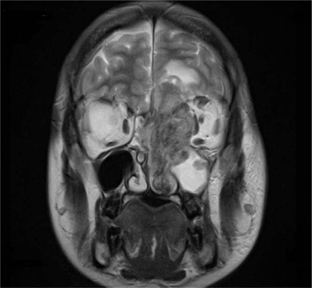
Fig. 1.
MRI, T2-weighted, coronal plane, demonstrates a mass filling the left nasal cavity, sphenoid sinus, and ethmoid air cells. The tumor remodels the left maxillary sinus and extends intracranially.
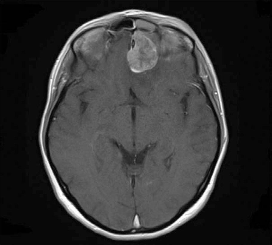
Fig. 2.
Axial contrast-enhanced T1-weighted MRI demonstrates an enhancing mass extending into the anterior cranial fossa.
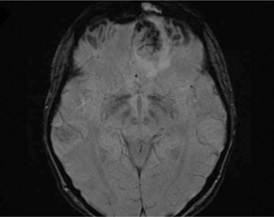
Fig. 3.
SWI MRI, axial plane shows a heterogeneous mass with intratumoral low-signal foci, consistent with hemosiderin deposition.
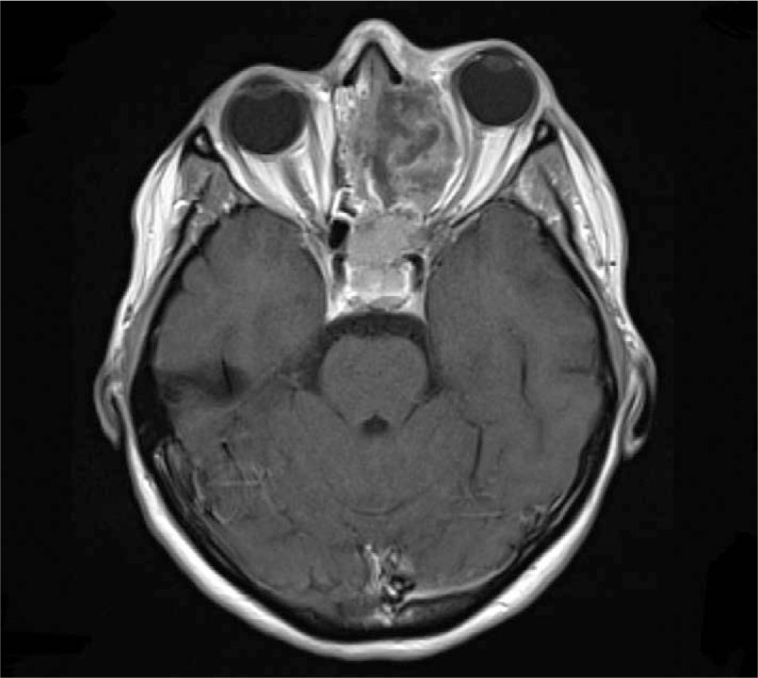
Fig. 4
Contrast-enhanced T1-weighted MRI, axial plane shows a heterogeneous mass in the left orbit. The mass displaces orbital contents, causing proptosis of the left eye.
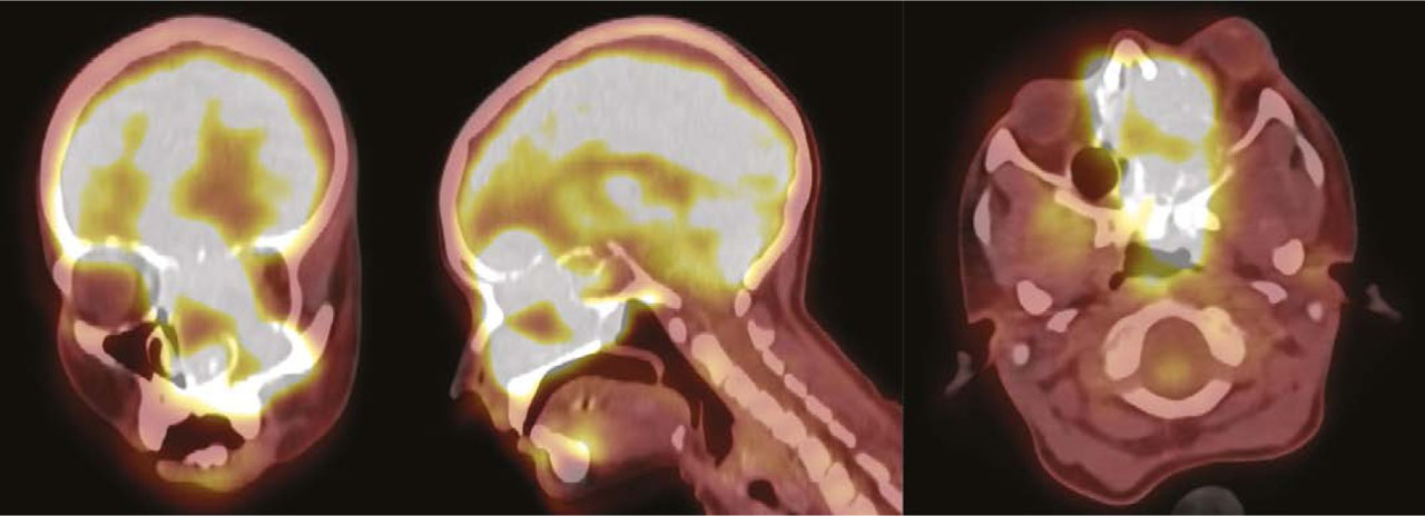
Fig. 5
[18F] FDG PET/CT—shows increased tracer uptake in the primary lesion in the paranasal sinuses. This includes the left maxillary and sphenoid sinuses, extending to nearby bones and the left orbit.
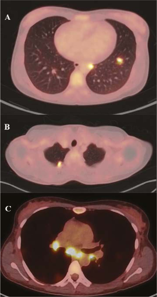
Fig. 6
[18F] FDG PET/CT shows increased radiotracer uptake in the lungs (A), cervical lymph nodes (B), and mediastinal lymph nodes (C), all consistent with metastatic lesions.
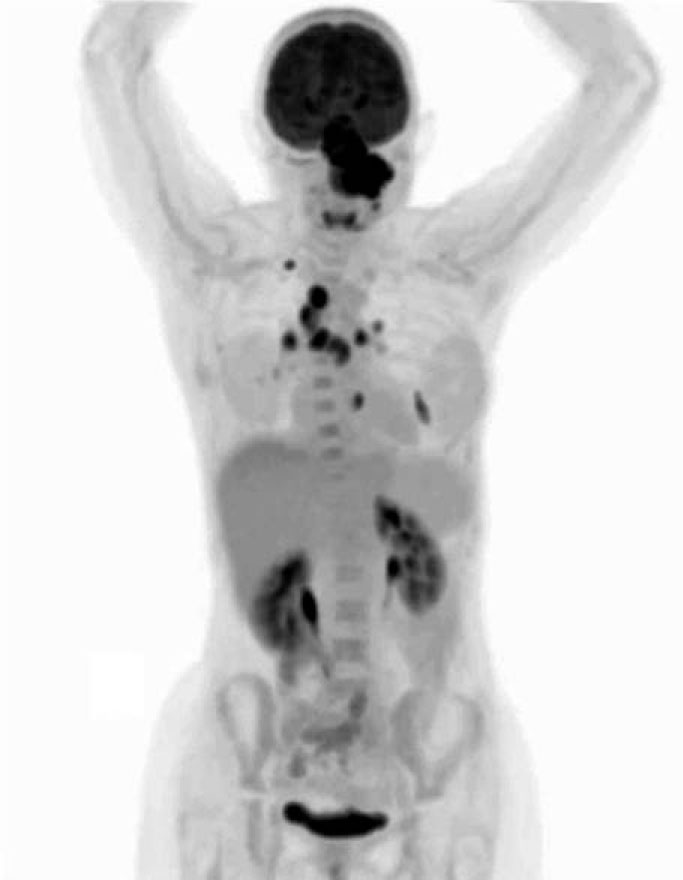
Fig. 7
[18F] FDG PET/CT, Maximum Intensity Projection (MIP) image, shows a hypermetabolic mass extending into the anterior cranial fossa. Metastatic involvement is visible in the lymph nodes and lungs. Physiologic FDG uptake appears in the brain, salivary glands, and urinary tract.