![Multimodal Imaging with MRI and [18F] FDG PET/CT in the Diagnosis and Assessment of Highly Aggressive NUT Carcinoma: A Case Study Cover](https://paradigm-files.s3.eu-central-1.amazonaws.com/BGBL/cover-image.jpg)
Introduction
In the current World Health Organization classification, NUT carcinoma is described as a highly aggressive, poorly differentiated malignancy associated with chromosomal rearrangements involving the NUTM1 gene [1].
NUT carcinoma is a rare malignancy with a median survival of approximately 6.5 months.
The NUTM1 gene is located on the long arm of chromosome 15 at the 15q14 locus. It forms fusion oncoproteins following chromosomal translocations. These fusion proteins inhibit normal cellular differentiation while simultaneously promoting cellular proliferation [2]. This mechanism likely explains the exceptionally rapid progression and poor prognosis of this cancer.
NUT carcinoma is sometimes described as a poorly differentiated squamous cell carcinoma; however, it is not yet officially recognized as a subtype of squamous cell carcinoma [1].
NUT carcinoma predominantly arises in midline organs, including the chest, head, and neck, although it may also occur in other anatomical locations [3].
The former name of the tumor, “midline NUT carcinoma”, stemmed from its tendency to arise within midline structures [4]. Although this term is no longer used, it highlights the tumor’s frequent presentation pattern. Notably, NUT carcinoma remains underrecognized and is often misdiagnosed in clinical practice [1]. The lack of clinically specific symptoms often leads to a diagnosis at an advanced stage, when symptoms related to primary tumor infiltration predominate [3]. This may partly account for the short median survival.
In clinical practice, NUT carcinoma is rarely suspected at the time of the initial presentation. Diagnostic imaging may raise suspicion by revealing a rapidly enlarging mass in a characteristic location. However, a definitive diagnosis is established through immunohistochemical analysis [3].
Case Report
A 32-year-old woman was admitted to the Department of Otorhinolaryngology, Head and Neck Surgery at the Central Clinical Hospital of the Medical University of Warsaw due to a tumor located in the left ethmoid region.
The patient initially presented with nonspecific symptoms, including recurrent epistaxis from the left nasal cavity and visual disturbances affecting the left eye, prompting an outpatient diagnostic evaluation. Head CT revealed a left-sided anterior ethmoid tumor extending through the nasal septum to the right side, just below the olfactory groove. The patient was subsequently referred to the Department of Otorhinolaryngology, where further imaging studies and a biopsy of the nasal cavity mass were performed.
To assess possible dura mater infiltration and central nervous system involvement, an MRI was performed. The examination revealed an extensive soft-tissue mass partially filling the left maxillary and sphenoid sinuses. The lesion almost completely occupied the ethmoid air cells and the nasal cavity and extended intracranially. The mass measured approximately 40 × 65 × 70 mm.
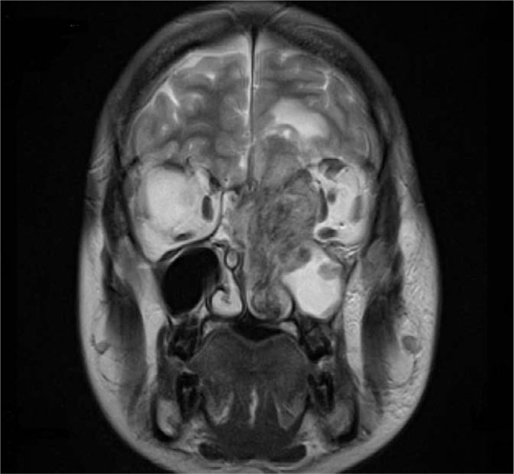
Fig. 1.
MRI, T2-weighted, coronal plane, demonstrates a mass filling the left nasal cavity, sphenoid sinus, and ethmoid air cells. The tumor remodels the left maxillary sinus and extends intracranially.
The tumor extended into the anterior cranial fossa, predominantly on the left side. Minimal extension beyond the falx cerebri into the right side could not be excluded. The lesion was in direct contact with the brain and caused edema of the brain tissue. However, a thin rim of cerebrospinal fluid surrounding most of the lesion suggested compression and deformation of the brain rather than direct parenchymal infiltration at this stage of evaluation.
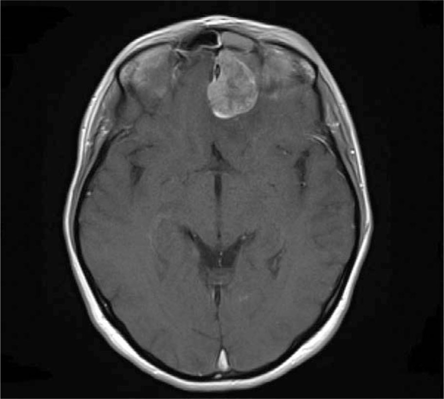
Fig. 2.
Axial contrast-enhanced T1-weighted MRI demonstrates an enhancing mass extending into the anterior cranial fossa.
A small portion of the tumor extended into the inferior orbital fissure and pterygopalatine fossa. Extension into the left orbit resulted in secondary exophthalmos of the left eye.
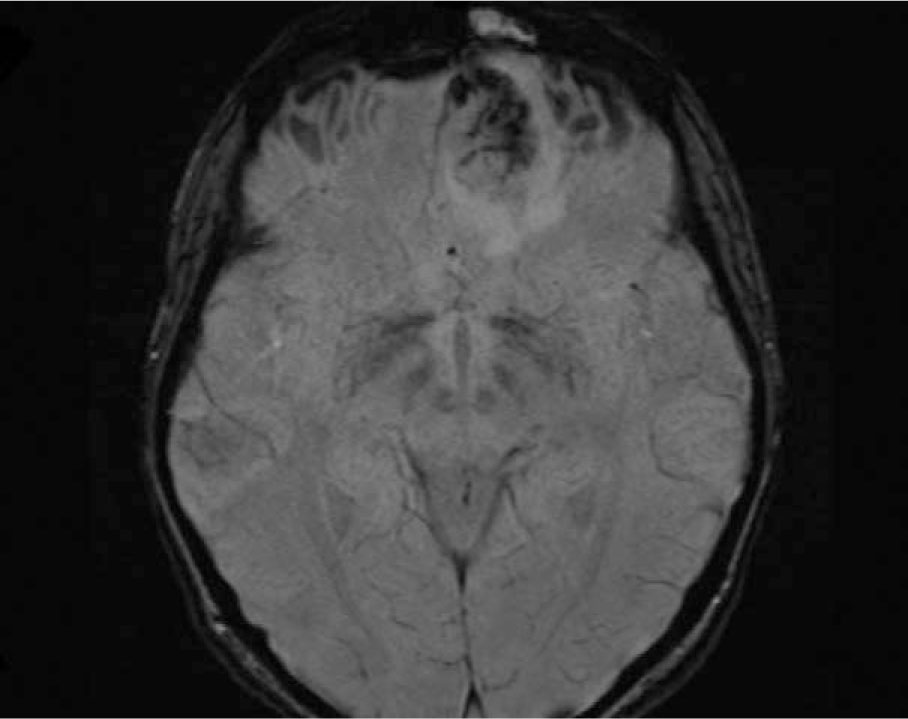
Fig. 3.
SWI MRI, axial plane shows a heterogeneous mass with intratumoral low-signal foci, consistent with hemosiderin deposition.
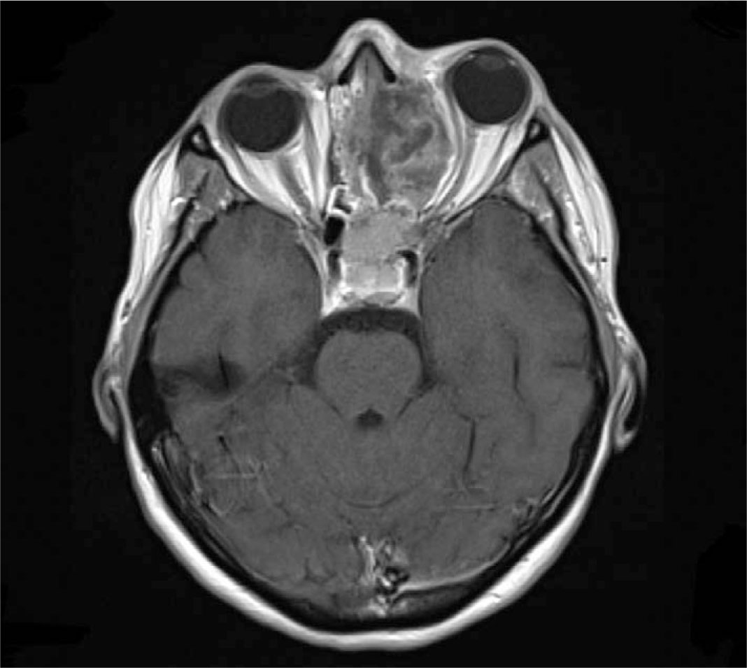
Fig. 4
Contrast-enhanced T1-weighted MRI, axial plane shows a heterogeneous mass in the left orbit. The mass displaces orbital contents, causing proptosis of the left eye.
A biopsy of the tumor was performed. Histopathological analysis revealed a poorly differentiated carcinoma suspicious for NUT carcinoma. [18F] FDG PET/CT demonstrated a metabolically active primary lesion involving the paranasal sinuses, including the left maxillary and sphenoid sinuses. The mass infiltrated the ethmoid, sphenoid, frontal, and maxillary bones.
The tumor also invaded the left orbit and destroyed its medial wall, resulting in secondary proptosis of the left eye. The lesion extended into the anterior cranial fossa and infiltrated the frontal lobe to a depth of approximately 2 cm, with associated edema.
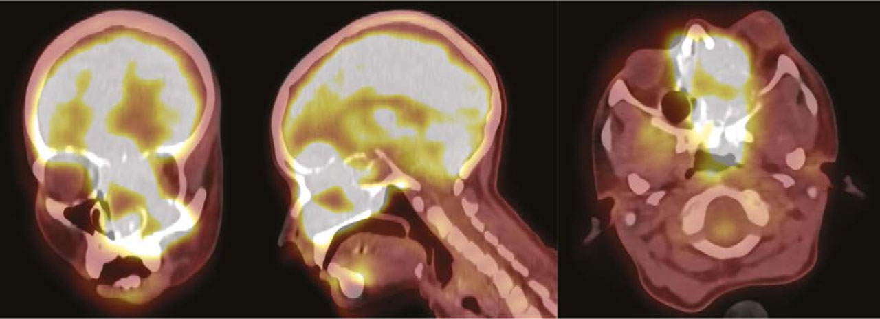
Fig. 5
[18F] FDG PET/CT—shows increased tracer uptake in the primary lesion in the paranasal sinuses. This includes the left maxillary and sphenoid sinuses, extending to nearby bones and the left orbit.
Areas of increased deoxyglucose metabolism indicated metastatic lesions in the lungs as well as in the cervical and mediastinal lymph nodes, consistent with disseminated disease.
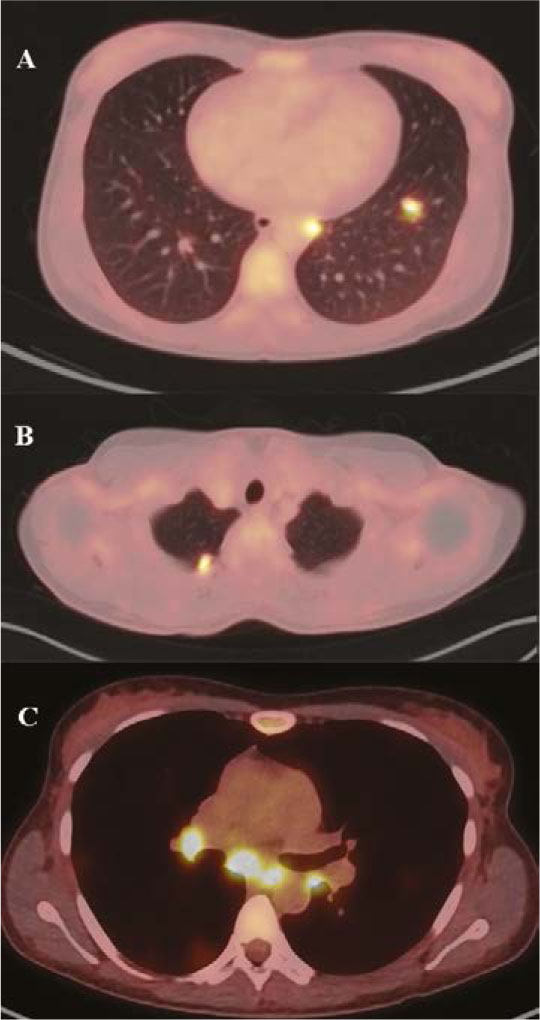
Fig. 6
[18F] FDG PET/CT shows increased radiotracer uptake in the lungs (A), cervical lymph nodes (B), and mediastinal lymph nodes (C), all consistent with metastatic lesions.
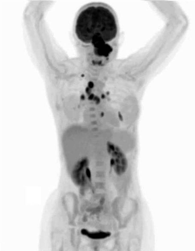
Fig. 7
[18F] FDG PET/CT, Maximum Intensity Projection (MIP) image, shows a hypermetabolic mass extending into the anterior cranial fossa. Metastatic involvement is visible in the lymph nodes and lungs. Physiologic FDG uptake appears in the brain, salivary glands, and urinary tract.
The advanced local extent of the tumor, its spread beyond the paranasal sinuses, and the presence of distant organ metastases rendered the patient ineligible for surgical intervention. Consequently, a referral to the National Institute of Oncology in Warsaw for systemic therapy was made.
A repeat biopsy and immunohistochemical staining confirmed the diagnosis of NUT carcinoma. Histology showed a poorly differentiated non-small cell carcinoma with an immunoprofile matching NUT carcinoma. The immunohistochemical profile supported epithelial differentiation (CKAE1/3 positivity) [5] and squamous differentiation (p40 and p63 positivity) [6]. Markers of neuroendocrine differentiation, including chromogranin and synaptophysin, were negative [7]. The Ki-67 proliferation index was approximately 80%, indicating a highly proliferative neoplasm [8].
Ultimately, the diagnosis was established by immunohistochemical detection of NUT protein overexpression using the NUT-specific monoclonal antibody C52/B1 [9]. Diffuse speckled nuclear staining was observed in the majority of tumor cells, confirming the diagnosis of NUT carcinoma [3].
Systemic therapy was initiated; however, the disease progressed rapidly. Severe epistaxis necessitated transfusion of leukocyte-depleted packed red blood cells. Ongoing disease progression and the lack of a therapeutic response led to the discontinuation of curative treatment. The patient was subsequently referred to hospice care for symptom management, including morphine administration and parenteral nutrition, and died shortly after cessation of oncological treatment.
MRI vs PET/CT
Currently, no studies have directly compared the diagnostic performance of MRI with that of PET/CT in the evaluation of NUT carcinomas, due to the extremely rare occurrence of this cancer.
The most relevant data derive from a multicenter study evaluating imaging modalities for the detection of recurrent sinonasal squamous cell carcinoma. PET/CT demonstrated higher sensitivity than MRI for the recurrence detection (95.2% vs. 72.4%), whereas MRI exhibited higher specificity (97.1%) than CT (90.8%). Both modalities showed comparably high negative predictive values: 97.6% for PET/CT and 97.9% for MRI. Although these findings cannot be directly extrapolated to NUT carcinoma, they illustrate the high diagnostic utility of both methods. The study highlighted the role of PET/CT in identifying metabolically active disease, as demonstrated in the present case, whereas MRI proved particularly valuable for assessing structural involvement. These findings suggest that both modalities should be used complementarily during diagnostic evaluation.
The utility of multimodal imaging is further supported by the relatively low sensitivity of PET/CT for the detection of muscle and perineural involvement, which limits its use as a standalone method for local staging [10].
Literature data further support the combined use of both modalities in assessing lymph node involvement in patients with head and neck cancers, with reported sensitivities of 80.4% for both methods and specificities of 85.7% for MRI and 87.3% for PET/CT. Both modalities showed negative predictive values of approximately 84.5% [11].
Treatment
Currently, no established standard treatment exists for NUT carcinoma. Management is generally individualized and multidisciplinary, comprising radical surgery, radiotherapy, and systemic therapy. Furthermore, enrollment in prospective clinical trials is encouraged whenever feasible [3].
The molecular basis for targeted therapy is the recurrent BRD4::NUTM1 fusion, in which fragments of the BRD4 and NUTM1 genes combine to form the BRD4::NUTM1 fusion gene encoding an oncogenic chimeric protein [2–3]. This fusion protein involves the BRD4 component of the BET family. In this setting, BET inhibitors target the BRD4::NUTM1 fusion protein, leading to reduced expression of oncogenic transcriptional drivers such as TP63, MYC, and SOX2 [3].
One of the newest BET inhibitors, currently being evaluated in two active clinical trials, is ZEN-3694. Available data suggest its efficacy both as a monotherapy and in combination with abemaciclib, a CDK4/6 inhibitor. The FDA (U.S. Food and Drug Administration) has granted a fast-track designation to ZEN-3694 in combination with abemaciclib (Verzenio) for patients with metastatic or unresectable NUT carcinoma who have received at least one prior line of chemotherapy [12]. A more experimental approach involves direct disruption of the BRD4::NUTM1 fusion gene using the CRISPR-Cas9 system. In a recent study published in Molecular Therapy Oncology, CRISPR-Cas9 disrupted the genetic profile of NUT carcinoma cells, effectively reducing their proliferative capacity while activating apoptotic pathways [13].
Overall, targeted therapy for NUT carcinoma remains investigational. BET inhibitor-based combinations, immunotherapy, and genome editing represent the most advanced therapeutic strategies to date; however, sufficient data to establish a standard of care are still lacking.
Discussion
This case exemplifies the rapid progression of NUT carcinoma described in the literature [1, 3–4] and underscores the decisive role of diagnostic imaging in guiding clinical management.
Imaging plays a central role in the evaluation of NUT carcinoma. CT, MRI, and PET/CT are particularly important for determining disease extent and assessing treatment response [14]. Although the tumor lacks pathognomonic radiological features, certain imaging characteristics may raise suspicion for NUT carcinoma. Typical findings include tumor distribution with a predilection for the ethmoid sinus and nasal cavity. Additional suggestive features include heterogeneous enhancement on MRI and orbital involvement. Extension beyond the skull base has also been frequently reported [15].
In the present case, diagnostic imaging was essential for identifying the aggressive, infiltrative growth pattern characteristic of NUT carcinoma. MRI demonstrated an extensive soft-tissue mass infiltrating the nasal cavity, ethmoid bone, and paranasal sinuses. Notably, evidence of extension into the anterior cranial fossa underscored the critical role of MRI in surgical planning and treatment decision-making for patients with NUT carcinoma [16].
[18F] FDG PET/CT is recommended for patients who may be candidates for surgical treatment [3] and is particularly valuable for assessing disease extent through the visualization of metastatic foci [16]. In the present case, PET/CT enabled the detection of metastatic lesions and the assessment of metabolic activity at all disease sites, including lesions not identified on prior imaging studies. The visualization of extensive metabolically active tissue extending from the paranasal sinuses confirmed the tumor’s high biological activity. The presence of metastatic lesions indicated disseminated advanced disease at the time of diagnosis, which ultimately precluded surgical treatment and led to referral for systemic therapy.
This case underscores the central role of diagnostic imaging in raising suspicion for NUT carcinoma and in precisely delineating tumor extent and its relationship to adjacent critical anatomical structures.