
Introduction
The management of impacted canines is among the most clinically challenging tooth displacements in orthodontic practice, with a prevalence of approximately 1–3% in the general population.1 Undetected or inadequately treated impaction carries risk of root resorption of adjacent incisors, follicular cyst formation, and prolonged arch instability, each of which may compromise long-term functional and aesthetic outcomes.2
Over the past decades, a substantial body of literature has addressed key aspects of impacted canine management, including interceptive treatment, surgical exposure techniques, orthodontic traction mechanics and prognostic factors influencing treatment outcomes.3,4,5,6,7 More recently, advances in three-dimensional imaging and digital planning have improved diagnostic accuracy and enhanced the ability to assess the spatial relationship between impacted canines and adjacent structures.8 Concurrently, the integration of temporary skeletal anchorage devices into traction biomechanics allows clinicians to effectively manage complex tooth movements.9 However, the extent to which these developments translate into improved clinical outcome remains uncertain.
Therefore, an updated review is essential to synthesise current evidence to support evidence-based clinical decision-making in the diagnosis and management of impacted canines.
Literature review
This literature review aimed to synthesise evidence published in the past decade (2014–2024) on the diagnosis and management of impacted canines. A comprehensive electronic search was conducted across four databases: PubMed, Scopus, ClinicalTrials.gov, and the Cochrane Library using the following search terms: ((impact* OR unerupt* OR ectopic*) AND (canine* OR cuspid*) AND (surgery OR surgical* OR expos*)) AND ((functional OR aesthetic OR health OR obliteration OR outcome) OR (piggyback OR nickel titanium OR NiTi OR magnet* OR removable appliance)). The search strategy and keywords used across difference databases is presented in Table I. Only fully published articles were included in this literature review. The review process was conducted in accordance with PRISMA principles, with predefined inclusion and exclusion criteria aligned to the study objectives.
Table I
Search Strategy and Keywords Used Across Different Databases
| Search engine | Keywords |
|---|---|
| PubMed | ((impact* OR unerupt* OR ectopic*) AND (canine* OR cuspid*) AND (surgery OR surgical* OR expos*)) AND ((functional OR aesthetic OR health OR obliteration OR outcome) OR (piggyback OR nickel titanium OR NiTi OR magnet* OR removable appliance)) |
| Scopus | ((impact* OR unerupt* OR ectopic*) AND (canine* OR cuspid*) AND (surgery OR surgical* OR expos*)) AND ((functional OR aesthetic OR health OR obliteration OR outcome) OR (piggyback OR nickel titanium OR NiTi OR magnet* OR removable appliance)) |
| ClinicalTrials.gov | ((impact* OR unerupt* OR ectopic*) AND (canine* OR cuspid*) AND (surgery OR surgical* OR expos*)) AND ((functional OR aesthetic OR health OR obliteration OR outcome) OR (piggyback OR nickel titanium OR NiTi OR magnet* OR removable appliance)) |
| Cochrane | ((impact* OR unerupt* OR ectopic*) AND (canine* OR cuspid*) AND (surgery OR surgical* OR expos*)) AND ((functional OR aesthetic OR health OR obliteration OR outcome) OR (piggyback OR nickel titanium OR NiTi OR magnet* OR removable appliance)) |
Inclusion criteria include fully published, peer-reviewed articles written in English; review articles (systematic reviews, narrative reviews, meta-analyses); studies involving human subjects; studies relevant to the topic of impacted canines; original research studies, including randomised controlled trials, cohort studies, case-control studies, and case reports and case series; and articles with clearly defined methodology and appropriate statistical analysis.
Exclusion criteria include articles not published in English; abstracts, conference proceedings, editorials, letters, and expert opinions; animal studies or in vitro studies; studies not directly relevant to the research topic; and articles with insufficient methodological details or incomplete data.
A total of 605 records were identified, including 604 from databases and one from trial registers. After removal of 94 duplicate records, 511 records remained for title and abstract screening, of which 442 were excluded. Of the 69 reports sought for retrieval, seven could not be obtained. Subsequently, 62 full-text articles were assessed for eligibility; of these, six were excluded due to predefined reasons. Finally, 56 studies were included in the qualitative synthesis. The PRISMA Flow Chart shows the process for article selection is presented in Figure 1.
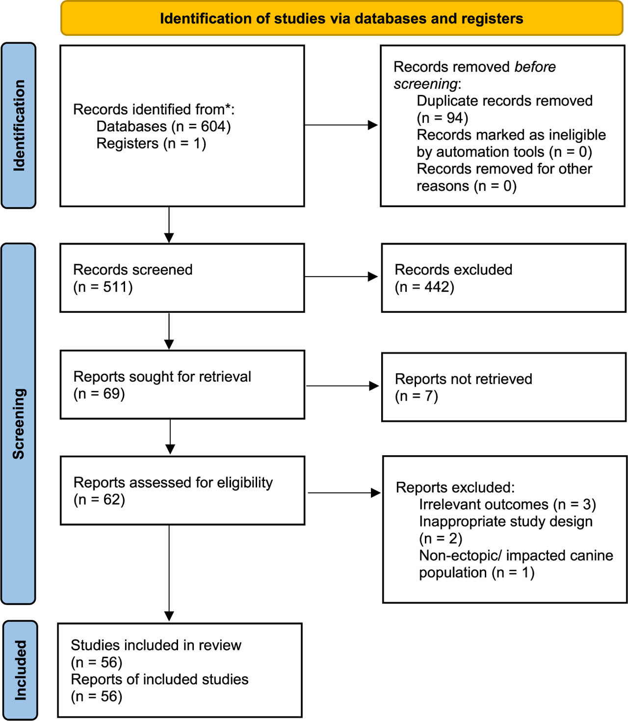
Figure 1.
PRISMA diagram.
Diagnosis and imaging
Various radiographic techniques have been described to aid in the diagnosis and treatment planning of impacted canines, including panoramic radiography and parallax methods. However, these conventional two-dimensional (2D) imaging modalities may be limited in their ability to determine the bucco-palatal position of the impacted tooth and its relationship to adjacent structures.
Cone Beam Computed Tomography (CBCT) provides three-dimensional (3D) imaging that allows precise localisation of impacted canines and their relationship with anatomical structures, including the maxillary sinus, cortical bone, and adjacent teeth.10 It further enables detection of associated pathology, especially canine ankylosis and root resorption of adjacent teeth; these findings may alter management decisions and prevent unnecessary extraction.2,6 From a perspective of treatment planning, CBCT assists in determining the likelihood of spontaneous eruption following interceptive extraction of primary canine and guides the planning for surgical exposure should interceptive treatment fails.8
Despite these diagnostic advantages, routine use of CBCT is not recommended because of the higher radiation dose compared to conventional radiographs.11 Current recommendations, consistent with the ALARA principle, reserve CBCT for selected cases where conventional radiographs fail to provide sufficient information.11
The KPG index, introduced by Kau et al., provides a quantitative method for assessing treatment difficulty of impacted canines. The impacted canines are classified as easy (0–9), moderate (10–14), difficult (15–19), or extremely difficult (≥20).12 Validation studies have demonstrated moderate correlation between predicted and actual treatment outcomes in easy and moderately difficult cases, weak correlation in difficult cases, and strong correlation in extremely difficult cases.13
Emerging developments include three-dimensional reconstruction from panoramic radiographs and the application of deep learning for detection of root resorption of impacted canine.14,15 However, these technologies remain at an early investigational stage, with prospective clinical studies required to establish their clinical impact.
Early interceptive treatment
Extraction of primary canine
Interceptive extraction of the primary canine is a commonly used early intervention aimed at facilitating the eruption of palatally displaced permanent canines.4,16,17 The rationale for removal of deciduous predecessor is it modifies eruptive guidance of permanent canine, eliminate physical obstruction, and potentially create space within the dental arch.4,17
Several systematic reviews have reported that extraction of primary canines in minimally crowded arches increases the likelihood of spontaneous eruption of the palatally ectopic permanent canines in 50–69% of cases.16,17 However, a Cochrane review by Benson et al. concluded that the certainty of evidence remains low due to small sample sizes, high risks of bias, and potential reporting errors across existing trials.1 Furthermore, there is also no robust evidence demonstrating interceptive extraction significantly reduces the need for subsequent surgical intervention.1
Despite this, there is broad consensus that the initial severity of displacement is the most critical prognostic indicator for interceptive extractions. Favourable prognostic indicators where spontaneous eruption after interceptive extraction is more likely include impacted canines located in a favourable location at sector 1-2,1,18 exhibits a mild mesial inclination of less than 30°,16,18 and at pre-pubertal age.18 In contrast, severely displaced canines, such as those located in sector 4-5 with alpha angles exceeding 30°, are less likely to respond to interceptive treatment.1,6,18
An observation period of 12 months is proposed by multiple studies after interceptive extractions.1,4 The probability of future spontaneous is low if canine is has not erupted beyond this time point.4
Adjunctive orthodontic interventions
These include rapid maxillary expansion (RME), transpalatal arches (TPA), or cervical-pull headgear that have been proposed to facilitate the eruption of displaced canines. Elangovan et al. reported that while some clinical trials showed increased rates of spontaneous eruption when these appliances are used either alone or in combination, many studies have small sample sizes, short follow-up periods and inconsistent randomisation. 16 Larger well-designed randomised controlled trials are required to establish the true effectiveness of these adjunctive interventions. Furthermore, interceptive treatment may prolong overall treatment duration and requires careful monitoring to determine whether additional intervention becomes necessary.
Surgical management of impacted canines
Open vs closed surgical exposure
Two main surgical techniques are routinely used to uncover palatally displaced canines. The closed technique involves raising a full mucoperiosteal flap to expose the crown, bonding an orthodontic attachment, and subsequently repositioning and suturing the flap so the tooth remains covered during initial traction. In open technique, a window of bone and soft tissue is removed from around the tooth and a dressing is placed to cover the exposed area. The tooth is either left to erupt or moved using orthodontic attachment.
Current evidence suggests that both techniques are highly effective in achieving canine eruption, with no significant differences in success rates.19,20,21 A Cochrane review by Parkin et al. concluded, with low certainty, that there are no differences in outcomes in open versus closed surgical technique in terms of success of treatment, complications, aesthetics, periodontal outcomes, and patient-centred outcomes.3 The finding on periodontal outcomes is consistent with a subsequent systematic review specific on periodontal outcomes.7
Some findings by Cochrane review by Parkin et al. was inconsistent with a systematic review by Cassina et al. conducted at similar period. Cassina et al. reported that open surgical exposure is associated with shorter initial alignment duration compared to closed technique, possibly due to lack of a repositioned flap over the exposed canine.19 Cassina et al. also concluded lower odds of ankylosis in open exposure technique;19 however, this conclusion was derived from a single non-randomised prospective study,22 limiting the strength of this finding to support a definitive advantage of one surgical approach.
Subsequent to the Cochrane review, Björksved et al. conducted a randomised-controlled trials and found no significant differences in surgery time, post-operative complications, and cost.23,24 Nevertheless, Björksved et al. reported patients in closed exposure group experienced greater pain and discomfort during active orthodontic traction phase.24
Overall, the available evidence does not clearly favour either technique. Given the low certainty of existing evidence and heterogeneity across studies, the choice between open and closed exposure therefore remains guided by clinician preference, surgical experience, and case-specific anatomical considerations. 3 A summary of evidence comparing open and closed surgical exposure across eruption efficacy, periodontal outcomes, complications, and patient-centred outcomes are presented in Table II.
TABLE II
Summary of evidence comparing open and closed surgical exposure for impacted maxillary canines
| Outcome | Summary findings |
|---|---|
| Eruption success | Both techniques achieve comparable eruption rates. No evidence supports superiority of either approach |
| Alignment duration | Conflicting. One systematic review favours open exposure. Discrepancy between reviews attributable to differences in study inclusion. Evidence remains inconclusive |
| Periodontal parameters | No clinically significant difference in probing depth, attachment level, or keratinised tissue width between the techniques |
| Gingival recession | Closed technique theoretically favoured through flap repositioning preserving keratinised gingiva, but this is not confirmed by clinical outcome data |
| Pain and discomfort | No difference in systematic reviews. One randomised-controlled trial reported greater pain and discomfort during active orthodontic traction in closed exposure |
| Aesthetic | No significant difference overall. One retrospective study reported darker crown colour following closed exposure |
| Surgery time and cost | No significant difference in operative time or cost between techniques |
Modified surgical techniques and emerging approaches
Alternative surgical approaches have also been described. The tunnel technique, originally introduced by Crescini et al. is a modified closed-eruption procedure for buccal canines impacted high in the midalveolar region.25 The procedure involves guiding the impacted tooth downward through the socket of the extracted deciduous canine. The primary goals of this technique are to preserve the buccal bone plate and prevent poor aesthetic outcomes, such as an elongated clinical crown or reduced labial bone support. Evidence supporting this method remains limited, case reports and clinical trials are scarce.
More recently, CO2 and diode laser technologies has been integrated into the management of palatally impacted canines have as a minimally invasive alternative to conventional approach. In addition to facilitating soft tissue removal, laser technology has been proposed to exert photobiomodulation effect, where specific wavelengths activate subcellular photoreceptors and stimulates tissue regeneration and healing.
In a randomised controlled trial, Impellizzeri et al. reported a greater tooth movement in patients treated with CO2 or diode laser compared to conventional surgery, suggesting that the use of laser may enhance or stimulate the spontaneous eruption of impacted canine.26 From a surgical perspective, the photothermal effect simultaneously coagulates small vessels during incision, resulting in minimal intraoperative bleeding and improved surgical visibility. Patients treated with CO2 and diode lasers experience have also been reported to experience reduced post-operative pain and swelling, along with faster healing and functional recovery.26 However, the study is based on studies with small sample size, restricting the strength of conclusions supporting their routine clinical use.
Orthodontic alignment of impacted canines
Mechanics and appliances
Various orthodontic mechanics have been described for the traction and alignment of impacted canines, with selection guided by canine's initial position, space availability and anchorage requirements. Orthodontic appliances and auxiliaries employed include includes nickel-titanium (NiTi) closed-coil springs for continuous light force for controlled movement, power chains and elastic threads for gradual traction forces, gold chains, Kilroy springs, Ballista springs, miniscrews, strong rigid buccal arms, and magnets.3,25,27,28,29
Kilroy springs deliver constant force via a vertical loop and have been shown through finite element analyses to produce higher von Mises stress and greater displacement compared to Ballista springs and NiTi closed-coil springs, although they also tend to exert greater stress on adjacent teeth.27,28 Piggyback wires are considered a suitable alternative for canines displaced 2–4 mm from the occlusal plane, as they produce lower reactionary forces on adjacent teeth.30
Biomechanics of traction
The magnitude and direction of force application are critical determinants for successful orthodontic alignment of impacted canines.31 Excessive force risks root resorption, pulpal necrosis, or damage to the periodontal ligament, particularly in buccally impacted canines.7,32 Buccally impacted canines present greater challenges in managing adverse periodontal effects and require careful consideration of surgical technique interventions, proper marginal gingival positioning with adequate attachment, inflammation control, and the application of controlled orthodontic forces.32 When positioned close to the incisor roots, buccally directed force risks root damage and may impede tooth movement; therefore, initial traction should be directed occlusally and distally before redirecting the tooth buccally into its final position.25 When ankylosis is suspected, traction force should be reduced, a periapical radiograph should be obtained to assess for osteoid tissue, and referral to a periodontist if indicated.33
Temporary anchorage devices (TADs)
The use of TADs, or miniscrews, in the management of impacted canines is growing in popularity. A systematic review and meta-analysis by Alharbi et al. reported that there is limited data on whether TADs are superior in terms of overall treatment duration, adverse effect, and number of visits; however, there is moderate-quality evidence that TADs provide a superior anchorage preservation compared to conventional methods such as TPA and headgear.34
Beyond anchorage control, miniscrews have demonstrated efficacy in minimising lateral incisor root resorption during canine disimpaction. In a clinical controlled trial, Heravi et al. reported significantly less root resorption with TAD-assisted approach compared to conventional techniques.35 The protocol involves inserting two bracket-head miniscrews, measuring 1.4 mm in diameter and 8 mm in length, to the mesial and distal to the maxillary second premolar, right angle to the palate and 5 mm from the embrasure.
Heravi et al. argued that the conventional technique, where initial arch alignment was done before active canine traction, carries several disadvantages. 35 First, bracket placement on the lateral incisors shifts their root apices closer to the resorptive follicle of the impacted canine, particularly s torque is expressed through rectangular wires. Some clinicians mitigate this by remaining in round wires during the aligning phase; however, this represents a compromise rather than a solution. The use of rectangular archwires in conventional technique, despite able to neutralise reaction forces, carries risk of anchorage loss and extended treatment duration.35 Therefore, it is crucial to move the impacted canine away from the root of the adjacent tooth prior to bracket placement.
Alternative treatments strategies
Autotransplantation
Autotransplantation is a viable treatment option in situations where orthodontic traction is not a practical solution, for example, in high alpha angle (>30°) with high sector, a high vertical position, and dilacerated roots.36,37 A study with mean follow-up of 21 years reported a survival rate of 67.7% for autotransplanted canines, with mean survival time of 15.8 years.37 Saccomanno et al., in their systematic review, identified younger age, female sex, proper root canal treatment, and effective post-operative care as factors associated with improved outcomes. Insufficient bone support, gingival recession, interdental soft tissue defects, and deep periodontal pocketing were reported as cause of failure.36,37 Risks include damage to the periodontal ligaments, cementum, and darkening of tooth requiring full-coverage restoration.36,37,38 As individual outcomes remain difficult to predict, patients should be counselled for potential failure and consequences prior to proceeding with autotransplantation treatment.36
Impacted mandibular canines
Mandibular canine impaction is a rare clinical entity, with a reported prevalence ranging from 0.008% to 1.29%, while transmigration occurs in approximately 0.12% to 0.98% of the population.39 Clinical management typically involves either surgical extraction for deeply impacted cases or surgical exposure for those in a favourable position for guided eruption.39 Unlike the maxillary arch, where interceptive expansion is well-documented, there is a distinct lack of evidence and clinical consensus regarding interceptive protocols for mandibular canines, which are often severe by the time of detection.39,40
In a single-centre cross-sectional study, Bolooki et al. identified that canine angulation, the horizontal position of the root apex, the vertical height of the crown, and the degree of overlap serve as the primary predictors for surgical decision-making.40 Specifically, surgical exposure is favoured for angulations of 0°– 15°, crown heights between the CEJ and mid-root, and minimal horizontal overlap. In contrast, surgical extraction is preferred for angulations >45°, positions beneath the root apex, or transmigration across the dental midline.40 Despite identifying these predictors, the study did not evaluate long-term success rates, and its findings highlight the need for multi-centre prospective research to standardise mandibular management protocols.
Complications and biological considerations
Root resorption
Root resorption of adjacent teeth is a major concern, particularly in cases of severe canine impaction.41,42,43 Palatally impacted canines positioned in closer proximity to lateral incisors (greater mesial sector) or those with a horizontal position (higher alpha angle) are associated with increased pocket depth, gingival recession and reduced width of keratinized tissue following interceptive treatment, surgical exposure, and orthodontic traction.6
Delayed diagnosis and treatment lead to prolonged contact and sustained pressure on the lateral incisor roots, thereby risks root resorption and failed orthodontic alignment.42,43 Early detection through CBCT enables accurate assessment of impaction severity and resorption risk,42 while judicious selection of orthodontic mechanics can further mitigate force transmission to adjacent dentition.43
Periodontal outcome
Excessive force can lead to loss of periodontal attachment;44 whereas light, continuous force induces relatively long-lasting levels of cytokines necessary for continuous periodontal remodelling.45 Although a light force of 20–30 g is generally considered sufficient to mechanically erupt an impacted canine, clinical practice frequently involves forces that exceed this threshold, which may result in greater CEJ-to-alveolar crest distances on the mesial side of the impacted tooth and interproximal bone loss.44
Closed surgical exposure has been favoured on theoretical grounds due to better preservation of keratinized gingiva, reduced risk of gingival recession, and a lower likelihood of plaque accumulation and inflammation. However, both Cochrane reviews and systematic reviews have concluded that available evidence is insufficient to demonstrate difference in periodontal parameters between open and closed techniques,3,7 a finding corroborated by multiple retrospective studies examining long-term periodontal outcome.31,46 One retrospective study proposed piezoelectric surgery as means to expose canine without damage to the periodontium; however, outcomes were compared against the contralateral spontaneously erupted canines, rather than conventional surgical bur.47 Taken together, current evidence does not support superiority of either exposure techniques on periodontal ground alone.
Grisar et al., in their systematic review, concluded that the mesial sector and vertical position of impacted canine can influence periodontal outcomes.6 Buccally impacted canines are suggested to present greater risk of adverse periodontal outcomes,6,32 yet multiple studies report no clinical significant difference in periodontal parameters between palatally and buccally impacted canines following closed management,31,44 and neither open nor closed surgical exposure has been shown to be superior for the management of buccally impacted canines.32
Pulp vitality
Pulpal health of orthodontically-treated impacted canines remains an under-reported outcome,46 despite significant tooth movement involved in their traction. The extensive movement subjects the pulp to prolonged mechanical stress, risking neurovascular compromise and pulp necrosis. While some evidence suggests altered pulp testing scores post-treatment,48 there is a paucity of data linking specific mechanical strategies to pulpal vitality.
For example, the piggyback technique warrants scrutiny in this regard. A 0.014” NiTi wire engaged during initial stage is associated with greatest tooth displacement and least predictable tooth displacement. A review of available literature identified three studies reporting the technique in sufficient detail, none of which documented pulpal outcome.49,50,51 Singh et al. started the piggyback mechanic following crossbite correction;51 Jowett et al. commenced piggyback treatment after the archwire progression from 0.018” NiTi to 0.018” or 0.020” AJW alongside closed exposure;50 Singh et al. described piggyback traction initiation following confirmation of adequate mesiodistal space, though this report involved a permanent central incisor rather than a canine.52 None of these studies specified the timing of initial piggyback archwire placement in weeks, nor did report any pulpal outcomes.
Long term outcomes and stability
Post-treatment aesthetics and functional stability are important determinants of treatment success. Aesthetic concerns following impacted canine treatment include gingival margin discrepancies, which can lead to asymmetry in the smile, and gingiva colour or texture differences between the exposed canine and adjacent teeth. Evidence regarding aesthetic outcome for closed versus open exposure remains divided: one study reported darker colour following closed exposure,46 while another found no significant difference between surgical techniques.21
Functional stability depends on proper occlusion to avoid secondary malocclusion or relapse. Long-term follow-ups are essential to assess the stability of the aligned canine and monitor for any periodontal or orthodontic relapse.46 Retention strategies, whether fixed or removable retainers, is indicated particularly where space closure has been undertaken. Patient-reported outcomes, including oral health-related quality of life, should be incorporated as outcome measures alongside clinical indices to provide a complete picture of treatment success.24,53
Clinical Implication
Based on the available evidence, the following clinical decision algorithm is proposed to guide diagnosis and management of impacted maxillary canines (Figure 2).
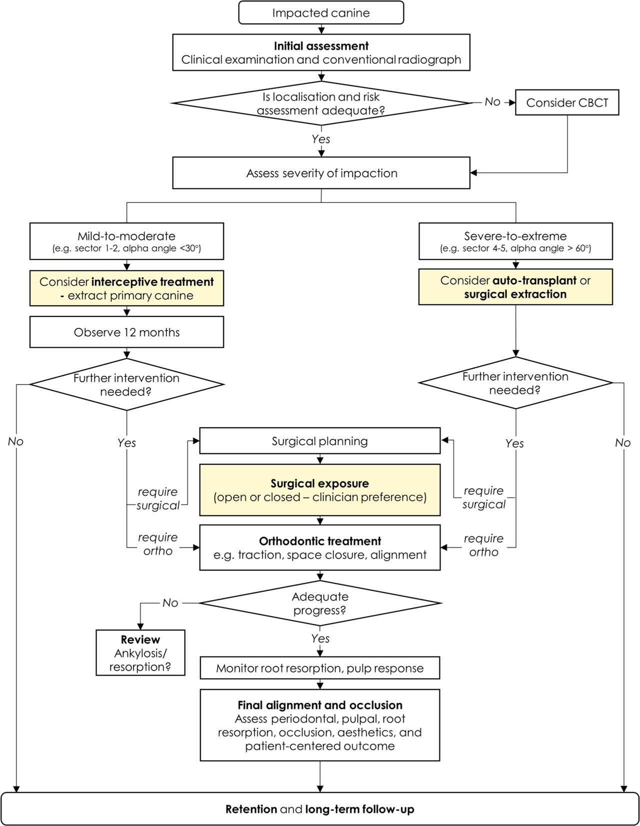
Figure 2.
Proposed algorithm for diagnosis and management of impacted maxillary canines.
Conclusion
The management of impacted canines remains a complex and multidisciplinary challenge requiring accurate diagnosis, appropriate case selection, and coordinated orthodontic–surgical intervention. CBCT has improved diagnostic accuracy, particularly in detecting root resorption and assessing spatial relationships; but its use should remain selective in accordance with radiation safety principles.
Interceptive extraction of the primary canine show moderate success in carefully selected cases; however, the overall certainly of evidence is low, and severity of displacement remains the most reliable prognostic factor. Similarly, both open and closed surgical exposure are effective in facilitating eruption, but current evidence does not demonstrate clear superiority of either approach.
Orthodontic traction mechanics and anchorage strategies, including TADs, offer biomechanical advantage, although their impact on treatment duration remains insufficiently established. Root resorption and periodontal health remain key determinants of long-term success, while pulpal outcomes remain under-reported.
Current evidence is limited by methodological heterogeneity and small sample sizes. Future research should prioritise high-quality trials, standardised outcomes, and long-term follow-up to guide evidence-based management.