
Introduction
Over the last few decades, many interceptive orthopaedic treatment approaches have been suggested to correct skeletal Class III malocclusion, but their efficacy and efficiency vary. This is due to the uncertainty of mandibular growth timing, direction and amount.1 However, the prognosis is improved if treatment is carried out during the prepubertal growth phase, because of maxillary sutures' patency.2,3 Such interceptive treatment aims to correct the skeletal discrepancy, or improve it enough to allow for orthodontic camouflage in the future and avoid orthognathic surgery.2,3
A facemask (FM) on a tooth-supported framework has been traditionally used for the interceptive treatment of skeletal Class III malocclusion. Due to its tooth-borne nature, forces transmitted through the appliance produce dental and skeletal effects. Unfavourable dental adverse effects are crowding and proclination of maxillary anterior teeth, retroclination of mandibular anterior teeth, maxillary molar mesialization, and posterior rotation of the mandible increasing the lower anterior facial height.4,5
To overcome the side effects seen in FM therapy, De Clerck et al.6 introduced skeletal anchorage, using maxillary and mandibular miniplates, to maximize the skeletal effects and minimize dentoalveolar compensation. This approach also allowed for full-time maxillary protraction via Class III elastics.6 This treatment approach requires the eruption of the permanent mandibular canines before initiation. To facilitate an earlier start to maxillary protraction, alternative methods such as the hybrid-hyrax mentoplate have been proposed.7,8 Although the systematic review by Feng et al.9 showed that Bone-Anchored Maxillary Protraction (BAMP) resulted in greater maxillary advancement with fewer dental side effects than tooth-borne appliances, a more recent systematic review by Cornelis et al.10 showed conflicting results. The authors reported that BAMP and conventional FM therapy appear to provide a comparable maxillary protraction effect.10 However, BAMP showed better vertical control compared to conventional FM therapy.10 Notably, the randomized controlled trial (RCT) with a 3-year follow-up by Mandall et al.11 showed promising results, demonstrating a 27% reduction in the perceived need for orthognathic surgery at the follow-up observation in the BAMP group compared to the control group. Still, the insertion of miniplate or mentoplate requires a relatively invasive surgical procedure that involves raising a flap, generally performed by an oral surgeon or periodontist.7,12
In recent years, miniscrew-assisted maxillary protraction (MAMP) has emerged as a less invasive alternative, using miniscrews and intraoral appliances to apply Class III mechanics.12,13,14,15 While initial studies have demonstrated the effectiveness of MAMP in achieving favourable skeletal changes, there is limited evidence directly comparing its outcomes to those of bone-anchored Class III elastics (BAC3E). Therefore, the present study aimed to assess the dental, skeletal, and soft tissue effects of MAMP combined with computer-aided design/manufacturing (CAD/CAM) appliances, and to compare these outcomes with both untreated Class III controls and the well-established BAC3E protocol. Additionally, we sought to evaluate the complications associated with the M AMP protocol.
Materials and methods
This prospective pilot case-control study was approved by the University of Melbourne Human Research Ethics Committee (Reference Number: 2023-24688-37780-4). Consecutive patients were recruited from the University of Melbourne Dental Clinic (MDC), Royal Dental Hospital Melbourne (RDHM), and private orthodontic practices in Victoria, Australia. The eligibility criteria were as follows: (1) patients in their pre-peak growth phase according to the cervical vertebral maturation method (CS1-CS3); (2) Class III skeletal pattern with horizontal maxillary deficiency (SNA<80°); (3) anterior crossbite or incisor end-to-end relationship; (4) erupted mandibular permanent first and second premolars, or mandibular permanent canines and first premolars; (5) absence of craniofacial or cleft lip/palate deformities. All participants and their guardians received comprehensive information regarding the study's content, associated risks, and potential benefits verbally and via a Participant Information Sheet. Written informed consent was obtained from all participants and their guardians.
The group treated with MAMP comprised 11 consecutive patients. A positive control group consisting of 11 individuals treated with BAC3E was retrieved from Cevidanes et al.16 upon contacting the corresponding author. Additionally, a retrospective negative control group of 11 untreated subjects with skeletal Class III malocclusion was obtained from the Department of Orthodontics at the University of Florence, Italy. Those two retrospective control groups were matched to the treated group in terms of age, gender, and vertical skeletal pattern. Vertical growth patterns determine the direction and extent of skeletal changes during treatment. For example, dolichofacial patients may exhibit downward and backward mandibular rotation, which can compromise treatment outcomes. This is less likely to occur in brachyfacial patients. Matching vertical patterns ensures a fair comparison of growth modifications achieved by different treatment modalities.
In the MAMP protocol, the intervention involved placing two miniscrews (2.0x 9 mm PSM Medical Solutions, Tuttlingen, Germany) in the anterior maxilla, 2-3 mm lateral to the median palatal suture, distal to the third palatal rugae, and perpendicular to the palatal bone. In the mandible, two miniscrews (2.0x 7 mm PSM Medical Solutions, Tuttlingen, Germany) were placed in the interradicular areas, one on each side between the permanent first and second premolar or between the permanent canine and first premolar depending on the root divergence and the teeth erupted. Mandibular miniscrews were placed 1-2 mm above the mucogingival junction with an insertion angle of approximately 60° to the occlusal plane. The miniscrews were placed under local anaesthesia using a contra-angle implant driver with a maximum insertion torque of 25 Ncm. Following this, intra-oral scans were taken (TRIOS; 3Shape, Copenhagen, Denmark) and sent to a dental technician who designed the maxillary and mandibular frameworks based on the prescription described below, with computer-aided design (Figure 1).
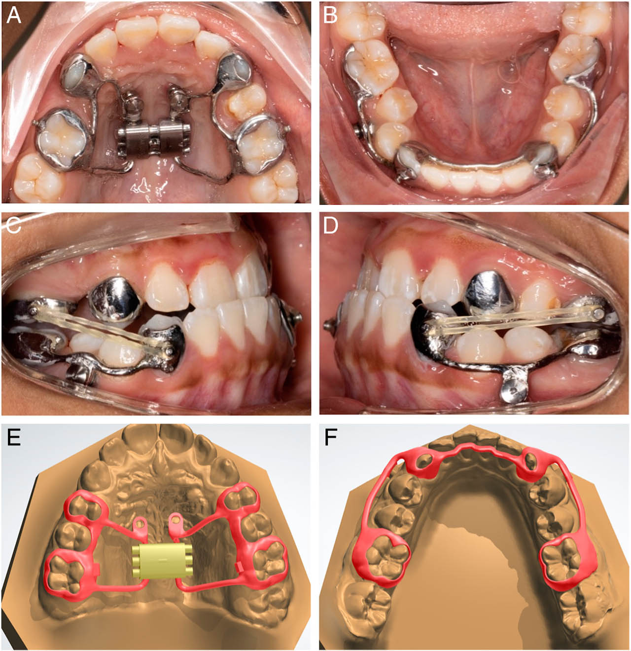
Figure 1.
(A) and (B), Intra-oral maxillary and mandibular frameworks. An alternative maxillary framework design is presented in this patient, modified for the eruption status and hypomineralization of the premolar; (C) and (D), Class III elastics between traction buttons in the same patient; (E) and (F), Typical CAD-CAM design of the maxillary and mandibular frameworks, in another patient.
The maxillary framework joined metal bands around the first premolar and permanent first molar bilaterally. Two housings for the insertion of fixation screws were incorporated into the framework. In the mandibular arch, the framework comprised three separate parts: two buccal parts and one lingual bar, mimicking the design proposed by Gera et al.12 The buccal parts covered the buccal surfaces of the permanent canines and first molars, bilaterally. Similar to the maxillary framework, the buccal parts featured two housings for the insertion of fixation screws. The final metal framework design was produced through a selective laser melting process (computer-aided manufacturing) using the material Remanium Star (Dentaurum, Ispringen, Germany). The mandibular framework was initially printed in one piece and subsequently sectioned into three separate parts following the printing process. Traction buttons were then welded to the buccal surfaces of metal bands on the maxillary permanent first molars, facilitating the attachment of Class III elastics to the mandibular framework. A Hyrax screw (10 mm - Forastadent, Pforzheim, Germany) was incorporated only in patients with transverse maxillary deficiency. Traction buttons were also welded to the anterior buccal surfaces of the mandibular framework for attachment of Class III elastics. The frameworks were then electro-polished, and the bonding surfaces were sandblasted.
The dental surfaces were acid-etched with 37% phosphoric acid gel for 30 seconds. After rinsing and drying, Rely X Unicem (3M ESPE, St. Paul, Minn, USA) was used to cement the appliance and light-cured for 30 seconds. Fixation screws were then placed to secure the frameworks over the miniscrews. When an expansion screw was part of the design, it was activated one turn a day (0.25 mm) until the desired expansion was achieved. Patients were instructed to wear Class III elastics full-time and were replaced at least three times a day. The initial force applied for maxillary protraction was 100 gm on each side and was progressively increased up to 300 gm on each side after eight weeks. Patients were monitored every month after the frameworks had been placed. Maxillary protraction was stopped after clinically achieving at least 2 to 3 mm positive dental overjet. All clinical steps were monitored by a single clinician (DL). All complications were recorded throughout treatment.
To evaluate treatment changes, skeletal and dental parameters were measured using lateral cephalograms taken at two time points: before treatment (T0) and after treatment (T1). All cephalometric landmarks and measurements were adopted from previous studies.16 One single operator (DL) digitally traced all the lateral cephalograms using Quick Ceph Studio (Sarasota, Florida, USA), and subsequent measurements of cephalometric variables were retrieved from the same software. To measure the overjet (OJ) and overbite (OB) changes between pre- and post-treatment, measurements were obtained from intra-oral scans imported into OrthoAnalyzer (3Shape Ortho System, Copenhagen, Denmark). Soft tissue changes of the profile were evaluated on the superimposition of lateral cephalograms. To assess the intra-examiner reliability, all the lateral cephalogram measurements were repeated by the same operator (DL) after a two-week interval.
Statistical analysis was conducted using IBM SPSS Statistics for Windows (Version 29; IBM Corp, Armonk, NewYork, USA), with the significance level set to P<0.05. Continuous variables were expressed as their mean values and standard deviations. The normality of variables was assessed using the Shapiro-Wilk test. As normality was confirmed, an ANOVA with Tukey's post-hoc tests were used to analyse the differences between the treatment and control groups. Within the MAMP treatment group, differences between time points were compared using paired t-tests. Intra-class correlation coefficients (ICC; two-way mixed model, absolute agreement) and Bland-Altman analyses were employed to assess measurement reliability.
Results
The intraclass correlation coefficients (ICC) for the cephalometric variables indicated excellent intra-observer agreement of the repeated measurements with ICCs>0.80 (Supplementary Table I). Bland Altman plots for SNA, ANB, SN-ML, U1-PP, and L1-ML (Appendix) show most differences fall within the limits of agreement, confirming good measurement reliability.
Table I presents the baseline characteristics, the mean duration of treatment/observation for each group, as well as the dentofacial characteristics at T0 for the treated and control groups. In the MAMP group, seven patients required maxillary expansion with a Hyrax screw.
Table I.
Baseline characteristics and pre-treatment cephalometric variables of included subjects
| Variables | MAMPMean (±SD) | MAMPMin | MAMPMax | BAC3EMean (±SD) | BAC3EMin | BAC3EMax | ControlMean (±SD) | ControlMin | ControlMax |
|---|---|---|---|---|---|---|---|---|---|
| Gender (F/M) | 7/4 | 7/4 | 7/4 | ||||||
| Age (years) | 11.7 (±0.7) | 11.8 (±0.9) | 11.8 (±0.7) | ||||||
| Duration of treatment/observation (months) | 9.6 (±1.9) | 12.1 (±1.4) | 12.0 (±0.0) | ||||||
| SNA (°) | 79.0 (±2.7) | 71.2 | 80.6 | 80.7 (±3.4) | 74.5 | 83.8 | 80.0 (±3.5) | 73.1 | 84.6 |
| Co-A (mm) | 76.6 (±5.8) | 66.1 | 86.8 | 86.0 (±3.1) | 81.8 | 91.8 | 78.5 (±4.5) | 70.8 | 86.6 |
| SNB (°) | 81.2 (±3.4) | 71.8 | 83.8 | 80.7 (±3.2) | 75.4 | 86.4 | 81.2 (±3.4) | 75.6 | 88.1 |
| Co-Gn (mm) | 110.8 (±8.0) | 97.3 | 123.3 | 113.2 (±3.4) | 105.4 | 118.2 | 107.1 (±6.5) | 101.1 | 121.9 |
| ANB (°) | −2.2 (±1.5) | −0.5 | −5.4 | 0.1 (±1.7) | −2.6 | 3.1 | −1.2 (±3.0) | −8.0 | 1.6 |
| Wits (mm) | −3.7 (±1.3) | −2.0 | −5.7 | −6.7 (±2.6) | −3.7 | −11.5 | −4.7 (±2.7) | −1.8 | −10.0 |
| Mx-Md differential (mm) | 34.2 (±3.2) | 29.1 | 38.8 | 27.2 (±3.7) | 22.8 | 33.6 | 26.0 (±4.8) | 23.1 | 36.0 |
| SN-ML (°) | 35.4 (±3.9) | 27.2 | 42.4 | 33.6 (±3.7) | 27.3 | 39.9 | 33.8 (±5.3) | 22.4 | 39.7 |
| PP-ML (°) | 25.1 (±3.7) | 17.8 | 30.5 | 24.7 (±4.1) | 19.6 | 32.1 | 25.6 (±5.6) | 14.3 | 32.2 |
| ANS-Menton (mm) | 58.6 (±5.0) | 48.6 | 66.1 | 63.9 (±4.5) | 58.1 | 72.4 | 59.6 (±5.7) | 50.9 | 69.6 |
| U1-PP (°) | 118.3 (±6.8) | 107.3 | 130.2 | 116.5 (±6.9) | 106.1 | 133.5 | 115.0 (±9.2) | 99.3 | 126.9 |
| L1-ML (°) | 85.0 (±6.3) | 73.3 | 93.0 | 88.0 (±8.0) | 73.5 | 101.4 | 82.4 (±7.1) | 72.4 | 93.8 |
The treatment effect within the MAMP group is shown in Table II. Statistically significant differences between T1 and T0 were found for the maxillary skeletal variables (SNA±2.5±1.1°, P<.001; Co-A 2.4±1.1 mm, P<.001) but not for the mandibular skeletal variables (SNB −0.1±0.7°, P=.578; Co-Gn 0.8±1.5 mm, P=.100). Vertically, a significant decrease in SN-ML angle was observed (−0.6±0.8°, P<.05). Dentoalveolar changes showed statistically significant retroclination of the maxillary incisors (U1-PP −4.7±4.6°, P<.01) and proclination of the mandibular incisors (L1-ML 1.6±2.3°, P<.05). The OJ improved significantly by 3.7 mm (P<.001), while the overbite did not show any significant changes (−0.2 mm, P=.802).
Table II.
T0-T1 treatment effect and comparisons for MAMP, BAC3E, and negative controls. (Statistics: ANOVA and post-hoc Tukey tests)
| Variables | MAMP (n=11) | BAC3E (n=11) | Negative control (n=11) | P-value between all groups | |||
|---|---|---|---|---|---|---|---|
| Maxillary skeletal | Mean (±SD) | P-value | Mean (±SD) | P-value vs. MAMP | Mean (±SD) | P-value vs. MAMP | |
| SNA (°) | 2.5 (±1.1) | P<0.001* | 2.7 (±2.3) | P=0.970 | 0 (±0.7) | P<0.001* | P<0.001* |
| Co-A (mm) | 2.4 (±1.1) | P<0.001* | 5.4 (±2.0) | P<0.001* | 1.3 (±0.8) | P=0.194 | P<0.001* |
| Mandibular skeletal | |||||||
| SNB (°) | −0.1 (±0.7) | P=0.578 | −0.4 (±1.5) | P=0.750 | 0.3 (±0.5) | P=0.553 | P=0.220 |
| Co-Gn (mm) | 0.8 (±1.5) | P=0.100 | 1.7 (±0.5) | P=0.248 | 2.6 (±1.6) | P=0.008* | P=0.001* |
| Maxillary/Mandibular | |||||||
| ANB (°) | 2.7 (±1.5) | P<0.001* | 3.1 (±1.7) | P=0.711 | −0.3 (±0.4) | P<0.001* | P<0.001* |
| Wits (mm) | 4.0 (±1.5) | P<0.001* | 4.2 (±3.4) | P=0.970 | −0.6 (±0.8) | P<0.001* | P<0.001* |
| Mx-Md Differential (mm) | −1.5 (±1.7) | P=0.012* | −3.7 (±1.8) | P=0.011* | 1.3 (±1.3) | P<0.001* | P<0.001* |
| Vertical skeletal | |||||||
| SN-ML (°) | −0.6 (±0.8) | P=0.027* | −0.8 (±2.3) | P=0.947 | −0.2 (±1.1) | P=0.821 | P=0.653 |
| PP-ML (°) | 0.0 (±1.3) | P=0.945 | 1.6 (±2.7) | P=0.098 | 0.3 (±0.8) | P=0.890 | P=0.095 |
| ANS-Menton (mm) | 1.7 (±1.1) | P<0.001* | 1.6 (±2.6) | P=0.978 | 1.3 (±0.9) | P=0.872 | P=0.882 |
| Sagittal dentoalveolar | |||||||
| U1-PP (°) | −4.7 (±4.6) | P=0.006* | −3.7 (±4.2) | P=0.783 | 0.4 (±1.6) | P=0.008* | P=0.008* |
| L1-ML (°) | 1.6 (±2.3) | P=0.042* | 3.8 (±5.4) | P=0.321 | −0.3 (±1.8) | P=0.438 | P=0.037* |
| Dental measurements | |||||||
| OJ (mm) | 3.7 (±2.4) | P<0.001* | |||||
| OB (mm) | −0.2 (±2.8) | P=0.802 | |||||
Changes between T1 and T0 (Table III) showed statistically significant differences among the three groups regarding the effects of active treatment. While the improvement in the SNA between the MAMP and BAC3E groups was not statistically significantly different (2.5±1.1° versus 2.7±2.3°, respectively, P=.970), it was significantly better in the MAMP group compared to the negative controls (2.5±1.1° versus 0±0.7°, respectively, P<.001). However, regarding midfacial length (Co-A), the MAMP group showed significantly less improvement than the BAC3E group (2.4±1.1 mm versus 5.4±2.0 mm, respectively, P <.001).
Table III.
Distribution of the complications noted in the MAMP group
| Nature of complication | N (%) |
|---|---|
| Miniscrews failure | |
| Maxillary | 0 (0) |
| Mandibular | 7 (31.8) |
| Debonding of appliance | |
| Maxillary | 1 (9.1) |
| Mandibular | 5 (45.5) |
| Intrusion of mandibular molars | 5 (45.5) |
Regarding the sagittal position of the mandible, there were no statistically significant differences between MAMP and BAC3E groups (SNB, −0.1±0.7° versus −0.4±1.5°, respectively, P=.750). However, significant differences in Co-Gn changes were observed in the MAMP group compared to the negative control group (0.8±1.5 mm versus 2.6±1.6 mm, respectively, P <.01).
In terms of intermaxillary relationship, when the MAMP group was compared to the negative control group, all three parameters showed statistical significance (ANB, 2.7±1.5° versus −0.3±0.4° P<.001; Wits, 4.0±1.5 mm versus −0.6±0.8 mm, P<.001; maxillomandibular differential, −1.5±1.7 mm versus 1.3±1.3 mm, P<.001). However, the BAC3E group showed greater improvement compared to the MAMP group, with a statistically significant difference in reduction in the maxillomandibular differential (BAC3E, 3.7±1.8 mm; MAMP, 1.5±1.7 mm, P<.05).
Vertically, no significant changes were observed between the three groups (SN-ML, P=.653; PP-ML, P=.095; ANS-Me, P=.882).
For the MAMP group, the maxillary incisors showed a slight retroclination of 4.7°, while the mandibular incisors showed a slight proclination of 1.6°. The changes in the maxillary incisor angulation were significant when compared to the negative control group (P<.05).
The eleven patients in the MAMP group had a total of 44 miniscrews placed, 22 of which were palatal and 22 were mandibular. No miniscrew failures were noted in the maxilla (Table III). However, seven out of 22 miniscrews failed in the man dible. Regarding debonding of appliances, only one out of 11 patients experienced debonding of the maxillary appliance, while five out of 11 patients had their mandibular appliance either debonded actively due to miniscrew failure, or debonded unintentionally due to bond failure. Intrusion of mandibular molars was noted in five out of 11 patients.
In terms of soft tissue changes, the superimposition of lateral cephalograms in the MAMP group showed an enhanced soft tissue profile with improved upper lip projection, maxillary advancement, and good control of mandibular growth (Figure 2).
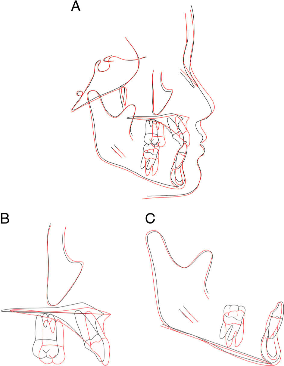
Figure 2.
Pre- (black) and post-treatment (red) lateral cephalogram superimposition of a subject in the MAMP group.
Discussion
The present study demonstrates that MAMP, combined with CAD/CAM appliances, is an effective early intervention for Class III patients, producing significant maxillary advancement with minimal dentoalveolar side effects. While the skeletal improvements were slightly less pronounced than those achieved with BAC3E, likely due to the indirect anchorage and shorter treatment duration, MAMP still resulted in clinically meaningful changes compared to untreated controls. MAMP and BAC3E can be carried out successfully in late mixed to early permanent dentition if the ideal timing for FM therapy is missed.6,12,15 The delayed treatment timing for MAMP and BAC3E is attributed to the necessity for maxillary bone maturation and the eruption of permanent mandibular canines and/or premolars, which is essential for facilitating the placement of skeletal anchorage.6,17,18 Despite the later initiation of treatment, both MAMP and BAC3E are conducted during the pre-peak growth phase, which continues to enable a positive response in the maxillary structures.3,19 Although there is a risk of reduced skeletal effect, treatment at a later stage offers the advantage of a shorter post-orthopaedic period of facial growth, thereby decreasing the likelihood of mandibular catch-up growth.13
Regarding the skeletal changes observed, the MAMP group showed a significant increase in SNA (2.5°) and Co-A (2.4 mm). This is more than the increase seen with negative controls but is less than the increase observed with BAC3E (SNA, 2.7°; Co-A, 5.4 mm). The difference between MAMP and BAC3E could be related to the longer treatment duration (MAMP: 9.6 months; BAC3E: 12.1 months) and direct anchorage utilized in BAC3E. Skeletal anchorage has been shown to improve skeletal outcomes. Previous studies have reported that the use of skeletal anchorage with FM can result in a greater increase in treatment outcomes compared to tooth-borne FM.20,21
The treatment outcomes reflecting sagittal mandibular skeletal (SNB, Co-Gn) for MAMP and BAC3E demonstrated their effectiveness in controlling mandibular growth, as evidenced by the lack of significant differences in the sagittal position of the mandible. This is particularly important for patients presenting with mandibular prognathism. These findings are supported by previous studies12,13,14,15,22, confirming that early intervention with MAMP and BAC3E effectively controls sagittal mandibular growth. MAMP showed no clinically significant vertical changes in the mandible. The present findings agree with recent MAMP studies, that reported insignificant posterior or anterior rotation of the mandible, indicating effective vertical control.12,13,15 This is an important consideration, especially for hyperdivergent patients. Insignificant anterior rotation of the mandible was seen in BAC3E patients. Elnager et al.23 observed a slight anterior rotation of the mandible, possibly attributed to forces directly applied to miniplates.
Minimal dental changes were noted in the maxillary and mandibular incisors following MAMP. The maxillary incisors exhibited slight retroclination (U1-PP, −4.7°), while the mandibular incisors showed slight proclination (L1-ML, 1.6°). These results are consistent with Gera et al.12, who reported similar findings and suggested that these contribute to the mild increase in OJ, compared to what is typically seen with conventional FM therapy, due to the proclination of maxillary incisors and retroclination of mandibular incisors. However, the present results differ from the MAMP study by Kamel et al.13, where both maxillary and mandibular incisors demonstrated proclination. The authors reported that maxillary incisor proclination could be due to the inherent flexibility of the protraction arm in the maxillary framework leading to anchorage loss.13 The maxillary framework utilized in the present study was 3-dimensionally (3D) printed, ensuring high rigidity to minimize the risk of anchorage loss and unwanted dental side effects. The minimal dental effects associated with MAMP have positive clinical implications, particularly for patients with initial proclination of maxillary incisors and retroclination of mandibular incisors due to dental compensations. Although not statistically significant, the lesser proclination of the mandibular incisors seen with MAMP compared to BAC3E could be related to miniscrew failure, leading to some anchorage loss.
Soft tissue changes in patients from the MAMP group showed an improvement in upper lip projection. Additionally, a restriction in the projection of the lower lip and soft tissue pogonion was observed. This may be due to effective control of sagittal mandibular growth and minimal rotation of the mandible. Kamel et al.13 reported similar findings, with a significant forward movement of the upper lip alongside backward movement of the mandibular soft tissue with MAMP.
Regarding the complications associated with MAMP, the present study found that palatal miniscrews were stable with a survival rate of 100% (n=22). In contrast, mandibular miniscrews exhibited a lower survival rate (n=15, 68.2%) compared to those placed in the palate. These findings agree with those reported by Arqub et al.24, who documented a high survival rate of over 90% for palatal miniscrews and a lower 12-month survival rate of 75.5% for mandibular inter-radicular miniscrews. The higher survival rate for palatal miniscrews is due to the bone quality and the presence of keratinized tissue.24 Typically, the use of two or more palatal miniscrews allows for a larger surface area to distribute loads, thereby enhancing stability. Conversely, the thicker cortical bone in the mandible requires greater insertion torque, which can cause bone overheating and local necrosis, ultimately increasing the risk of failure.25 Additionally, root proximity is a significant factor, as a smaller distance between the miniscrew and adjacent roots could increase the likelihood of failure.26 The narrower vestibule in the mandible compared to the maxilla further complicates oral hygiene, making it more difficult for patients to maintain cleanliness in the area.27 All miniscrew failures in the mandibular region were unilateral. Despite this, the rigid CAD/CAM framework used in the present study allowed the mandibular arch to function effectively as a unit with the presence of just one miniscrew. A higher rate of appliance debonding was noted in the mandible compared to the maxilla. This could be due to the three-piece mandibular framework being inherently more prone to debonding under masticatory load. A potential solution to mitigate this issue is to fabricate a one-piece mandibular framework, which could reduce the incidence of debonding but might be more challenging to manage clinically. This approach necessitates the direct placement of miniscrews after inserting the framework. However, if debonding occurs, both the miniscrews and the framework would need to be removed entirely, adding complexity to the procedure. Intrusion of the mandibular molar was noted in some cases (n=5, 45.5%) following MAMP (Figure 3). One possible explanation for this phenomenon is that the application of force on the button of the permanent mandibular canine was positioned anteriorly to the axis of rotation of the mandibular framework, which passes through the two miniscrews. Consequently, given the presence of a vertical force component, rotational movement of the buccal bar around the miniscrew occurred, resulting in the mandibular molar intrusion. This problem can be overcome by designing a traction hook slightly posteriorly between the mandibular first and second premolar, closer to the axis of rotation (the miniscrew). Fortunately, this undesirable side effect resolved quickly, with molar eruption occurring within four weeks after framework removal.
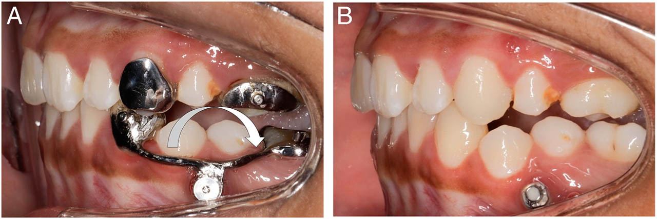
Figure 3.
(A) Intrusion noted on tooth 36; (B) Four weeks after framework removal.
The notion that maxillary expansion may loosen and stimulate a cellular response in the circumaxillary suture, potentially resulting in a more favourable response to the protraction forces, remains a topic of debate. Some studies have suggested that FM therapy alone produced the same orthopaedic effect.28 This was backed further by a recent systematic review and meta-analysis, which concluded that the sole rationale for rapid maxillary expansion (RME) in FM patients is transverse maxillary deficiency.29 Regarding the Alternate Rapid Maxillary Expansion and Constriction (Alt-RAMEC) protocol, Masucci et al.30 compared the modified Alt-RAMEC/FM protocol with the RME/FM protocol, reporting that the former exhibited greater maxillary advancement (SNA +1.2°). However, the results from Masucci et al.30 differ from those of a recent cone beam computed tomography study conducted by Fischer et al.31, which investigated the effects of a 6-week Alt-RAMEC/FM protocol compared to the RME/FM protocol. Fischer et al.31 found that both treatment groups showed favourable orthopaedic changes, with no statistically significant difference observed between the two protocols. Therefore, in this study, only patients with a transverse maxillary deficiency were prescribed expansion screws.
When evaluating the different treatment protocols, some clinical aspects should be considered. For FM therapy, a wear time of 14–16 hours has been suggested to achieve the desired skeletal effects.32 FM is bulky and less comfortable than the intraoral Class III elastics in MAMP and BAC3E, which are acceptable to be worn full-time. BAC3E necessitates a total of eight surgical procedures for both placement and removal, with young patients typically expressing a preference for general anaesthesia during such interventions.17 On the other hand, MAMP only requires the miniscrews to be placed under local anaesthesia and is generally better tolerated by patients. Furthermore, there is no risk of miniscrews being ankylosed due to significant bony overgrowth, which can sometimes happen with miniplates.17 The skeletal anchorage utilized in both MAMP and BAC3E can remain in situ for the distalization of the maxillary molars subsequent to maxillary protraction.16 This serves to facilitate the resolution of crowding if required during the second phase of treatment with fixed appliances. The delayed initiation of MAMP and BAC3E means that patients do not have an extended retention period and can transition to phase two treatment relatively quickly.16
The findings of this study are limited by a pilot sample size and short duration of observation, which only covers the immediate post-treatment phase. To assess the stability of MAMP, long-term follow-up data extending beyond the pubertal growth spurt is necessary to determine whether the treatment can effectively reduce the need for surgery in some cases. The use of historical controls introduces potential issues related to secular trends and differences in data acquisition. Furthermore, the BAC3E sample and untreated controls consisted of European patients, while the present MAMP-treated sample reflects the multicultural nature of Australia, comprising a mix of Asian and Caucasian patients. Although efforts were made to match the groups based on key characteristics, the possibility of confounding factors cannot be entirely eliminated. Limitations of 2D cephalometric analysis include projection errors, landmark identification issues, and measurement inaccuracies.33,34 Despite these limitations, the present study provides valuable insights into the effectiveness of MAMP as a less invasive alternative to BAC3E for early Class III intervention. The promising results warrant further investigation with larger, prospective trials, and longer follow-up periods to confirm the findings and assess the long-term stability of the treatment outcomes.
Conclusions
The MAMP protocol, combined with the use of precisely fabricated frameworks utilizing CAD/CAM technology, offers an effective early intervention for skeletal Class III patients. This minimally invasive alternative to BAC3E enables true skeletal changes without dental compensations while ensuring effective vertical control. The absence of posterior rotation of the mandible is an advantage, particularly in hyperdivergent patients. The insignificant difference in the sagittal position of the mandible indicates effective control of mandibular growth.