
1. Introduction
Aquaculture is a significant provider of highly nutritious protein, fostering food security and rural communities globally (Guillen et al., 2025; Haque & Mahmud, 2025). Globally, Oreochromis niloticus (Nile tilapia), as the second most farmed fish species, holds significant economic importance due to its massive annual production volume (FAO, 2026). O. niloticus has become a particularly valuable species due to its swift growth, effective feed conversion, and environmental adaptability, thereby becoming a cornerstone in the aquaculture industry (El-Sayed & Fitzsimmons, 2023). Nonetheless, there are several challenges facing and impeding aquaculture expansion, and environmental pollutants are one of them (Narwal et al., 2025). Water pollution is a severe worry to aquaculture, as chemicals such as heavy metals (HMs) and industrial effluents hinder fish well-being and survivability (Kakade et al., 2023). Long-term contact with these contaminants increases disease vulnerability and retards growth, which ultimately compromises aquaculture sustainability and productivity (Mustafa et al., 2024).
Copper (Cu) and nickel (Ni) are two HMs that often coexist in the environment. Each metal has its distinctive toxicological implications (Mebane et al., 2020). Cu is a crucial trace element for organisms’ development, but too much exposure to it can have detrimental consequences (Malhotra et al., 2020). Although traditionally classified as a non-essential metal, Ni exhibits biological effects and toxicity that depend on its bioavailability, dose-response relationships, and environmental conditions (Nys et al., 2025; Zhou et al., 2025). In aquatic ecosystems, both metals are present at low levels naturally through natural rock weathering, but anthropogenic activities can increase their concentrations above background levels (Morariu et al., 2025; Oladimeji et al., 2025). Recent toxicological studies highlight that metal mixtures can produce synergistic or antagonistic effects distinct from single-metal exposures, necessitating more comprehensive real-world monitoring (Majid et al., 2025; Smii et al., 2025). Consequently, their elevated levels can impact aquatic organisms (Timpano et al., 2022). The toxicity of HM in fish is primarily mediated through excess reactive oxygen species (ROS) release, leading to oxidative damage, hormonal dysregulation, and compromised intestinal integrity (Shahjahan et al., 2022; Yin et al., 2025), alongside the inhibition of antioxidant defences (Bhat et al., 2025).
Intestinal health is essential for optimal growth and feed utilization in fish, as the intestine is the main site of digestion, nutrient absorption, and immune defence. Healthy intestinal tissues ensure efficient enzyme secretion and nutrient uptake, supporting growth and metabolism (Muneeb et al., 2026). Conversely, intestinal damage induced by environmental stressors, such as metal co-exposure, disrupts these processes, impairing absorptive capacity and leading to growth retardation (Taslima et al., 2022). To elucidate the underlying mechanisms of Cu–Ni co-exposure toxicity, we evaluated key molecular markers that reflect intestinal health status. Specifically, we assessed intestinal absorptive capacity by analysing the expression of specific nutrient transporters: solute carrier family 15 member A1a (slc15a1a), solute carrier family 15 member A2 (slc15a2), and solute carrier family 2 member 1b (slc2a1b) (Kombat et al., 2024). Furthermore, we evaluated pro-inflammatory genes such as interleukin-1 beta (il1b) and v-rel avian reticuloendotheliosis viral oncogene homolog A (rela) to monitor the nuclear factor kappa B (NF-κB)-mediated stress response and inflammation (Wu et al., 2026). These markers are important as they reliably reflect intestinal homeostasis and physiological health in the context of metal-induced toxicity (Wang et al., 2023; Yin et al., 2025).
Previous studies have shown that single Cu and Ni exposure impedes growth and fish survivability (Gao et al., 2023; Naz et al., 2023; Wang et al., 2020). Cu exposure induced oxidative stress, hemato-biochemical disruptions, and delayed growth in O. niloticus (Shokr, 2020). Meanwhile, Ni-exposed O. niloticus showed immunosuppression and pathological alterations (El-Houseiny et al., 2022; Ahmed et al., 2025). However, studies on the combined effects of these metals in fish are limited. For instance, while Pavione and Val (2025) reported that Cu–Ni co-exposure induced neurotoxicity and metabolic disturbances in tambaqui (Colossoma macropomum), such interactive effects remain unexplored in O. niloticus.
Recently, there has been an urgent need for nutritional practices to reduce the negative consequences of waterborne pollutants. In this sense, applications of aquafeed supplements have drawn attention, especially with growing concerns over chemical additives (Abdel Rahman et al., 2025; Hu et al., 2025). Modern nutraceutical approaches increasingly focus on sustainable agro-byproducts, as they offer an eco-friendly strategy to alleviate oxidative stress and support gut health in aquaculture (Lourenço et al., 2025; Vahdat et al., 2026). Citrullus lanatus (watermelon) represents one of the most extensively consumed fruits in the world and an abundant, affordable fruit in Egypt (Al-Sayed & Ahmed, 2013). Watermelon rind (WMR), as a by-product, is a rich source of prebiotics, pectin, and fibre. Also, it has a high content of citrulline, amino acids, and vitamins (Manivannan et al., 2020; Ben Romdhane et al., 2024). Due to the presence of carboxyl (pectin) and hydroxyl (cellulose) functional groups in WMR, it can serve as an effective HM-binding agent (Reddy et al., 2014; Onyango et al., 2025). In addition, WMR contains a considerable quantity of phenolic compounds that exhibit strong hydroxyl radical-scavenging, antioxidant, and antibacterial activity (Gu et al., 2023; Rezagholizade-Shirvan et al., 2023). Dietary WMR has previously shown benefits for growth and immunity of O. niloticus (Doan et al., 2021).
Nevertheless, the mitigating potential of WMR against the cumulative toxic effects of Cu–Ni co-exposure, particularly concerning intestinal gene expression, remains unexplored. Therefore, this study aims to investigate the toxicity mechanism of Cu–Ni co-exposure and the potential alleviating effect of WMR on growth, digestive function, and intestinal health markers in O. niloticus.
2. Material and methods
2.1. Preparation of toxicants
Copper chloride (CuCl2 · 2H2O) and nickel chloride (NiCl2 · 6H2O) obtained from Alpha Chemika, Mumbai, India, were utilized as a source of Cu and Ni. For this trial, the nominal concentrations of these metal salts were set at 3.35 mg/L and 3.60 mg/L, respectively. These concentrations were selected as 1/10 of the reported 96-h lethal concentration 50 for O. niloticus (Abdel-Hamid et al., 2021, for Cu; Abou-Hadeed et al., 2008, as cited in El-Houseiny et al., 2022, for Ni), respectively. These chemicals were dissolved in deionized water to generate a stock solution, which was then adjusted to the necessary nominal levels immediately before use.
2.2. Formulation of investigated diets
The watermelons were bought from a local market in Zagazig, Egypt. The rinds (comprising the peel and white mesocarp) were separated, accounting for approximately 30–40% of the total fruit weight (Rico et al., 2020; Ashoka et al., 2022). The rinds were then oven-dried at 60°C for 2 days until a constant weight was achieved, resulting in a moisture content of 13.50% determined according to standard methods (AOAC, 2005). Subsequently, the dried material was crushed into fine powder (WMR) and incorporated into the basal diet at 0 and 40 g/kg to prepare control and WMR diets (Doan et al., 2021). The feed components were pelleted in a meat mincer, pelletized into 2-mm pellets, and then forced-air dried for a whole day. Until they were needed, the pellets were then kept at 4°C. O. niloticus’s nutritional needs were taken into consideration when developing the tested diets’ components in line with NRC (2011) guidelines. Moreover, the investigated diets were chemically analysed adopting the AOAC (2005) methodology as presented in Table 1.
Table 1
Diet formulations and chemical composition of the tested diets
| Control diet | WMR diet | |
|---|---|---|
| Ingredients (g/kg) | ||
| Fish meal 66% CP | 80 | 80 |
| Soybean meal 48% CP | 300 | 300 |
| Corn gluten meal 60% CP | 120 | 120 |
| Corn oil + Cod liver oil | 40 | 40 |
| Premix* | 30 | 30 |
| Wheat bran | 80 | 80 |
| Ground yellow corn | 350 | 310 |
| WMR | 0 | 40 |
| Total | 1,000 | 1,000 |
| Chemical analysis (%) | ||
| Nitrogen-free extract** | 49.42 | 48.58 |
| Crude fibre | 5.36 | 5.87 |
| Crude lipids | 7.21 | 7.10 |
| Ash | 6.94 | 7.40 |
| Crude protein | 31.07 | 31.05 |
| Gross energy (kcal/kg) | 4,468 | 4,405 |
WMR: watermelon rind.
* Each 1 kg of premix contained: Vitamins: vitamin K3 (1.50 g), vitamin E (7 g), vitamin D3 (2,000,000 IU), vitamin A (8,000,000 IU), nicotinic acid (20 g), vitamin B1 (0.70 g), pantothenic acid (7 g), vitamin B2 (3.50 mg), biotin (50 mg), folic acid (0.70 mg), vitamin B12 (7 mg); Minerals: manganese sulphate (5.30 g), copper sulphate (2.70 g), cobalt sulphate (70 mg), zinc sulphate (4 g), iron sulphate (20 g), sodium selenite (70 mg), calcium iodine (0.34 g), and carrier (CaHPO4·2H2O up to 1 kg).
**Nitrogen-free extract = 100 – (% crude fibre + % crude lipids + % crude protein + % ash).
2.3. Fish housing and experimental scheme
Fingerlings of O. niloticus were acquired from the Fish Research Unit at Zagazig University, Egypt. Fish were kept in 150-L glass aquariums with proper ventilation for a duration of 15 days, with 10 fish kept in each tank. In addition to that, each tank was provided with chlorine-free water and compressed air for adequate oxygenation. Throughout the acclimation and experimental phases, fish were hand-fed to apparent satiation three times daily at 08:00, 13:30, and 16:00, ensuring adequate feed intake (FI) while minimizing waste. Moreover, water’s physical and chemical characteristics were monitored daily in line with the APHA (2005) guideline. The results highlighted that the values of 6.00 ± 0.90 mg/L, 28.80 ± 0.07°C, 7.10 ± 0.05, and 0.03 ± 0.001 mg/L were for dissolved oxygen, temperature, pH, and unionized ammonia. During the trial, a 12-h dark: 12-h light photoperiod was maintained. The initial weight (IW) of the fish was 37.30 ± 0.20 g.
A total of 160 fingerlings were randomly allocated into four different sets, each with 40 fish (4 replicates per set; 10 fish per replicate). The treatments were control, WMR, Cu + Ni, and WMR + Cu + Ni. The control and WMR groups were fed basal diets supplemented with 0 g/kg and 40 g/kg of WMR, respectively, without heavy metal exposure. Meanwhile, the Cu + Ni and WMR + Cu + Ni groups were fed the control and WMR diets, respectively, and co-exposed to nominal concentrations of copper chloride (3.35 mg/L) and nickel chloride (3.60 mg/L). On day 0, the feeding schedule and toxicant exposure began concurrently and lasted for 45 days. This study was conducted under a semi-static renewal system; to maintain optimal water quality and prevent waste accumulation, daily siphoning of fish wastes was performed. Furthermore, a 50% partial water exchange was carried out twice weekly. Physicochemical measurements were taken before and after each renewal. To maintain the target nominal concentrations, the appropriate volume of metal stock solutions was added immediately following each partial water exchange. The Institutional Animal Care and Use Committee of Zagazig University certified the study strategy for the use and handling of animals (ZU-IACUC/2/F/275/2026).
2.4. Growth performance determination and sampling
Fish were weighed at the end of the trial (final weight, FW). Growth performance parameters, including weight gain (WG), specific growth rate (SGR), FI, and feed conversion ratio (FCR), were calculated for each replicate tank using these formulas:
Mortality was recorded daily, and FCR was adjusted for biomass changes as needed. After a 45-day trial, 8 fish from each treatment group were randomly sampled for equal representation across replicate tanks (n = 2/replicate tank). Intermediate sampling was omitted to minimize stress and maintain experimental stability, with all analyses reflecting stable, long-term responses. The previous method (Neiffer & Stamper, 2009) involved anesthetizing fish with benzocaine solution (100 mg/L water). After inserting anticoagulant-free syringes into the caudal peduncle to induce bleeding, the blood was then left to coagulate (4°C for 6 h). Serum was collected using a centrifugation of specimens for 10 min at 1,198 × g to analyse growth-regulating hormones. Following blood collection, the whole fish were sacrificed humanely using a high dose of benzocaine solution (300 mg/L water) following AVMA (2007) recommendations. Intestinal tissues were then harvested and used for the analysis of digestive enzymes (n = 2/tank, total = 8/group), histological examination (n = 1/tank, total = 4/group), antioxidant indices (n = 2/tank, total = 8/group), and gene expression (n = 2/tank, total = 8/group).2.5. Determination of growth regulatory hormones and digestive enzymes
The regulatory hormones for growth, including growth hormone (GH; Cat. No. MBS044656) and leptin (Cat. No. MBS280395), were measured in the serum using MyBioSource, Inc., San Diego, USA. Intestinal specimens were used to estimate amylase, lipase, and trypsin following the techniques of Bernfeld (1955), Worthington (1993), and Khangembam et al. (2012), respectively, by applying a spectrophotometer, Jenway 6305 (Staffordshire, UK). Moreover, the Bradford method (Kruger, 2009) was employed to measure the quantity of total protein in the samples to evaluate enzyme activity per gram of tissue.
2.6. Intestinal histomorphometric investigations
Applying routine histology methods (Suvarna et al., 2018), tissue portions from the intestinal tract (0.50 cm in length) were collected in 10% neutral-buffered formalin solution and fixed for 24 h. After that, the tissues were carefully washed with distilled water to remove leftover formalin. Tissues were then dehydrated in a graded ethanol series (70–100% with 1 h for each phase), cleared in xylene, and infiltrated and embedded in paraffin. Subsequently, samples were sectioned at approximately 5-μm-thickness crosswise employing a Leica® microtome RM2125RTS (Leica Biosystems, Germany). Finally, the sections were stained with haematoxylin and eosin. Microscopic inspection was performed on 50 random fields per group (distributed across 4 fish/treatment) at 100× under an AmScope microscope. In this investigation, mucosal fold width (MW), mucosal fold height (MH), and mucosal surface area (MSA) were quantified based on the method of Wilson et al. (2018). To ensure independent biological replication, data from these fields were averaged for each fish, and the mean values were used for statistical analysis (n = 4 per group).
2.7. Intestinal antioxidants/oxidants measures
Based on the prior approach (Fernandez-Botran et al., 2002), the intestinal homogenates were prepared to measure the antioxidant defences, including glutathione peroxidase (GPx), superoxide dismutase (SOD), and total antioxidant capacity (TAC), as well as lipid peroxide index [malondialdehyde (MDA)]. In this sense, the specimens were first diluted with sodium chloride solution (NaCl, 0.9%), and then they were homogenized in phosphate-buffered saline at pH 7.50. Centrifugation was conducted to extract the supernatant to assess these indices for 15 min at 3,000 rpm.
The techniques of Paglia and Valentine (1967), Nishikimi et al. (1972), Koracevic et al. (2001), and Ohkawa et al. (1979) were applied to estimate GPx, SOD, TAC, and MDA. In addition, colorimetric kits from Biodiagnostic, Egypt, were utilized for the detection of GPx (Cat. No. GP2524), SOD (Cat. No. SD2521), TAC (Cat. No. TA2513), and MDA (Cat. No. MD2529). Furthermore, the total protein content in the supernatants was quantified (Kruger, 2009). All indices were normalized to total protein content and expressed as U/mg protein for GPx and SOD, ng/mg protein for TAC, and nmol/mg protein for MDA.
2.8. Gene expression analysis
The expression of the nutrient transporters (slc15a1a, slc15a2, and slc2a1b) and inflammatory genes (il1b and rela) was assessed using specific primers (Table 2). After a 45-day experiment, intestinal specimens (50 mg) were used to extract total RNA using QIAzol from QIAGEN (Germany). Quantitative real-time PCR (RT-qPCR) was performed using a Rotor-Gene Q2 plex real-time thermal cycler from QIAGEN. With the Thermo Scientific NanoDropR ND-1000 UV–Vis spectrophotometer (Waltham, USA) at 260–280 nm, the effectiveness of the extraction method was examined. The cDNA was created using a reverse transcriptase kit from Applied Biosystems (Foster City, USA).
Table 2
Gene primers of O. niloticus for RT-qPCR amplification
| Target gene | Primer sequences | TM | Efficiency | Size | Accession no. |
|---|---|---|---|---|---|
| gapdh | GATAATGGCAAACTTGTCGTCG | 58.33 | 99.53 | 205 | NM_001279552.1 |
| ACATTGGAGCATCGGGTGAG | 60.11 | ||||
| slc15a1a | ACGATTGGACAGGTTGCGAT | 60.04 | 101.5 | 133 | XM_005452882.2 |
| TGCCCAGAGCAATGAGGAAG | 60.03 | ||||
| slc15a2 | CTGCCTGTTTGCACCTGAAC | 59.97 | 99.36 | 85 | XM_005475385.4 |
| CAGACACTGGCTGCACTACA | 59.97 | ||||
| slc2a1b | CACTGTGGTGTCGCTGTTTG | 59.97 | 99.5 | 172 | NM_001279727.1 |
| TGCCACGAAGGCAAAAATGG | 59.97 | ||||
| il1b | CTCATGTCTGTCCGCTACCC | 59.90 | 98.54 | 103 | KF747686.1 |
| TGAAGCTTCTGTAGCGTGGG | 60.04 | ||||
| rela | GAGAAAGCGGACAGGAGACAT | 59.79 | 93.68 | 133 | XM_025905614.1 |
| AGCAGCTTGTGGAGGCTTG | 60.60 |
TM: melting temperature; gapdh: glyceraldehyde-3-phosphate dehydrogenase. slc15a1a: solute carrier family 15, member 1a; slc15a2: solute carrier family 15, member 2; slc2a1b: solute carrier family 2, member 1b; il1b: interleukin-1 beta; rela: v-rel avian reticuloendotheliosis viral oncogene homolog A.
The cycling protocol consisted of an initial denaturation step at 95°C for 10 min, followed by 40 cycles of denaturation, annealing, and extension at 95°C for 10 s, at 60°C for 15 s, and at 72°C for 15 s, respectively. The glyceraldehyde-3-phosphate dehydrogenase (gapdh) gene was chosen as a standard gene since it remained steady during all treatments. Following amplification, a melting curve assessment was done to guarantee that each amplifier was running at maximum efficiency. The relative gene expression levels were calculated using the 2–ΔΔCT method (Livak & Schmittgen, 2001), using the gapdh gene as the internal reference. The resulting fold change values were then subjected to statistical analysis.
2.9. Statistical analysis of data
All indices were statistically analysed using the replicate tank (n = 4 tanks per group) as the independent biological replicate. The normality and homogeneity of variance for the assessed parameters were verified using the Shapiro-Wilk and Levene tests, respectively. The data were then tested using two-way ANOVA in SPSS (Ver. 21, IBM Corp., Armonk, NY, USA). Treatment means were compared using Tukey’s HSD post hoc test, with significance set at P < 0.05. Results were presented as mean ± standard error (SE). Furthermore, GraphPad Prism (Ver. 10, San Diego, CA, USA) was used to visualize the gene expression findings.
3. Results
3.1. Growth indices
Table 3 highlights that the individual and interaction effects of WMR supplementation and Cu + Ni exposure were significant for growth metrics (P < 0.01). The FW, WG, SGR, and FI were considerably enhanced in the WMR group relative to the control, while the FCR was significantly decreased. On the other hand, the Cu + Ni group showed a marked decline in the FW, WG, SGR, and FI, alongside an elevated FCR in contrast to the control. However, there was a significant improvement in the FW, WG, SGR, and FI, and a lowering of the FCR in the WMR + Cu + Ni group compared with the Cu + Ni group.
Table 3
Effect of copper (Cu) and nickel (Ni) co-exposure and/or dietary watermelon rind powder (WMR) supplementation on growth metrics of O. niloticus
| Parameters | Control | WMR | Cu + Ni | WMR + Cu + Ni | P-value of WMR | P-value of Cu + Ni | P-value of interaction |
|---|---|---|---|---|---|---|---|
| IW (g) | 37.30 ± 0.36 | 37.20 ± 0.50 | 37.33 ± 0.48 | 37.37 ± 0.41 | 0.95 | 0.82 | 0.88 |
| FW (g) | 75.63 ± 0.77b | 109.40 ± 0.67a | 54.07 ± 1.13 d | 65.07 ± 1.03c | <0.01 | <0.01 | <0.01 |
| WG (g) | 38.33 ± 0.47b | 72.20 ± 0.32a | 16.74 ± 0.66 d | 27.70 ± 0.63c | <0.01 | <0.01 | <0.01 |
| SGR (%/day) | 1.57 ± 0.01b | 2.40 ± 0.02a | 0.82 ± 0.02 d | 1.23 ± 0.01c | <0.01 | <0.01 | <0.01 |
| FI (g) | 77.63 ± 0.97b | 92.15 ± 1.21a | 65.36 ± 1.92c | 70.44 ± 0.99b | <0.01 | <0.01 | <0.01 |
| FCR | 2.03 ± 0.03c | 1.28 ± 0.01 d | 3.91 ± 0.06a | 2.54 ± 0.05b | <0.01 | <0.01 | <0.01 |
IW: initial weight; FW: final weight; WG: weight gain; SGR: specific growth rate; FI: feed intake; FCR: feed conversion ratio. The data were analysed by two-way ANOVA (n = 4 replicate tanks per group), where the tank served as an independent experimental unit. Values having various superscripts in the same row differ significantly (Tukey’s HSD test; P < 0.05). Control and WMR groups: fish fed on control and WMR diets, respectively, without Cu–Ni co-exposure. Cu + Ni and WMR + Cu + Ni groups: fish fed on control and WMR diets, respectively, and exposed to Cu–Ni toxicity.
3.2. Mortality (%), growth-regulating hormones, and digestive enzymes
Two-way ANOVA revealed significant individual effects of WMR supplementation, Cu + Ni exposure, as well as their interaction on mortality and biochemical parameters (P < 0.05, Table 4), except for the individual effect of WMR on trypsin and the interaction effect on GH. No mortality was observed in the control or WMR groups, whereas Cu + Ni exposure induced significant mortality, which was remarkably mitigated by WMR supplementation (P < 0.01). Raw counts per replicate tank are provided in Supplementary Table S1 to demonstrate inter-tank variation. Regarding metabolic and digestive parameters, the WMR group exhibited considerably enhanced amylase, lipase, and trypsin compared to the control. In contrast, the Cu + Ni group showed elevated leptin levels and suppressed digestive enzyme activities relative to the control. Notably, the WMR + Cu + Ni group revealed decreased leptin and enhanced digestive enzyme activities compared with the Cu + Ni group.
Table 4
Effect of copper (Cu) and nickel (Ni) co-exposure and/or dietary watermelon rind powder (WMR) supplementation on mortality (%), growth-regulating hormones, and digestive enzyme activities of O. niloticus
| Parameters | Control | WMR | Cu + Ni | WMR + Cu + Ni | P-value of WMR | P-value of Cu + Ni | P-value of interaction |
|---|---|---|---|---|---|---|---|
| Mortality (%) | 0.00 ± 0.00c | 0.00 ± 0.00c | 25.00 ± 2.89a | 7.50 ± 2.50b | <0.01 | <0.01 | <0.01 |
| GH (ng/mL) | 15.19 ± 0.33 | 18.73 ± 0.31 | 10.50 ± 0.55 | 12.69 ± 0.63 | <0.01 | <0.01 | 0.18 |
| Leptin (ng/mL) | 9.88 ± 0.32c | 10.86 ± 0.34c | 18.57 ± 0.38a | 15.92 ± 0.23b | 0.02 | <0.01 | <0.01 |
| Amylase (U/g protein) | 97.10 ± 0.39b | 101.40 ± 0.49a | 71.82 ± 0.70 d | 92.39 ± 0.94c | <0.01 | <0.01 | <0.01 |
| Lipase (U/g protein) | 38.63 ± 0.17b | 43.23 ± 0.95a | 26.21 ± 1.20c | 34.96 ± 1.01b | <0.01 | <0.01 | 0.04 |
| Trypsin (U/g protein) | 45.08 ± 0.75b | 56.02 ± 1.11a | 25.08 ± 1.48 d | 40.59 ± 0.25c | 0.18 | <0.01 | 0.04 |
GH: growth hormone. The data were analysed by two-way ANOVA (n = 4 replicate tanks per group), where the tank served as an independent experimental unit. Values having various superscripts in the same row differ significantly (Tukey’s HSD test; P < 0.05). Control and WMR groups: fish fed on control and WMR diets, respectively, without Cu–Ni co-exposure. Cu + Ni and WMR + Cu + Ni groups: fish fed on control and WMR diets, respectively, and exposed to Cu–Ni toxicity.
3.3. Intestinal histopathology and histomorphometry
Normal histomorphological features of the intestine, including the submucosa, lamina propria, and muscular layer, were observed in the control and WMR groups (Figure 1a and b). On the other hand, the Cu + Ni group revealed necrotic and desquamated epithelial lining of the mucosa, accompanied by intense inflammatory cell aggregates in the lamina propria and submucosa (Figure 1c). Furthermore, an improvement in the structural integrity of intestinal layers was observed in the WMR + Cu + Ni group, despite the presence of some denuded mucosal fold epithelium and an edematous intestinal wall (Figure 1d).
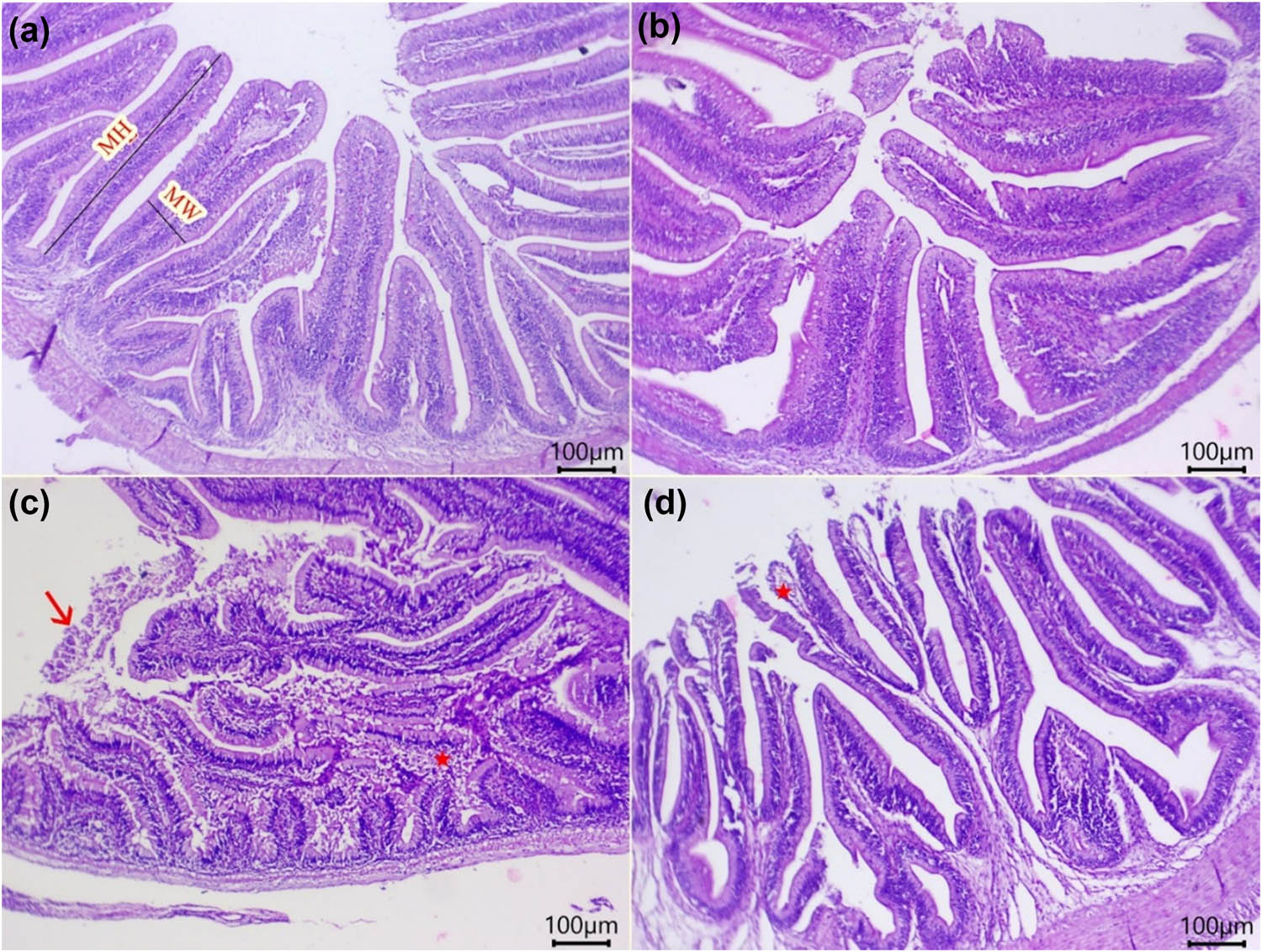
Figure 1
Representative photomicrograph (H&E) of O. niloticus intestine following copper (Cu) and nickel (Ni) co-exposure and/or dietary watermelon rind powder (WMR) supplementation. (a and b) Intestine of the control and WMR groups, showing normal histomorphology of mucosal folds, lamina propria, muscular layer, and submucosa. (c) Intestine of the Cu + Ni group, showing necrotic, desquamated epithelial lining mucosa (arrow), along with intense inflammatory cell aggregates in the lamina propria and submucosa (star). (d) Intestine of the WMR + Cu + Ni group, showing minor denudation of the mucosal fold epithelium and an edematous intestinal wall (star). MW: mucosal fold width. MH: mucosal fold height. Control and WMR groups: fish fed on control and WMR diets, respectively, without Cu–Ni co-exposure. Cu + Ni and WMR + Cu + Ni groups: fish fed on control and WMR diets, respectively, and exposed to Cu–Ni toxicity. Scale bar: 100 µm.
Moreover, the intestinal morphometrics, including MW, MH, and MSA, were significantly influenced by the individual effects of both WMR and Cu + Ni exposure (P < 0.01; Table 5). The WMR supplementation substantially increased MW, MH, and MSA over the control (P < 0.01). Conversely, Cu + Ni exposure caused a marked decline in these parameters relative to the control (P < 0.01). Regarding the interaction effect, while it did not significantly alter MW (P = 0.75) and MH (P = 0.31), it exerted a significant influence on MSA (P < 0.01). Notably, WMR inclusion in the Cu + Ni group significantly enhanced MSA compared to the Cu + Ni group alone.
Table 5
Effect of copper (Cu) and nickel (Ni) co-exposure and/or dietary watermelon rind powder (WMR) supplementation on intestinal morphometries of O. niloticus
| Parameters | Control | WMR | Cu + Ni | WMR + Cu + Ni | P-value of WMR | P-value of Cu + Ni | P-value of interaction |
|---|---|---|---|---|---|---|---|
| MW (µm) | 87.28 ± 1.44 | 111.00 ± 0.43 | 62.00 ± 0.91 | 85.00 ± 1.29 | <0.01 | <0.01 | 0.75 |
| MH (µm) | 533.80 ± 1.46 | 672.90 ± 13.95 | 405.80 ± 10.67 | 522.60 ± 11.68 | <0.01 | <0.01 | 0.31 |
| MSA (mm2) | 0.05 ± 0.001b | 0.07 ± 0.002a | 0.02 ± 0.00c | 0.04 ± 0.00b | <0.01 | <0.01 | <0.01 |
MW: mucosal fold width; MH: mucosal fold height; MSA: mucosal surface area. The data were analysed by two-way ANOVA (n = 4 replicate tanks per group), where the tank served as an independent experimental unit. Values having various superscripts in the same row differ significantly (Tukey’s HSD test; P < 0.05). Control and WMR groups: fish fed on control and WMR diets, respectively, without Cu–Ni co-exposure. Cu + Ni and WMR + Cu + Ni groups: fish fed on control and WMR diets, respectively, and exposed to Cu–Ni toxicity.
3.4. Antioxidant–oxidant response
The individual effects of the WMR supplementation and Cu + Ni exposure, as well as their interaction, were significant for GPx, SOD, TAC, and MDA (P < 0.01; Table 6). The WMR group showed a notable increase in GPx, SOD, and TAC, and a decline in MDA levels compared with the control. Conversely, the GPx, SOD, and TAC in the Cu + Ni group were significantly diminished, and MDA levels were increased relative to the control. Compared to the Cu + Ni group, incorporation of WMR into the diet of Cu + Ni-exposed fish remarkably improved GPx, SOD, and TAC, and decreased MDA.
Table 6
Effect of copper (Cu) and nickel (Ni) co-exposure and/or dietary watermelon rind powder (WMR) supplementation on antioxidant status of O. niloticus
| Parameters | Control | WMR | Cu + Ni | WMR + Cu + Ni | P-value of WMR | P-value of Cu + Ni | P-value of interaction |
|---|---|---|---|---|---|---|---|
| GPx (U/mg protein) | 67.14 ± 0.36b | 74.98 ± 1.92a | 41.83 ± 0.65c | 62.44 ± 0.90b | <0.01 | <0.01 | <0.01 |
| SOD(U/mg protein) | 75.92 ± 1.00b | 82.67 ± 1.06a | 52.04 ± 0.74 d | 67.96 ± 0.82c | <0.01 | <0.01 | <0.01 |
| TAC(µmol/mg protein) | 2.80 ± 0.02b | 4.67 ± 0.09a | 1.46 ± 0.17 d | 2.31 ± 0.05c | <0.01 | <0.01 | <0.01 |
| MDA(nmol/mg protein) | 1.60 ± 0.08c | 1.47 ± 0.08c | 8.57 ± 0.18a | 6.04 ± 0.17b | <0.01 | <0.01 | <0.01 |
GPx: glutathione peroxidase; SOD: superoxide dismutase; TAC: total antioxidant capacity; MDA: malondialdehyde. The data were analysed by two-way ANOVA (n = 4 replicate tanks per group), where the tank served as an independent experimental unit. Values having various superscripts in the same row differ significantly (Tukey’s HSD test; P < 0.05). Control and WMR groups: fish fed on control and WMR diets, respectively, without Cu–Ni co-exposure. Cu + Ni and WMR + Cu + Ni groups: fish fed on control and WMR diets, respectively, and exposed to Cu–Ni toxicity.
3.5. Expression of nutrient transporters and inflammatory genes
Figures 2 and 3 highlight that the individual and interaction effects of WMR supplementation and Cu + Ni exposure were significant for slc15a1a, slc15a2, slc2a1b, il1b, and rela genes (P < 0.01). The WMR group showed significantly upregulated slc15a1a, slc15a2, and slc2a1b genes compared with the control. Nevertheless, a marked downregulation in slc15a1a, slc15a2, and slc2a1b and upregulation of il1b and rela were obvious in the Cu + Ni group compared with those in the control. Relative to the Cu + Ni group, the expression of slc15a1a, slc15a2, and slc2a1b was upregulated, and the expression of il1b and rela was decreased in the WMR + Cu + Ni group.
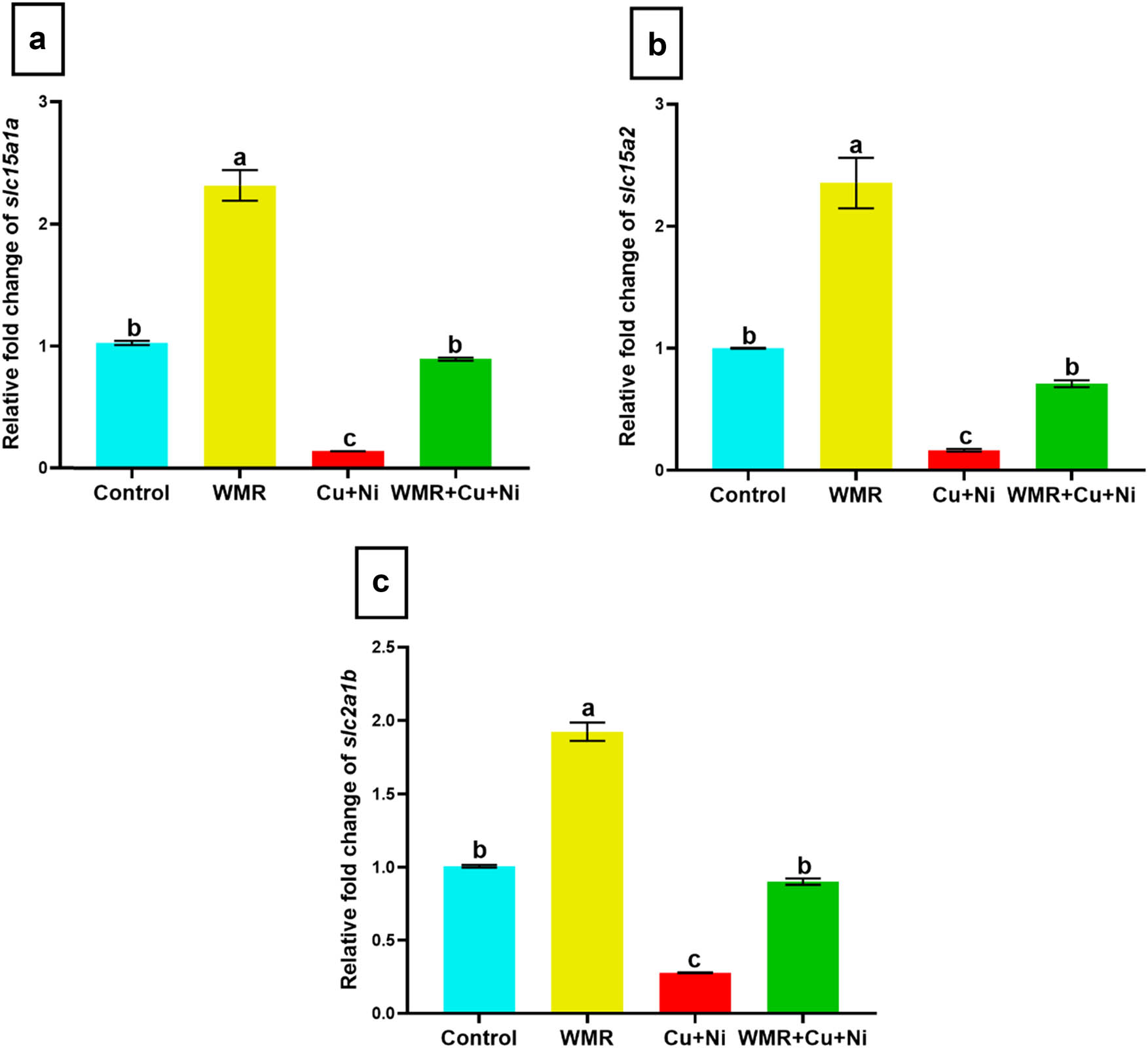
Figure 2
Effect of the interaction between copper (Cu) and nickel (Ni) co-exposure and dietary watermelon rind powder (WMR) supplementation on intestinal nutrient transporter genes of O. niloticus. (a) slc15a1a: solute carrier family 15, member 1a. (b) slc15a2: solute carrier family 15, member 2. (c) slc2a1b: solute carrier family 2, member 1b. The data were analysed by two-way ANOVA (n = 4 replicate tanks per group), where the tank served as an independent experimental unit. Values on the bars (means ± SE) with various superscripts differ significantly (Tukey’s HSD test; P < 0.05). Control and WMR groups: fish fed on control and WMR diets, respectively, without Cu–Ni co-exposure. Cu + Ni and WMR + Cu + Ni groups: fish fed on control and WMR diets, respectively, and exposed to Cu–Ni toxicity.
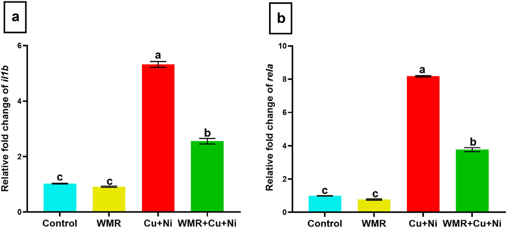
Figure 3
Effect of the interaction between copper (Cu) and nickel (Ni) co-exposure and dietary watermelon rind powder (WMR) supplementation on intestinal inflammatory genes of O. niloticus. (a) il1b: interleukin-1 beta. (b) rela: v-rel avian reticuloendotheliosis viral oncogene homolog A. The data were analysed by two-way ANOVA (n = 4 replicate tanks per group), where the tank served as an independent experimental unit. Values on the bars (means ± SE) with various superscripts differ significantly (Tukey’s HSD test; P < 0.05). Control and WMR groups: fish fed on control and WMR diets, respectively, without Cu–Ni co-exposure. Cu + Ni and WMR + Cu + Ni groups: fish fed on control and WMR diets, respectively, and exposed to Cu–Ni toxicity.
4. Discussion
The present study investigated the alleviating effects of dietary WMR on the growth and gut health of O. niloticus co-exposed to waterborne Cu and Ni toxicity. Cu + Ni co-exposure suppressed O. niloticus growth (lower WG and SGR, higher FCR), consistent with previous reports on O. niloticus following single exposure to Cu (Shokr, 2020) and Ni (Ahmed et al., 2025; El-Houseiny et al., 2022; Sheethal et al., 2024). These adverse consequences result from metabolic stress that redirects energy allocation away from growth towards detoxification and tissue repair (Jiang et al., 2015; Sreedevi et al., 1992).
The observed reduction in digestive enzyme activities (amylase, lipase, and trypsin) likely impairs nutrient digestion and limits the availability of energy for growth (Xie et al., 2019; Lee et al., 2023). These inhibitory effects are likely mediated by oxidative stress and intestinal damage, which diverts energy towards detoxification (Kharkan et al., 2023; Pavione & Val, 2025; Wang et al., 2023). In addition, this work indicates that Cu and Ni co-exposure disrupted endocrine growth regulation by modifying GH and leptin signalling. We observed decreased GH and elevated leptin levels in the metal-exposed fish. GH stimulates somatic growth via the GH/IGF axis, and its inhibition by pollutants negatively affects cell proliferation and differentiation (Bersin et al., 2024; Canosa & Bertucci, 2023; Caputo et al., 2021).
Leptin regulates fish appetite and energy homeostasis and is primarily produced by adipose tissue (Picó et al., 2022). Beyond its satiety role, leptin in teleosts influences immune, reproductive, and osmoregulatory functions (Copeland et al., 2011; Ohga et al., 2021). Its elevation in our study likely signals a metabolic shift towards survival over growth, leading to the observed reduction in FI (Liu et al., 2018). Moreover, the rise in leptin may act as a counter-regulatory response to declining GH levels, reflecting an antagonistic interaction (Gong et al., 2022). Such endocrine disruptions are consistent with the toxic stress response documented in O. niloticus under Ni exposure (El-Houseiny et al., 2022) and other teleost species under Cu exposure (Zebral et al., 2018). These findings confirm that such hormonal alterations play a central role in metal-induced growth impairment.
Dietary WMR effectively counteracted Cu and Ni-induced growth inhibition by enhancing digestive enzyme activity, endocrine control, and FI. Our findings align with previous studies in O. niloticus showing that WMR and C. lanatus seed-enriched diets promote growth and reduce FCR (Doan et al., 2020, 2021; Iheanacho et al., 2018). These benefits stem from WMR’s nutrient profile, including prebiotic fibres, phytochemicals, vitamins, and carbohydrates, which optimize digestion and gut microbiota. Furthermore, bioactive compounds like citrulline act as antioxidants and immune stimulants, reducing the metabolic cost of stress and redirecting energy towards somatic growth (Gorbanalinejad & Nobakht, 2017; Tarazona-Díaz et al., 2011), thereby improving overall performance.
Cu and Ni co-exposure induced significant intestinal histopathological and morphometric damage, likely driven by oxidative stress, ROS-mediated DNA damage, and lipid/protein oxidation (Das & Saikia, 2024; Pavione & Val, 2025). Comparable intestinal histopathological changes have been observed in Cu-exposed tiger pufferfish (Takifugu rubripes) (Liu et al., 2024) and Ni-exposed O. niloticus (Ahmed et al., 2025), corroborating these findings. Dietary WMR mitigated Cu + Ni-induced intestinal damage, likely through its antioxidant, radical-scavenging, and anti-inflammatory properties. These protective effects are attributed to WMR’s rich bioactive profile, including citrulline, lycopene, flavonoids, vitamin C, alkaloids, and beta-carotene (Bahrudin et al., 2025; Rahman et al., 2013). By suppressing oxidative stress, these constituents mitigate tissue damage, a finding supported by earlier studies in lead-exposed rats (Michael et al., 2021). This role of WMR in maintaining gut integrity is further corroborated by its efficacy in enhancing intestinal histoarchitecture in rohu (Labeo rohita) (Vijayan et al., 2024).
Cu and Ni co-exposure significantly suppressed nutrient transporters (slc15a1a, slc15a2, and slc2a1b). The slc15 family is critical for peptide reabsorption, and its downregulation suggests impaired uptake and stunted growth (Orozco et al., 2017; Vacca et al., 2022). Similarly, slc2a1b (glut1b) facilitates glucose uptake to meet the energetic demands of the intestinal epithelium (Zhang et al., 2023). Thus, its downregulation suggests both restricted energy (glucose) uptake and systemic metabolic disruption in the gut (Kang et al., 2021; Wei et al., 2025). These molecular changes likely result from metal-induced oxidative stress, which compromises transporter structure and function (Jiang et al., 2015; Kumar et al., 2023).
Collectively, these hormonal and transcriptional changes highlight an integrated metabolic response, where energy is diverted from growth towards stress adaptation. This indicates that metal-induced growth suppression results from the combined effects of endocrine dysregulation and impaired intestinal metabolic function. This aligns with previous findings in O. niloticus, where Cu exposure disrupted slc family expression (Huang et al., 2015). Notably, dietary WMR partially restored nutrient transporter expression, likely mediating its growth-enhancing effects through the same antioxidant mechanisms observed in other models (Agbana, 2023).
Metal co-exposure (Cu and Ni) induced oxidative stress in O. niloticus, evidenced by depleted antioxidant defences (GPx, SOD, TAC) and elevated MDA levels. This redox imbalance, which correlated with a high mortality rate (25% in the Cu + Ni group), highlights the failure of the fish’s antioxidant system to counteract the ROS generated by Cu and Ni. Mechanistically, Cu promotes ROS through redox cycling and Fenton-like reactions (Jiang et al., 2014), while Ni triggers ROS via the Ni2+/Ni3+-mediated Fenton/Haber–Weiss pathway (Guo et al., 2015; Salnikow et al., 1994). These results corroborate the oxidative damage and physiological impairment documented in O. niloticus under individual Cu or Ni exposure (Abdel-Hamid et al., 2021; Ahmed et al., 2025).
Conversely, dietary WMR exerted pronounced antioxidant effects in O. niloticus, significantly boosting GPx, SOD, and TAC levels while reducing MDA, which likely contributed to the lower mortality rate (7.50% in the WMR + Cu + Ni group). These findings align with reports of WMR enhancing antioxidant defences and mitigating lipid peroxidation in L. rohita (Vijayan et al., 2024). Such potent antioxidant characteristics are attributable to WMR’s rich profile of phenolic acids (vanillic, p-hydroxybenzoic, gallic, and chlorogenic acids) and flavonoids (hesperetin and catechin), which effectively neutralize metal-induced oxidative stress (Alsuwayt, 2025; Mushtaq et al., 2015).
Cu and Ni co-exposure induced a pro-inflammatory state in O. niloticus, characterized by the upregulation of il1b and rela. This response is likely mediated by metal-induced ROS acting as signalling molecules for inflammatory pathways (Guo et al., 2019; Wu et al., 2026). Our findings corroborate previous reports of disrupted inflammatory mechanisms in O. niloticus under Ni stress (El-Houseiny et al., 2022), as well as similar observations in other teleosts under Cu exposure (Wang et al., 2023). Notably, dietary WMR suppressed these metal-induced inflammatory pathways in this study. The anti-inflammatory efficacy of WMR has been previously reported in topmouth gudgeon (Pseudorasbora parva) (Wu et al., 2021). It is largely attributed to WMR’s rich bioactive profile, including flavonoids, phenolics, citrulline, and arginine, which effectively modulate inflammatory signalling and nitric oxide pathways (Nahar et al., 2024; Priastomo et al., 2025).
In this study, Cu–Ni co-exposure impaired growth, digestive, and antioxidant functions while inducing intestinal damage and inflammation, all of which were effectively mitigated by WMR supplementation. As a study limitation, the reported exposure concentrations were nominal, based on the mass of metal salts added to the tanks. Since analytical verification was not performed, the findings should be interpreted as the biological response to these nominal levels.
5. Conclusion
The present study demonstrates that exposure to a combined mixture of Cu and Ni compromises intestinal integrity and physiological function, ultimately leading to reduced growth in O. niloticus. This growth inhibition is likely mediated through the disruption of the nutrient transporters (slc15a1a, slc15a2, and slc2a1b) and hormonal control of growth (GH and leptin). Moreover, Cu and Ni negatively influence digestive function, weaken antioxidant defences, and upregulate inflammatory pathways (il1b and rela). Conversely, WMR supplementation effectively alleviates Cu- and Ni-induced intestinal damage, restores nutrient transport balance, and improves growth performance, possibly by enhancing antioxidant status. As a result, WMR may serve as a functional dietary strategy to protect O. niloticus exposed to waterborne contaminants.
Acknowledgments
This work was supported by the Deanship of Scientific Research, Vice Presidency for Graduate Studies and Scientific Research, King Faisal University, Saudi Arabia [Grant No. KFU265830]. The authors thank the Aquatic Animal Medicine Department, Faculty of Veterinary Medicine, Zagazig University, for their kind help during the experimental procedures. We want to express our sincere gratitude to Dr. Mohammed S. Sobh, Department of Pathology, Faculty of Veterinary Medicine, Zagazig University, for his valuable efforts in performing the histopathological examination.
Funding information
This work was supported by the Deanship of Scientific Research, Vice Presidency for Graduate Studies and Scientific Research, King Faisal University, Saudi Arabia [Grant No. KFU265830].
Author contributions
Conceptualization, A.N.A.R., A.Y., S.T.E., R.E.I.; Data curation, A.N.A.R., T.K.; Formal analysis, A.N.A.R., T.K.; Investigation, A.N.A.R., A.Y., S.T.E., T.K., A.T.M., M.A., R.T.M., H.A.H., A.A., R.E.I.; Methodology, A.N.A.R., R.E.I.; Resources, A.N.A.R., A.Y., S.T.E., T.K., A.T.M., M.A., R.T.M., H.A.H., A.A., R.E.I.; Validation, A.N.A.R; Visualization, A.N.A.R., R.E.I.; Writing – original draft, A.N.A.R.; Writing – review & editing, all authors. All authors have read and agreed to the published version of the manuscript.
Conflict of interest statement
The authors have no relevant financial or non-financial interests to disclose.
Data availability statement
Data are available upon reasonable request from the corresponding author.