
1. Introduction
Cardiovascular disease (CVD) remains the leading cause of death worldwide, with approximately 20.5 million deaths recorded globally in 2021. The global burden is projected to increase substantially, with the annual CVD-related mortality estimated to reach 35.6 million by 2050, driven largely by population growth, ageing, and persistent cardiovascular risk factors (Chong et al., 2025; Di Cesare et al., 2024). Pathologically, CVD development is characterized by obesity, dyslipidaemia, endothelial dysfunction, and elevated blood pressure, which are often associated with negative lifestyle practices such as a poor diet and smoking (Olas, 2020; Pimenta et al., 2018). Dyslipidaemia, defined by elevated levels of total cholesterol (TC), low-density lipoprotein (LDL) cholesterol, and triglycerides (TGs), alongside decreased high-density lipoprotein (HDL) cholesterol (Angelis-Pereira et al., 2013), is a primary risk factor for atherosclerosis and the subsequent development of CVD (Katsiki et al., 2016; Kopin & Lowenstein, 2017).
The vascular endothelium is a single-cell monolayer forming the first barrier between the bloodstream and vascular smooth muscle (Malik et al., 2018; Mehta et al., 2014). Under physiological conditions, endothelial cells maintain vascular homeostasis by balancing vasodilatory mediators, such as nitric oxide (NO), and vasoconstrictive factors. However, in obesity and related metabolic disorders, this homeostasis is disrupted, resulting in impaired endothelial-dependent vasodilation, increased inflammatory cytokine production, and excessive oxidative stress (Bagi et al., 2012; Dinh et al., 2014; Mlinar et al., 2007; Pimenta et al., 2018; Vasquez et al., 2019). Elevated levels of tumour necrosis factor-α (TNF-α) and interleukin-6 (IL-6) promote endothelial activation and leukocyte adhesion, whereas the overproduction of reactive oxygen species reduces NO bioavailability, thereby accelerating vascular dysfunction (Bagi et al., 2012; Mlinar et al., 2007; Vasquez et al., 2019). Addressing these mechanisms is essential for preventing downstream cardiovascular complications.
CVD and its associated metabolic risk factors are generally managed via pharmacological therapy and lifestyle modification; however, long-term adherence, cost, and treatment-associated side effects limit their overall effectiveness (Dixon et al., 2020; Peluzio et al., 2021; Vieira et al., 2021). Consequently, there is growing interest in adjunct or alternative therapeutic strategies, including probiotic interventions, which have demonstrated potential benefits in metabolic regulation (Ettinger et al., 2014; Sun & Buys, 2015; Vasquez et al., 2019). Kefir, a fermented milk product rich in lactic acid bacteria, acetic acid bacteria, and yeasts, has attracted substantial scientific attention owing to its diverse microbial composition and reported bioactivity (Azizi et al., 2021; Yilmaz & Arslan, 2022).
A growing body of preclinical evidence supports the cardiometabolic benefits of kefir and its constituent probiotic strains in improving lipid profiles and suppressing inflammation (Akar et al., 2021; Chen et al., 2016; Kim et al., 2017; Qaisrani et al., 2025). In rodent models, kefir supplementation has been shown to improve dyslipidaemia and hepatic lipid metabolism (Angelis-Pereira et al., 2013; Choi et al., 2017). Several in vivo studies have also demonstrated that kefir enhances endothelial-dependent vasodilation and restores NO bioavailability in hypertensive or high-fat diet (HFD) animals, partly through the upregulation of endothelial NO synthase (eNOS) (Friques et al., 2015). The standardized AB-Kefir probiotic is a commercial product containing selected Lactobacillus and Bifidobacterium strains. Existing in vitro data and animal models provide mechanistic evidence, suggesting that probiotics enriched with Lactobacillus and Bifidobacterium can improve lipid profiles and vascular function through anti-inflammatory, antioxidant, and NO-preserving pathways. In animal models, these strains reduce serum TC, LDL, and TG levels while increasing HDL (Abdi et al., 2022). Lactobacillus and Bifidobacterium species protect endothelial cells by suppressing reactive oxygen species production, limiting lipid peroxidation, and downregulating pro-inflammatory signalling pathways, including TNF-α, IL-6, and nuclear factor-kappa B activation (Chen et al., 2022; Tseng & Chu, 2025). Additionally, these strains enhance the endogenous activity of antioxidant enzymes, such as superoxide dismutase (SOD) and catalase, while decreasing malondialdehyde (MDA) levels in oxidative stress models (Hao et al., 2023; Vitheejongjaroen et al., 2022).
Unlike conventional kefir, the AB-Kefir probiotic contains a standardized consortium consisting of six Lactobacillus species, one Bifidobacterium longum strain, and Streptococcus thermophilus, with a guaranteed viable count of 1011 CFU/g (supplied by SYNBIO TECH INC., Kaohsiung, Taiwan). Using this standardized formulation ensures reproducibility of the probiotic composition and facilitates future translational studies. The previous work on this specific probiotic mixture primarily focused on obesity, glucose metabolism, and hepatic steatosis (Chen et al., 2021); however, its effects on endothelial dysfunction, vascular reactivity, dyslipidaemia, vascular inflammatory markers, and oxidative stress in HFD-induced obesity have not been investigated. We hypothesized that supplementation with AB-Kefir would ameliorate endothelial dysfunction and metabolic abnormalities in HFD-induced obese rats by preserving NO-dependent endothelial signalling and modulating vascular inflammatory and oxidative stress responses. Therefore, we evaluated the therapeutic potential of AB-Kefir in mitigating dyslipidaemia and endothelial dysfunction in HFD-induced obese rats. Specifically, this study assessed whether AB-Kefir improves circulating lipid profiles, restores endothelial-dependent vasodilation, and modulates key inflammatory and oxidative stress biomarkers, including TNF-α, IL-6, SOD, and MDA. By elucidating these mechanisms, this research contributes to a deeper understanding of how the AB-Kefir probiotic can serve as a promising dietary intervention for preventing obesity-related cardiovascular complications.
2. Material and methods
2.1. AB-Kefir probiotic description
AB-Kefir probiotic is manufactured and supplied by SYNBIO TECH INC. (Kaohsiung, Taiwan) (Mutalub et al., 2025). The formulation contains six lactic acid bacteria strains and one Bifidobacterium strain, with a total cell count of 1011 CFU/g (Chen et al., 2021). The updated taxonomic classifications of the constituent strains are Bifidobacterium longum BL986 (CGMCC 18743), Lactobacillus (L.) acidophilus LA1063 (CGMCC 18744), Limosilactobacillus fermentum (formerly L. fermentum LF26) (DSM 32784), Lactobacillus helveticus LH43 (DSM 32787), Lacticaseibacillus paracasei ssp. paracasei (formerly L. paracasei ssp. paracasei LPC12) (DSM 32785), Lacticaseibacillus rhamnosus (formerly L. rhamnosus LRH10) (DSM 32786), and Streptococcus thermophilus ST30 (DSM 32788) (Chen et al., 2021).
2.2. AB-Kefir probiotic preparation
AB-Kefir was received in powder form (1011 CFU/g). A stock working solution with a concentration of 2 × 10⁹ CFU/mL was prepared by suspending 1 g of the probiotic powder in 50 mL of phosphate-buffered saline. Rats were administered the freshly prepared solution at 1010 CFU/kg body weight (BW)/day, as previously described (Chen et al., 2021).
2.3. Animals
The experimental protocol was approved by the Institutional Animal Care and Use Committee of Universiti Sains Malaysia [Approval No. USM/IACUC/2020/(123)(1065)] and performed in accordance with the institutional guidelines, national regulations, and Animal Research: Reporting of In Vivo Experiments (ARRIVE) guidelines. Twenty-four male Sprague–Dawley rats (6 weeks old, weighing 150–175 g) were obtained from the Animal Research and Service Center, Universiti Sains Malaysia. The rats were housed in pairs within polypropylene cages (approximately 40 cm × 25 cm × 20 cm) according to their treatment groups. Cages were lined with wood shavings, which were replaced twice weekly. Rats were maintained under standard laboratory conditions of 25 ± 2°C and a 12-h light/dark cycle throughout the experiment. Environmental enrichment, including paper nesting materials and cardboard tubes, was provided to encourage species-specific behaviours and minimize psychological stress. After a 1-week acclimatization period, the rats were randomly allocated into three experimental groups (n = 8 per group): the normal control group (NOR; fed standard commercial pellets from Gold Coin Feedmills, Port Klang, Malaysia for 22 weeks), the obese group (OB; fed a modified HFD for 14 weeks to induce obesity, then maintained for an additional 8 weeks), and the obese + AB-Kefir group (OB + AB-Kefir; fed a modified HFD for 14 weeks, followed by AB-Kefir administration via oral gavage at 1010 CFU/kg BW/day for 8 weeks). To control for handling stress, the NOR and OB groups received an equivalent volume of vehicle (distilled water) via oral gavage daily during the 8-week intervention period. All groups continued their respective diets until week 22, and individual BWs were recorded on week 0, 14, and 22. The standard laboratory rodent maintenance diet (720 P, Gold Coin Feedmills, Port Klang, Malaysia) consisted of corn, soybean, grain by-products, animal protein, vegetable oil, salt, and calcium carbonate. This commercial formulation was not based on the AIN-93 purified diet specifications, but matched the diet used in our previous study (Mutalub et al., 2025). The modified HFD was prepared according to a protocol adapted from Azemi et al. (2021), comprising 50% standard commercial food pellets, 21% pure ghee, 10% beef tallow, 15% full-cream milk powder, and 4% white sugar, yielding a total fat content of 57.8% by weight, compared with 7.8% fat in the standard commercial pellets. The complete nutritional compositions of both diets are provided in Table 1.
2.4. Humane endpoints
Rats were monitored daily throughout the 22-week experimental period, with increased frequency if any clinical concerns were observed. The predefined humane endpoints approved by the institutional ethical committee included BW loss ≥20% from baseline or a failure to gain weight appropriate for age; marked reduction in food or water intake lasting more than 48 h; signs of severe lethargy, unresponsiveness to external stimuli, or an inability to ambulate sufficiently to access food or water; signs of pain or distress unresponsive to approved analgesics, including persistent vocalization, self-mutilation, hunched posture, or piloerection; respiratory distress; or any moribund state characterized by persistent hypothermia, profound weakness, or an inability to maintain sternal recumbency.
2.5. Anaesthesia
At the end of the 22-week experiment, all rats were anaesthetized via intraperitoneal injection using a combination of ketamine (100 mg/kg, Ketamine 10% Inj, Dutch Farm International BV, Holland) and xylazine (10 mg/kg, Xylazine 2% Inj, Dutch Farm International BV, Holland) prior to blood/tissue collection and euthanasia (Sotoudeh & Namavar, 2022). Following injection, rats were placed individually in clean, quiet holding cages and monitored continuously for the onset of anaesthesia. The depth of anaesthesia was verified using physiological indicators, including loss of the righting reflex, absence of a response to toe pinching, reduced respiratory rate, and absence of the corneal reflex. Only animals that demonstrated these signs of a deep surgical plane of anaesthesia were advanced to the blood collection and euthanasia stage.
2.6. Blood collection
Blood samples were obtained via cardiac puncture into anticoagulant-free vacutainer tubes and centrifuged at 1,500 × g (4°C) for 25 min (Kubota 4000, Tokyo, Japan) to isolate serum. Samples were stored at −80°C for subsequent biochemical analyses.
2.7. Euthanasia
Euthanasia was performed by administering a combination of xylazine (30 mg/kg) and ketamine (300 mg/kg) delivered intraperitoneally, while the rats remained fully anaesthetized (Underwood & Anthony, 2020). Death was confirmed using multiple physiological criteria to ensure the complete cessation of vital functions: absence of a heartbeat or palpable pulse, absence of spontaneous respiration for at least 2 min, absence of corneal and pedal reflexes, and the presentation of fixed, dilated pupils.
2.8. Thoracic aorta collection
Each rat was dissected, and the thoracic aorta was excised and placed in ice-cold oxygenated physiological solution [1.2 mM potassium dihydrogen phosphate (KH2PO4), 4.7 mM potassium chloride (KCl), 5.5 mM d-glucose, 118 mM sodium chloride (NaCl), 25 mM sodium hydrogen carbonate (NaHCO3), 1.18 mM (MgSO4·7H2O), and 2.0 mM calcium chloride dihydrate (CaCl2·2H2O)] for functional studies and biochemical analyses.
2.9. Lipid profile analysis and atherogenic index (AI) determination
Serum samples were analysed for TC, TG, LDL, and HDL using the colorimetric method with an Integra 800 automatic immunoanalyser (Roche Diagnostic Systems, Mannheim, Germany) at an external reference laboratory (BP Healthcare, Kota Bharu, Kelantan, Malaysia). The AI of the serum was determined using the following formula: AI = log (TG/HDL) (Kim et al., 2022).
2.10. Functional study
Vascular reactivity was assessed using a dual-chamber organ bath system (PanLab, Harvard Apparatus) equipped with isometric force transducers (MLT0201, ADInstruments, Dunedin, New Zealand) and logged via LabChart software (Version 8, ADInstruments, Dunedin, New Zealand). For functional assessments, the thoracic aorta was carefully dissected and cleared of perivascular adipose and connective tissues. Each aorta was cut into three to four separate, adjacent rings (each 3 mm in length). To prevent sequential tissue exposure and blocker effects, these separate rings were allocated in parallel to independent organ bath chambers; individual segments were never reused for multiple protocols. The rings were vertically suspended in organ bath chambers containing 20 mL of physiological solution (pH 7.4) maintained at 37°C and continuously bubbled with 5% CO2 and 95% O2. The segments were equilibrated for 1 h at a resting tension of 1.0 g. Smooth muscle viability was confirmed by contracting the rings with 60 mM KCl. Endothelium integrity was assessed by pre-contracting the rings with phenylephrine (1 µM, #PHR1017, Sigma-Aldrich, USA) followed by relaxation with acetylcholine (1 µM, #A6626, Sigma-Aldrich, USA). Endothelium-dependent relaxation was evaluated in phenylephrine-pre-contracted rings with cumulative doses of acetylcholine (1 nM to 10 µM). The effects of pharmacological blockers were studied by pre-incubating aortic rings for 30 min with 100 µM L-nitro-arginine methyl ester (l-NAME; N5751-5G, Sigma-Aldrich, USA, an eNOS inhibitor) or 0.1 µM wortmannin [W1628-5MG, Sigma-Aldrich, USA, a phosphatidylinositol-3-kinase (PI3K) inhibitor] (Friques et al., 2015; Kobayashi et al., 2004; Mokhtar et al., 2016). The rings were then subjected to phenylephrine, followed by incremental doses of acetylcholine, as described earlier.
2.11. Determination of inflammatory and oxidative stress markers in aortic tissue
The thoracic aorta was homogenized in phosphate-buffered saline at a ratio of 1:9 [tissue weight (g):phosphate-buffered saline (mL)] using a glass homogenizer on ice. The homogenate was centrifuged at 5,000 × g (4 °C) for 10 min to obtain the supernatant. Levels of the pro-inflammatory markers (TNF-α and IL-6) and oxidative stress markers (SOD and MDA) were quantified in duplicate using commercial enzyme-linked immunosorbent assay kits (Qayee Bio-Technology Co., Ltd., Shanghai, China) according to the manufacturer’s protocols: TNF-α (Cat No. QY-E10880), IL-6 (Cat No. QY-E11509), SOD (Cat No. QY-E11476), and MDA (Cat No. QY-E11488). To account for minor variations in tissue mass and extraction efficiency, the total protein concentration of each sample supernatant was determined in duplicate using a Protein Determination Kit (Cat No. 704002-480, Cayman Chemical, Ann Arbor, MI, USA) via a microplate-based Bradford method. Final biomarker concentrations were normalized to total protein content and expressed as pg/ml protein or ng/ml protein.
2.12. Statistical analysis
Data were expressed as means ± standard error of the mean (SEM) and analysed using GraphPad Prism version 10.0 (San Diego, CA, USA). The individual rat was defined as the true biological experimental unit (n = 8 per group) for all analyses. Because adjacent aortic rings from the same rat were distributed across separate, parallel treatment conditions, each rat contributed a single independent observation per treatment arm, preventing pseudoreplication. Aortic relaxation responses were expressed as percentages of the pre-contraction tension generated by 1 × 10⁻⁶ µM phenylephrine. To evaluate vascular efficacy, the maximal relaxation achieved was selected as the primary discrete endpoint. Statistical comparisons of maximal relaxation values and other biochemical parameters, including food intake, energy intake, lipid profile, oxidative stress, and inflammatory markers across experimental groups and blocker treatments, were conducted using a one-way analysis of variance (ANOVA) followed by Tukey’s post hoc test. For BW measurements obtained at multiple time points (week 0, 14, and 22), a two-way repeated-measures ANOVA was performed with time as the within-subject factor and experimental group as the between-subject factor, followed by Sidak’s multiple comparisons test. Prior to parametric analysis, the assumptions of normality of residuals and homogeneity of variances were validated using the Shapiro–Wilk test and the Brown–Forsythe test, respectively. A P-value <0.05 was considered significant.
3. Results
3.1. Impact of AB-Kefir probiotic treatment on body weight and fasting lipid profiles
BW progression is presented in Table 2. Two-way repeated-measures ANOVA revealed significant effects of time (P < 0.0001), treatment group (P = 0.0018), and time × treatment interaction (P < 0.0001). BW progressively increased from week 0 to week 22 in all experimental groups. Following 14 weeks of HFD feeding, OB groups exhibited significantly higher BW compared with the NOR group, confirming successful induction of obesity. Following 8 weeks of AB-Kefir probiotic formulation supplementation, the OB + AB-Kefir group demonstrated a different BW trajectory compared with OB rats, resulting in significantly lower final BW at week 22 (Table 2). The TC, LDL, and AI were significantly higher in the OB group than in the NOR group, whereas AB-Kefir probiotic treatment effectively improved these parameters in the OB + AB-Kefir group, achieving values similar to those of the NOR group. Furthermore, the HDL level, which was significantly reduced in the OB group, was restored to normal levels following AB-Kefir probiotic treatment (Table 3).
Table 2
Body weight progression, food consumption, and energy intake in all experimental groups
| Parameters | Groups | |||
|---|---|---|---|---|
| NOR | OB | OB + AB-Kefir | ||
| Body weight (g) | Week 0 | 159.4 ± 4.8 | 168.4 ± 5.1 | 160.3 ± 7.5 |
| Week 14 | 409.9 ± 10.7 | 498.0 ± 20.6a | 492.4 ± 20.0a | |
| Week 22 | 466.0 ± 15.7 | 596.6 ± 27.3a | 483.8 ± 16.9b | |
| Food intake | Total (g) | 3179.0 ± 92.7 | 2612.0 ± 106.9a | 2483.0 ± 80.6a |
| Mean (g/week) | 144.5 ± 3.6 | 118.7 ± 2.2a | 112.9 ± 1.7a | |
| Energy intake | Total (kcal) | 11030.0 ± 321.6 | 14078.0 ± 576.4a | 13383.0 ± 434.3a |
| Mean (kcal/week) | 501.4 ± 12.3 | 639.9 ± 11.9a | 608.3 ± 9.3a | |
Week 0 (before obesity induction), week 14 (after obesity induction), week 22 (final week of experiment). Data presented as mean ± SEM (n = 8). Body weight was analysed using two-way repeated-measures ANOVA followed by Sidak’s multiple comparisons test. Food intake and energy intake were analysed using one-way ANOVA followed by Tukey’s multiple comparisons test.
Table 3
Fasting lipid profile in all experimental rat groups
| Parameters | NOR | OB | OB + AB-Kefir |
|---|---|---|---|
| TC (mg/dL) | 65.00 ± 3.32 | 74.50 ± 1.71a | 61.83 ± 1.58b |
| TG (mg/dL) | 80.43 ± 8.76 | 125.60 ± 23.17 | 67.29 ± 8.88b |
| HDL (mg/dL) | 35.74 ± 3.27 | 13.27 ± 4.08a | 26.44 ± 1.16b |
| LDL (mg/dL) | 19.60 ± 1.50 | 30.50 ± 1.85a | 23.50 ± 1.23b |
| AI (log TG/HDL) | 0.37 ± 0.10 | 0.75 ± 0.11a | 0.36 ± 0.10b |
Data presented as mean ± SEM (n = 8). Statistical differences were analysed using one-way ANOVA followed by Tukey post hoc test.
a P < 0.05, indicated a significant difference compared to normal.
b P < 0.05, indicated a significant difference compared to the obese.
TC, total cholesterol; TG, triglyceride; HDL, high-density lipoprotein; LDL, low-density lipoprotein; AI, atherogenic index; NOR, normal control; OB, untreated obese; OB + AB-Kefir, obese treated with AB-Kefir.
3.2. Effect of AB-Kefir probiotic on acetylcholine-induced relaxation in the thoracic aorta
Aortic relaxation in response to acetylcholine was significantly impaired in the OB group, as represented by lower relaxation percentages than the NOR group. No significant difference in relaxation was observed between the OB + AB-Kefir and NOR groups. The OB + AB-Kefir exhibited significantly improved aortic relaxation, comparable to that of the NOR group (Figure 1a).
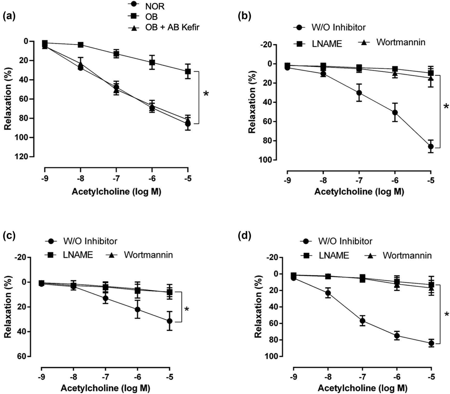
Figure 1
Aortic tissue relaxation responses to acetylcholine (1 nM to 0.1 µM) in all experimental rats without and with blockers. (a) Response in the three groups in the absence of pharmacological blockers. (b) Relaxation responses to acetylcholine without and with blockers in the normal group. (c) Relaxation responses to acetylcholine without and with blockers in the obese group. (d) Relaxation responses to acetylcholine without and with blockers in the OB + AB-Kefir group.
3.3. Impact of pharmacological inhibitors on acetylcholine-induced relaxation in the aortic tissue
The mechanisms underlying acetylcholine-induced endothelium-dependent relaxation were investigated by incubating the thoracic aorta with two pharmacological inhibitors: L-NAME and wortmannin. In the NOR group, the maximal relaxation induced by acetylcholine in the absence of inhibitors was 85.9 ± 6.5%. However, the presence of L-NAME and wortmannin significantly decreased the maximal relaxation to 9.5 ± 2.4% and 14.7 ± 3.4%, respectively (Figure 1b). In the OB group, the maximal acetylcholine-induced relaxation in the absence of inhibitors was 31.3 ± 7.6% (Figure 1c) and further reduced to 8.5 ± 2.3% and 8.7 ± 1.3% in the presence of L-NAME and wortmannin, respectively. In the OB + AB-Kefir group, the maximal acetylcholine-induced relaxation without inhibitors was 84.0 ± 4.6% (Figure 1d), diminishing to 12.9 ± 3.7% and 16.9 ± 3.4% with the addition of L-NAME and wortmannin, respectively.
3.4. Effect of AB-Kefir treatment probiotic on the levels of TNF-α and IL-6
The TNF-α level was significantly higher in the OB group than in both the NOR group (2,650 ± 297 pg/mL) and the OB + AB-Kefir group (2,825 ± 189 pg/mL) (Figure 2a). Conversely, no significant differences in IL-6 levels were detected among the three experimental groups (NOR: 555 ± 100 pg/mL, OB: 601 ± 95 pg/mL, and OB + AB-Kefir: 495 ± 49 pg/mL) (Figure 2b).
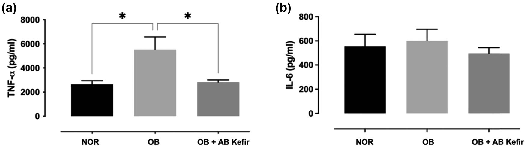
Figure 2
Level of inflammatory markers TNF-α (a) and IL-6 (b) in the thoracic aorta of experimental rats.
3.5. Effect of AB-Kefir probiotic treatment on SOD and MDA levels
The SOD level was significantly higher in both the NOR group (24.0 ± 2.3 ng/mL) and the OB + AB-Kefir group (24.9 ± 1.4 ng/mL) than in the OB group (11.7 ± 5.0 ng/mL). AB-Kefir probiotic treatment successfully prevented the reduction in SOD (Figure 3a). In contrast, no significant difference in MDA levels in the aorta was observed among the three experimental groups (NOR: 33.6 ± 0.9 ng/mL, OB: 32.4 ± 0.8 ng/mL, and OB + AB-Kefir: 33.9 ± 1.1 ng/mL) (Figure 3b).
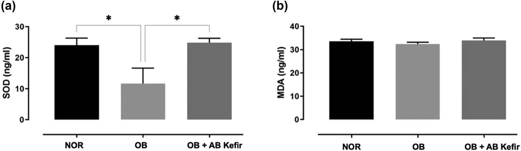
Figure 3
Oxidative stress markers SOD (a) and MDA (b) in the thoracic aorta of experimental rats.
4. Discussion
The findings of this study confirmed the hypothesis that dietary administration of AB-Kefir probiotics significantly improves serum lipid levels and endothelial function in HFD-induced obese rats. Chronic HFD feeding reliably induced hypercholesterolaemia and endothelial dysfunction, which are key features of obesity-related metabolic disorders. Dysregulated lipid metabolism is a well-known risk factor for CVD, and improving dyslipidaemia is a critical strategy for preventing CVD (Bourrie et al., 2020; Choi et al., 2017; Farag et al., 2020). In the present study, AB-Kefir intervention significantly reduced the BW of obese rats to levels comparable with those of the normal controls. This outcome suggests that the strains within the probiotic regulate energy balance and metabolism, potentially by modulating gut microbiota, which has been shown to influence lipid absorption and fat storage (Gao et al., 2019). Furthermore, the resulting lipid profiles highlight the therapeutic efficacy of AB-Kefir; TC, LDL, and AI were significantly elevated in obese rats but normalized following AB-Kefir treatment. Concurrently, the restoration of HDL levels in the OB + AB-Kefir group indicates an improvement in lipid metabolism, likely mediated by the probiotic’s capacity to upregulate cholesterol catabolism and excretion (Tung et al., 2017).
The lipid-modulating effect of kefir has been reported in previous studies. For example, Choi et al. (2017) observed that kefir administration attenuates dyslipidaemia by reducing blood TC, TG, and LDL while increasing HDL fractions in diet-induced obese mice. Similarly, Angelis-Pereira et al. (2013) demonstrated that kefir treatment decreased TG, LDL, and very LDL, along with an elevation in HDL. These results are consistent with the clinical findings reported by Abd-Alwahab & Al-Dulaimi (2018), showing decreased TC, TG, LDL, and very LDL levels alongside enhanced HDL in patients following kefir consumption. Notably, some conflicting data exist; Urdaneta et al. (2007) reported that 22 days of kefir supplementation did not significantly improve blood cholesterol levels in Wistar rats. Additionally, St-Onge et al. (2002) found no significant changes in serum TC, LDL, TG, or HDL levels after 4 weeks of kefir intake. These discrepancies may be attributed to differences in the microbial composition and concentration of the kefir used, as well as the relatively short treatment durations. Notably, St-Onge et al. (2002) observed an increase in propionic acid with kefir treatment. Propionic acid is a short-chain fatty acid that inhibits the incorporation of acetic acid into TG and plasma cholesterol, thus leading to reduced plasma TG and hepatic cholesterol. The AB-Kefir used in the present study contains six strains of Lactobacillus, a genus widely recognized for its high cholesterol-lowering capacity (Yilmaz & Arslan, 2022). A meta-analysis has shown that the administration of multiple strains produces higher hypocholesterolaemic efficacy than that of single strains (Sun & Buys, 2015).
Several interdependent mechanisms contribute to the hypocholesterolaemic activity of probiotics, including cholesterol adsorption and incorporation into bacterial cells (Miremadi et al., 2014), cholesterol deconjugation by bile salt hydrolase (Yusuf et al., 2020), and the conversion of cholesterol in the colon to coprostanol, which is excreted in the faeces (Lye et al., 2010; Shehata et al., 2016). Additionally, probiotics can suppress hepatic cholesterol synthesis through the action of bacterial fermentation end-products such as short-chain fatty acids (St-Onge et al., 2000), or by downregulating hydroxymethylglutaryl coenzyme A reductase, further decreasing cholesterol levels (Aggarwal et al., 2013). Although these mechanisms were not directly explored in the present study, AB-Kefir likely acts via one or more of these pathways. Further research is needed to elucidate the precise mechanism underlying the cholesterol-lowering effect.
In our study, AB-Kefir administration for 8 weeks significantly reduced pro-inflammatory cytokine TNF-α but did not affect IL-6 levels in the aortic tissue of obese rats. This localized suppression is consistent with the findings reported by Akar et al. (2021), who observed a significant downregulation of TNF-α and nuclear factor-kappa B protein expression in the hepatic tissue of kefir-treated metabolic syndrome rats. Moreover, a systematic review and meta-analysis found no significant differences in IL-6 levels following probiotics therapy (Samah et al., 2016). Our cytokine data partially align with the work of Yadav et al. (2019), who demonstrated that the consumption of probiotic L. fermentum MTCC 5898 in fermented milk reduced both TNF-α and IL-6 in high-cholesterol diet-fed rats. Clinical trials also partly support our findings; Bellikci-koyu et al., (2022) demonstrated reduced TNF-α, interferon-gamma, and IL-6 levels in metabolic syndrome patients following kefir intake, while Tenorio-Jimenez et al. (2018) observed decreased TNF-α, IL-6, and IL-8 levels in obese individuals with metabolic syndrome after 12 weeks of daily Lactobacillus reuteri V3401 supplementation. However, our findings contrast with those of Kim et al. (2017), who found that kefir administration in HFD-induced obese mice reduced circulating IL-6 but did not achieve a significant difference in TNF-α levels compared with untreated mice. Similarly, Hadisaputro et al. (2012) demonstrated that 30 days of kefir supplementation in diabetic rats did not significantly reduce TNF-α levels but decreased IL-6 and IL-1 levels. Finally, Bourrie et al. (2018) reported that none of the investigated kefir varieties, including commercial kefir, significantly affected inflammasome expression in HFD-fed mice, despite a pronounced reduction in plasma cholesterol.
The variability in the inflammatory responses to kefir intake may be attributed to differences in the baseline microbial composition, study sample characteristics, and treatment duration across studies (Ostadrahimi et al., 2015; Rosa et al., 2016). Furthermore, inflammatory changes may occur in adipose tissue, perivascular adipose tissue, and the gut, but not in the vasculature. Regulation of the gastrointestinal immune system is one of the primary mechanisms by which probiotic products exert distinct health benefits (Bourrie et al., 2016). While several studies reporting significant changes in IL-6 following kefir treatment evaluated adipose tissue or intestinal tissue samples, others used blood samples, whereas we focused on thoracic aortic tissue. The lack of significant differences in IL-6 levels in our study may also be related to the static MDA levels; kefir has been shown to reduce blood lipid peroxidation products, and peroxide molecules may positively upregulate the transcriptional generation of IL-6 (Judiono et al., 2014).
Oxidative stress plays a crucial role in the pathophysiology of CVD, with cardiometabolic disorders consistently linked to increased oxidative stress (Olas, 2020; Rosa et al., 2016). The detrimental effects of reactive oxygen species can be mitigated by dietary substances with robust radical scavenging or antioxidant activities (Rosa et al., 2017). In the present study, kefir treatment in obese rats significantly increased SOD but had no significant effect on MDA levels. This finding corroborates previous studies, where significant elevations in SOD activity were observed in hyperlipidaemic and diabetic rats after supplementation with L. acidophilus and Lacticaseibacillus casei, respectively, for 1–4 weeks (Harisa et al., 2009; Zhang et al., 2010). Similarly, Yadav et al. (2019) reported that the administration of probiotic L. fermentum MTCC enhanced endogenous SOD capacity and improved hyperlipidaemia in high-cholesterol diet-fed rats. Clinical investigations have also demonstrated increased SOD concentrations following probiotic consumption in type 2 diabetic patients (Asemi et al., 2013; Mazloom et al., 2013), and a meta-analysis did not detect a significant difference in systemic MDA levels despite increased SOD levels in type 2 diabetic patients following probiotic treatment (Samah et al., 2016). Rosa et al. (2016) found that kefir supplementation reduced MDA levels but decreased hepatic SOD levels in a rat model of metabolic syndrome. The variability in oxidative stress markers in response to kefir supplementation across different studies may be ascribed to differences in the probiotic strains and treatment durations. This present study suggests that the vascular benefits of the AB-Kefir probiotic are, at least in part, mediated through antioxidative defence pathways, as evidenced by the upregulation of aortic SOD activity.
Endothelial cells play a critical role in regulating vascular homeostasis, and endothelial dysfunction is often associated with metabolic disorders and CVD (Friques et al., 2015). The present study demonstrated diminished relaxation in response to acetylcholine in the aorta of obese rats compared with normal rats. After 8 weeks of AB-Kefir probiotic supplementation, the impaired endothelial function in obese rats was significantly improved, restoring their relaxation response to levels comparable with those of normal rats. This outcome aligns with the work of Friques et al. (2015), who reported improved acetylcholine-induced aortic vasodilation following 60 days of kefir administration in spontaneously hypertensive rats. Similarly, other studies have demonstrated improved vascular relaxation after supplementing obese rodents or hypertensive rats with probiotic strains such as Lactobacillus coryniformis or L. fermentum (Gómez-Guzmán et al., 2015; Toral et al., 2014). Clinical research has also shown enhanced vascular responsiveness in humans with metabolic diseases following probiotic treatment (Hariri et al., 2015). Meanwhile, recent studies have demonstrated that kefir intake attenuates endothelial dysfunction and improves arterial pressure control through the baroreflex mechanism, likely because of its antioxidant and anti-inflammatory properties (Brasil et al., 2018; Silva et al., 2020). Conversely, no significant improvement in endothelial dysfunction was observed in individuals with metabolic syndrome who received L. casei Shirota supplementation (Tripolt et al., 2013). The discrepancy may originate from differences in single-strain vs. multi-strain probiotics, as well as the absolute viable cell count.
Moreover, our study demonstrated that blocking the PI3K/Akt/eNOS pathway with L-NAME and wortmannin significantly decreased acetylcholine-induced aortic relaxation in normal controls. This finding confirms that the PI3K/Akt/eNOS pathway plays a significant role in the relaxation process. Crucially, even under complete pharmacological blockade, acetylcholine still induced residual vasorelaxation in normal rats, suggesting that other vasodilatory pathways contribute to the response (Akther et al., 2021). In untreated obese rats, L-NAME and wortmannin reduced acetylcholine-induced relaxations to a lesser extent, indicating that the PI3K/Akt/eNOS pathway partially contributes to the reduced relaxation response in obesity. Notably, in obese rats treated with the AB-Kefir probiotic for 8 weeks, pre-incubation with L-NAME and wortmannin almost completely blocked the improved vasodilation responsiveness, suggesting that the beneficial vascular effects of kefir in obese rats involve, at least in part, the functional restoration of endothelial NO availability and signalling. These findings are consistent with the previous work demonstrating increased NO levels following kefir treatment and a corresponding reduction in vascular relaxation after L-NAME blockade in spontaneously hypertensive rats (Friques et al., 2015). This provides evidence showing that NO is a key factor responsible for vasodilation in rat aortas, and the failure of NO signalling is a primary cause of endothelial dysfunction, characterized by impaired vasodilator response. Consequently, the vasoprotective action of kefir appears to be highly dependent on NO bioavailability.
Probiotic therapy has also been shown to enhance endothelial function in humans, both in vivo and in vitro. For example, soy milk fermented with L. plantarum or Streptococcus thermophilus upregulates NO generation and enhances eNOS activity in endothelial cells, indicating direct endothelial benefits (Cheng et al., 2013). Additionally, dietary supplementation with L. plantarum in stable coronary artery disease patients for 6 weeks improved endothelial function in both conduit and resistance vessels by increasing NO availability, as validated by brachial artery flow-mediated dilation measurements (Malik et al., 2018). While the mechanistic evidence in our study is based on pharmacological inhibition with L-NAME and wortmannin, future studies should incorporate direct molecular quantification of eNOS, Akt phosphorylation, and NO bioavailability.
5. Conclusions
AB-Kefir probiotic supplementation for 8 weeks significantly improved dyslipidaemia and endothelial dysfunction in HFD-induced obese rats. Mechanistically, AB-Kefir administration significantly reduced vascular TNF-α concentrations and increased SOD activity in the thoracic aorta, without significantly altering IL-6 or MDA profiles. These findings suggest partial modulation of the inflammatory and antioxidant pathways rather than generalized suppression. Overall, the investigated AB-Kefir probiotic may exert beneficial effects on multiple obesity-related complications, including lipid dysregulation, endothelial dysfunction, inflammation, and oxidative stress, which are the key contributors to CVD risk. Although the precise translational relevance of these rodent outcomes to humans remains to be determined, this study highlights the potential of the AB-Kefir probiotic as a functional food in managing metabolic and vascular complications associated with obesity.
5.1. Limitation of study
Although animals were randomized, cage effects cannot be completely excluded and represent a potential limitation common to rodent studies.
Funding information
This work was supported by a Universiti Sains Malaysia, Bridging with Project No.: R501-LR-RND003-0000001477-0000.
Authors contributions
Writing – review & editing, writing – original draft, visualization, validation, investigation, formal analysis, and data curation, Y.B.M.; Writing – review & editing, investigation, formal analysis, and data curation, A.K.A.; Visualization, validation, and conceptualization, A.H.G.R.; Writing – review & editing, writing – original draft, visualization, validation, and investigation, T.S.P.; Writing – review & editing, resources, L.C.C.; Writing – review & editing, writing – original draft, visualization, validation, supervision, resources, project administration, methodology, investigation, funding acquisition, formal analysis, data curation and conceptualization, S.S.M.
Conflict of interest statement
Lee Chia Chia from Synbio Tech Inc. (Kaohsiung, Taiwan) provided AB-Kefir for this study but did not contribute to data analysis. Her role was limited to providing expertise in the preparation and treatment of AB-Kefir for the rats. The other authors declare no conflict of interest.
Ethical approval
The experimental animal study protocols were approved by the Institutional Animal Care and Use Committee of Universiti Sains Malaysia with voucher number [USM/IACUC/2020/(123)(1065)].
Data availability statement
Data are available upon request.