
Table 1.
Nutrient composition of experimental diets
| Ingredients | % |
|---|---|
| Fish meal (65% CP) | 15 |
| Soybean meal (44% CP) | 35 |
| Yellow corn | 20 |
| Wheat bran | 7 |
| Wheat flour | 6 |
| Rice bran | 5 |
| Gluten | 5 |
| Fish oil | 3 |
| Soybean oil | 2 |
| Dicalcium phosphate | 1 |
| Vitamin and mineral premix 1 | 1 |
| Proximate profile | |
| Crude protein (%) | 32.12±0.14 |
| Crude lipids (%) | 8.2±0.11 |
| Fiber (%) | 3.42±0.16 |
| Ash (%) | 6.63±0.20 |
1 The feed was supplemented with essential minerals (325 mg Mn, 200 mg Fe, 25 mg Cu, 5 mg I, 5 mg Co/kg) and vitamins (3300 IU A, 410 IU D3, 2660 mg E, B-complex vitamins including 133 mg B1, 580 mg B2, 410 mg B6, 50 mg B12, 26.6 mg niacin, 2000 mg pantothenic acid, 9330 mg biotin, plus 4000 mg choline chloride, 330 mg inositol, and 9330 mg PABA per kg).
Table 2.
Sequences of primers used for quantitative real-time PCR (qRT-PCR) analysis
| Gene | Sequences (5′-3′) | Reference | Accession number |
|---|---|---|---|
| EF1A | F: TCAACGCTCAGGTCATCATC | Con et al. (2019) | XM_003458541 |
| R: ACGGTCGATCTTCTCAACCA | |||
| GHR1 | F: CAGACTTCTACGCTCAGGTC | El-Naggar et al. (2021) | AY973232.1 |
| R: CTGGATTCTGAGTTGCTGTC | |||
| IGF-1 | F: GTTTGTCTGTGGAGAGCGAGG | Y10830.1 | |
| R: GAAGCAGCACTCGTCCACG | |||
| GPx | F: CCAAGAGAACTGCAAGAACGA | El-Kassas et al. (2022) | DQ355022.1 |
| R: CAGGACACGTCATTCCTACAC | |||
| CAT | F: CCCAGCTCTTCATCCAGAAAC | Abdo et al. (2021) | JF801726.1 |
| R: GCCTCCGCATTGTACTTCTT | |||
| LYZ | F: AAGGGAAGCAGCAGCAGTTGTG | Esam et al. (2022) | XM_003460550.2 |
| R: CGTCCATGCCGTTAGCCTTGAG | |||
| C3 | F: GGTGTGGATGCACCTGAGAA | XM_013274267.2 | |
| R: GGGAAATCGGTACTTGGCCT |
Table 3.
Key growth performance indicators of Nile tilapia following a 75-day dietary trial
| Parameters | D1 | D2 | D3 | D4 | P-value |
|---|---|---|---|---|---|
| Initial body weight, g | 20±0.14 | 20±0.01 | 19.96±0.21 | 19.94±0.07 | 0.985 |
| Final body weight, g | 81.29±2.75 b | 65.27±2.53 c | 97.04±2.01 a | 78.56±2.75 b | 0.000 |
| Weight gain, g | 61.29±2.83 b | 45.28±2.53 c | 77.08±1.82 a | 58.62±2.77 b | 0.000 |
| Specific growth rate (SGR, %/day) | 1.87±0.05 b | 1.57±0.05 c | 2.11±0.02 a | 1.83±0.05 b | 0.000 |
| Feed intake, g (FI) | 122.86±1.13 b | 112.56±3.21 c | 138.79±1.99 a | 123.54±2.2 b | 0.000 |
| Feed conversion ratio (FCR) | 2.01±0.08 b | 2.5±0.15 a | 1.8±0.05 b | 2.12±0.13 b | 0.012 |
| Survival rate (SR, %) | 96.67±1.67 | 98.33±1.67 | 98.33±1.67 | 96.67±3.33 | 0.900 |
Table 4.
Biometric indices and intestinal morphology of Nile tilapia following a 75-day feeding trial
| Items | D1 | D2 | D3 | D4 | P-value |
|---|---|---|---|---|---|
| Hepatosomatic index (HSI, %) | 1.07±0.08 | 1.02±0.03 | 1.02±0.03 | 1.02±0.04 | 0.877 |
| Intestino-somatic index (ISI, %) | 2.53±0.13 | 2.68±0.18 | 2.37±0.03 | 2.39±0.09 | 0.306 |
| Visceral somatic index (VSI, %) | 3.82±0.16 | 3.93±0.21 | 3.57±0.05 | 3.61±0.13 | 0.344 |
| Fulton’s condition factor (K factor) | 2.16±0.11 | 2.11±0.07 | 2.02±0.03 | 2.12±0.04 | 0.569 |
| Villus length (µm) | 450.1±11.91 b | 225.4±4.4 c | 647.96±18.03 a | 416.27±5.38 b | 0.000 |
| Villus width (µm) | 153.41±4.17 b | 119.44±3.21 c | 224.91±6.36 a | 120.98±6.67 c | 0.000 |
| Goblet cells (cell/mm2) | 267±7 b | 146.33±10.81 c | 350.33±21.84 a | 233±7.64 b | 0.000 |
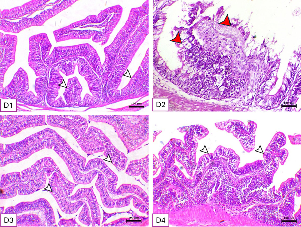
Figure 1.
The histomorphology of Nile tilapia intestine after a 75-day feeding experiment. Stain H&E. Bar = 100 µm. D1: basal diet free of supplements; D2: basal diet + zearalenone (1 mg/kg diet); D3: basal diet + sodium metasilicate (0.5 g/kg diet); D4: basal diet + zearalenone (1 mg/kg diet) + sodium metasilicate (0.5 g/kg diet). White arrow: normal villi and goblet cells. Red arrow: necrosis of the intestinal villi with marked sloughing of the mucosal lining.
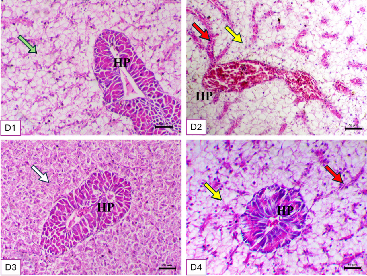
Figure 2.
The histomorphology of Nile tilapia liver after a 75-day feeding experiment. Stain H&E. Bar = 50 µm. D1: basal diet free of supplements; D2: basal diet + zearalenone (1 mg/kg diet); D3: basal diet + sodium metasilicate (0.5 g/kg diet); D4: basal diet + zearalenone (1 mg/kg diet) + sodium metasilicate (0.5 g/kg diet). Green arrow: normal pancreatic tissue (HP) and hepatocytes. Red arrow: congestion of the portal vein and blood sinusoids. White arrow: cytoplasmic eosinophilia. Yellow arrow: vacuolation of the hepatocytes.
Table 5.
Blood biomarker profiles of Nile tilapia following a 75-day feeding trial
| Items | D1 | D2 | D3 | D4 | P-value |
|---|---|---|---|---|---|
| Total protein (g/dL) | 4.29±0.25 a | 2.74±0.24 b | 4.64±0.12 a | 4.14±0.51 a | 0.013 |
| Albumin (g/dL) | 1.7±0.34 | 1.45±0.27 | 1.81±0.24 | 1.65±0.39 | 0.873 |
| Globulin (g/dL) | 2.59±0.16 a | 1.29±0.1 b | 2.82±0.19 a | 2.5±0.16 a | 0.000 |
| Total cholesterol (mg/dL) | 157.67±6.36 b | 211±10.02 a | 140±10.44 b | 158±10.12 b | 0.004 |
| Triglyceride (mg/dL) | 124.33±6.17 a | 72±5.51 c | 106.33±6.69 b | 98±2.65 b | 0.001 |
| High-density lipoprotein (mg/dL) | 45.13±6.92 | 43.65±4.39 | 48.02±3.78 | 41±5.59 | 0.822 |
| Alanine aminotransferase (IU/L) | 10.33±0.88 c | 70.33±1.76 a | 10.33±1.45 c | 22±1.15 b | 0.000 |
| Aspartate aminotransferase (IU/L) | 32±1.53 c | 113±3.21 a | 32.33±2.03 c | 48.67±1.45 b | 0.000 |
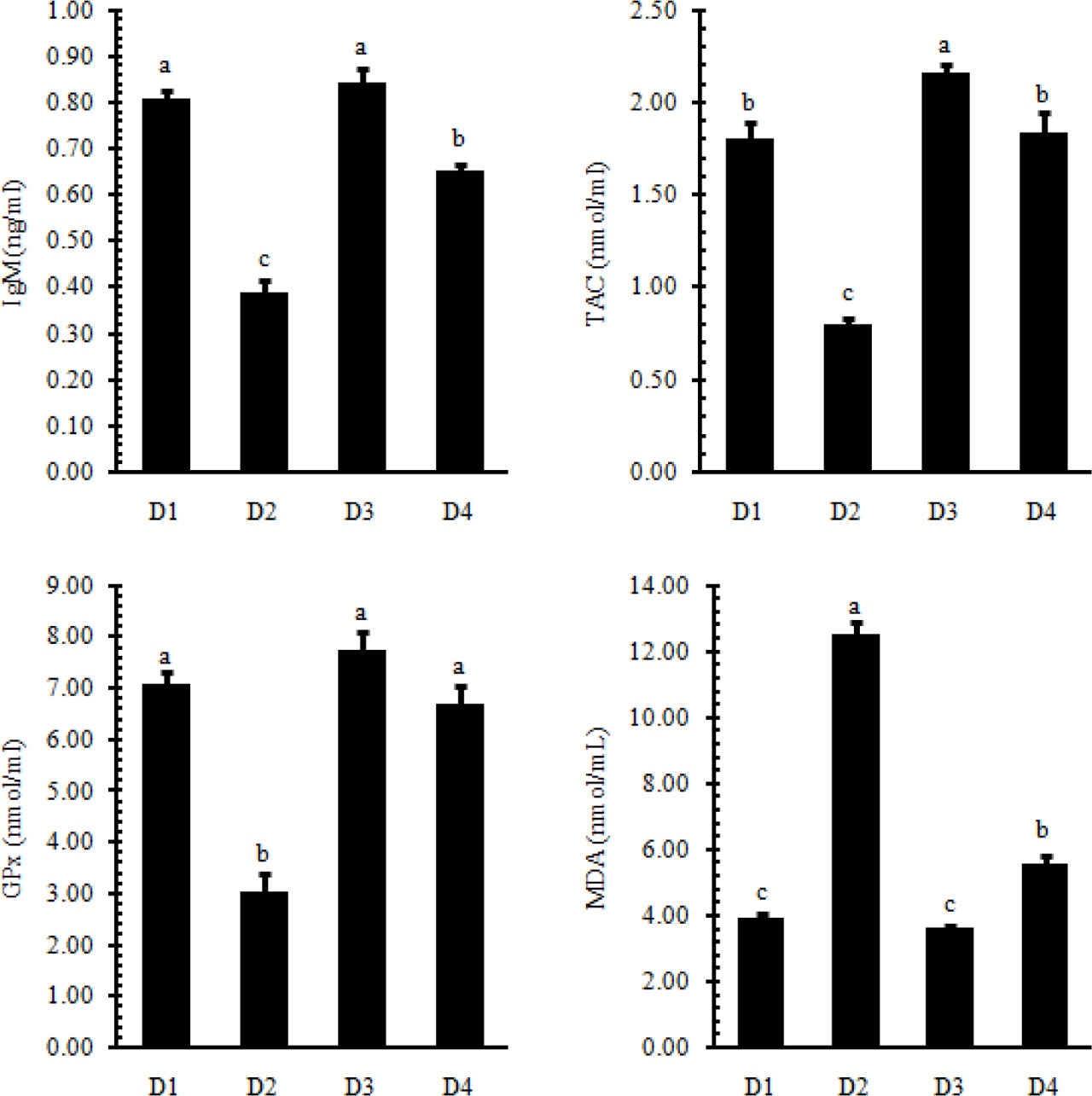
Figure 3.
Immunoglobulin M (IgM) and antioxidant biomarkers in Nile tilapia following 75 days of feeding.
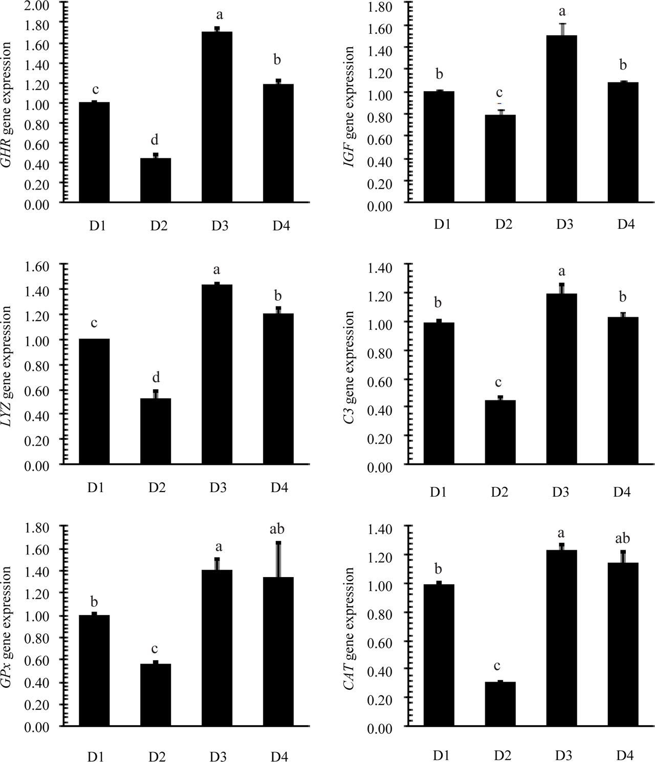
Figure 4.
Relative mRNA expression levels of growth hormone receptor (GHR), insulin-like growth factor (IGF), glutathione peroxidase (GPx), lysozyme (LYZ), catalase (CAT), and complement component 3 (C3) in the liver of Nile tilapia (Oreochromis niloticus) after a 75-day feeding trial. Data are expressed as mean ± standard error (SE). Different letters indicate statistically significant differences among treatment groups (P<0.05).