
Table 1.
Current status of the disease PTB across different counties
| Name of the animals | Region | Infection status | Economic loss | Strategy to counter the disease | References |
|---|---|---|---|---|---|
| 1 | 2 | 3 | 4 | 5 | 6 |
| Sheep and goats | Global distribution especially in Australia, Greece, Norway, Austria, USA, and Chile | Chronic bacterial disease having varying prevalence | Reduced productivity, mortality up to 20% annually in some Australian sheep flocks, costs of disease control, market restrictions | Vaccination Improved biosecurity measures Test-and-cull strategies | Windsor, 2015 |
| Cattle, goats, sheep, water buffalo, and deer | Latin American countries | Chronic gastrointestinal disease and possibility of Crohn’s disease link | No direct estimates for Latin America, U.S. losses range from $200 to 250 million annually along with production losses | Improved diagnostics (PCR, ELISA), enhanced management, and research on circulating strains | Espeschit et al., 2017 |
| Goats | Andalusia, Southern Spain | Chronic disease (22.54% true seroprevalence in goats, 87.5% seropositive herds) | Premature culling, reduced milk production, and high cost of control measures | Risk reduction measures, batch management, ventilation improvements, and potential control programmes | Barrero--Domínguez et al., 2019 |
| Boer goats (Capra hircus) | Missouri, USA | Endemic Animal-level prevalence: 1.4% Herd-level prevalence: 54.7% | Reduced milk yield Lower reproductive efficiency Increased mortality and premature culling risks | Management interventions depending on herd infection levels Surveillance for uninfected herds Long-term risk-based control in high-prevalence herds | Pithua and Kollias, 2012 |
| Goats (Capra hircus) producing milk | Global distribution (study conducted in Europe, America, Asia, and Brazil) | Detection in milk samples: ELISA: 1.1%–67.7% (wide variation among regions) PCR: 1.94%–37.74%, Combined global prevalence: 8.24% | Reduced productivity and marketability of goat milk Potential impact on consumer health due to MAP presence in milk | Use of sensitive diagnostic tests (ELISA and PCR) Control programmes targeting milk safety Improved hygiene practices during milk production | de Lacerda Roberto et al., 2021 |
| Cattle, sheep, and goats | Latin America and the Caribbean | Infection widespread in ruminants, occurrence of Johne’s disease characterised by diarrhoea, weight loss, and decreased productivity | Decreased milk production, weight loss, and increased mortality rates | Improved diagnostic methods like PCR and genotyping Implementation of control measures Development of vaccines tailored to specific MAP genotypes in the region | Correa-Valencia et al., 2021 |
| Sheep and goats | Austria | Moderate infection rate Seroprevalence for goats was 2.0% and for sheep, 0.7% Herd-level seroprevalence was 11.1% for goat herds and 8.9% for sheep flocks | Decreased productivity | Testing and culling MAP-positive animals Implementing preventive hygienic measures on farms Use of boot swab sampling for herd-level detection | Schrott et al., 2023 |
| Goats | Spain (experimental and field conditions) | Vaccinated and infected goats Vaccination triggers immune responses measured by interferon-gamma (IFN-ɣ) production | Significant impact due to its chronic nature and effects on livestock productivity | Vaccination Use of interferon-gamma release assays (IGRA) to evaluate immune responses post-vaccination or infection Development of new antigens to reduce cross-reactivity | Fernández et al., 2022 |
| Sheep | North of Portugal | 3.7% seroprevalence observed in sheep, with herd sizes of 31–60 showing the highest prevalence | Productivity loss and additional management costs | Limiting young stock’s contact with adults Improved hygiene practices Avoid spreading manure on grazing pastures during the same season | Quintas et al., 2021 |
Table 2.
Global incidence of the disease PTB
| Name of the animal | Distribution | Percentage of animals affected | Detection method | References |
|---|---|---|---|---|
| Sheep and goat | Lisbon | 27% of the flocks had seropositive animals | Serological examination | Mendes et al., 2004 a |
| Goats and sheep | Mexico | Widespread incidence in goats and sheep | PCR and bacterial culture | Estévez-Denaives et al., 2007 b; Claudia et al., 2016 b |
| Goats | Belgium | Seroprevalence was found to be 6% | ELISA | Lilenbaum et al., 2007 b |
| Dairy goats | France | Herd level apparent prevalence is 55.2%, estimated true prevalence 62.9% and at individual animal level they were 2.9 and 6.6%, respectively | Serological examination | Mercier et al., 2010 a |
| Sheep | Uttar Pradesh, India | 18% of 423 sheep showing paratuberculosis infection in an organized farm | Faecal smear examination | Paliwal et al., 1984 a |
| Goats | Maharastra, India | Prevalence of Johne’s disease was 8.7% | Serological examination | Srivastava and More, 1987 b |
| Goats | India | Overall incidence of Johne’s disease in goats to be 8.76% | post mortem examination | Kumar et al., 1988 a |
| Goats | North Indian states | 23.1% goats from Mathura (19.6%), Etawah (37.5%), Agra (40%) and Ajmer (21.9%) were affected | ELISA | Singh et al., 2010 b, c |
| Sheep | India | 7.4% prevalence of Johne’s disease | Faecal smear examination by Ziehl Neelsen (ZN) staining | Rahman, 1985 a |
| Goats | Odisha, India | Prevalence of 68.19% | ELISA | Biswal et al., 2018 a |
Table 3.
Different affected breed types in the disease PTB
| Name of the animal | Types of breeds | Percentage of animals affected | Detection method | References |
|---|---|---|---|---|
| Goats | Barbari and Sirohi breeds | Higher prevalence in Sirohi breed (40.5%) than in Barbari breed (20.6%) | Direct microscopy and an indigenous ELISA kit | Singh et al., 2007 c |
| Goats | Jakhrana, Sirohi and Marwari breeds | Jakhrana 10.5%, Sirohi 7.6%, and Marwari nil, respectively | Serological examination | Singh et al., 2007 b |
| Goats | Semi-arid zone breeds (Barbari and Jamunapari) and arid zone breeds (crossbred Rajasthani goats) | Semi-arid zone breeds were more susceptible to MAP infection, while arid zone breeds were comparatively resistant | Serological examination | Singh et al., 2009 b |
| Sheep | Local sheep breeds (Nilagiri, Sandyno) and exotic breed Dorset | Local sheep breeds were more vulnerable than exotic breed | Serological examination | Hemalatha et al., 2013 a |
| Goats | Mehsana and Surti breeds from the South Gujarat | Positivity rates were 9.2% FS examination, 21.9% DTH, 10.9% AGID, 12.5% IS900 PCR and 43.3% by indigenous ELISA test kit | Conventional, genetic, and serological examination | Barad et al., 2014 a |
| Goats | Gaddi goat breed | Sensitivity was found as 74.35% ELISA, 61.53% AGID, 46.15% IS900/IS1311 tissue PCR, 43.58% acid-fast staining of cytology and 35.89% tissue section | Different diagnostic methods | Thakur et al., 2019 a |
| Goats | Ganjam and Black Bengal and Raighar goat breeds | 42.30% in Ganjam breed, 35.93% in Black Bengal breed, while lowest in Raighar breed, about 29.63% | Serological examination | Mishra et al., 2024 a |
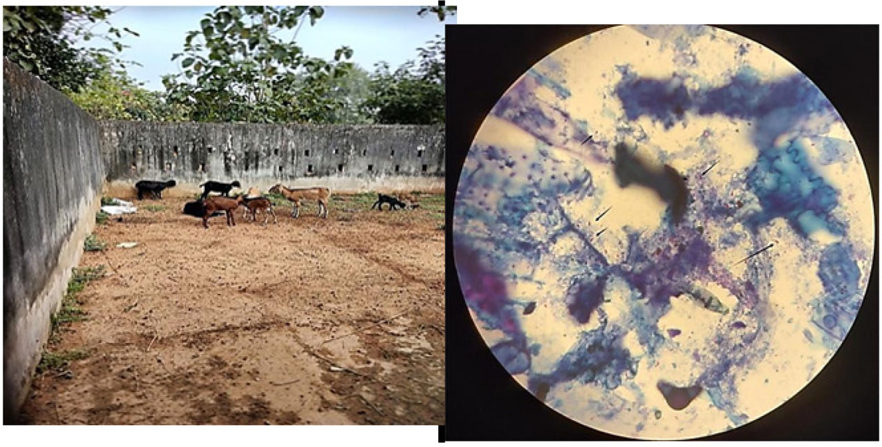
Figure 1.
Goats affected with PTB (left panel) and their faecal smear test (right panel). The left panel shows the isolated herd of goats affected with PTB in a study area of Odisha, India, and the right panel indicates a faecal smear showing acid-fast stained bacteria, as indicated by small black arrows.
Table 4.
Mode of transmission, pathogenesis and symptoms in animal
| Name of the animal | Mode of transmission | Pathogenesis | Symptoms detected | References |
|---|---|---|---|---|
| Goat (caprine species) | Ingestion of contaminated water, food, or milk from infected animals Intrauterine infection possible | Histopathology Ziehl-Neelsen staining Immunohistochemistry PCR (targeting IS900 sequence) Serological assays (e.g., PARACHEK® 2 Kit) | Subclinical stage: no visible symptoms Clinical stage: diarrhoea, severe weight loss, and poor body condition | Stefanova et al., 2024 b |
| Goat (caprine species) | Oral ingestion of contaminated faecal material, water, food, milk, or colostrum Potential intrauterine transmission | Enzyme-linked immunosorbent assay (ELISA), polymerase chain reaction (PCR) for MAP DNA detection, post-mortem necropsy and histopathological confirmation | Subclinical phase: MAP shedding in faeces without clinical symptoms Clinical phase: watery diarrhoea, weight loss, reduced milk production | Stefanova et al., 2024 a |
| Korean Black goats (Capra hircus aegagrus) | Transmission faecal–oral route, often occurring in ruminants, including goats, by ingesting contaminated feed or water | Chronic infectious enteric disease, leading to progressive intestinal inflammation and malabsorption | Weight loss, diarrhoea, reduced milk production, and poor body condition in infected animals | Lee et al., 2006 |
| Goat | Oral uptake through contaminated milk, water, feed, or the environment, ingestion of MAP shed in faeces or through contaminated tissues | MAP infects the host, primarily targeting the gastrointestinal tract, chronic granulomatous enteritis occurs, and in subclinical stages, bacteria proliferate intracellularly in macrophages, the pathogen can be shed intermittently in faeces. | Infected animals may not exhibit clinical signs initially, advanced cases show weight loss, diarrhoea, and reduced productivity | Jatav et al., 2022 |
| Cattle, sheep, goats | Oral ingestion of MAP through contaminated milk, water, feed, or environmental sources, inter-species transmission can occur, though less common | Chronic infectious granulomatous enteritis develops due to MAP infection, progression includes silent infection, subclinical stages, clinical disease, and advanced clinical disease stages. | Cattle: weight loss, diarrhoea, decreased milk production Sheep and goats: emaciation, anorexia, and severe disability | Borujeni et al., 2021 |
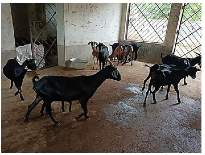
Figure 2.
MAP infected goats showing loss of body weight, emaciation.