
Caprine paratuberculosis (PTB), otherwise known as Johne’s disease in goats, is a persistent chronic enteropathic disease that affects domestic and wild ruminants ( Cunha et al., 2020; Idris et al., 2025; Islam et al., 2025), camelids, and only sporadically non-ruminants such as pigs, horses, carnivores, wild rabbits, and birds ( Curlik et al., 2020; Stanitznig et al., 2017), as well as humans ( Waddell et al., 2015). Caprine PTB is globally prevalent, including endemicity in India ( Kumar et al., 2007; Singh et al., 2013 b; Singh et al., 2014; Garg et al., 2015; Sonawane et al., 2016; Mukartal et al., 2017), and is a major concern in many developed countries, resulting in increased economic losses (Singh et al., 2013 a; Bruczyńska et al., 2023). This economic setback to the goat industry affects the meat, wool, and dairy sectors globally ( Sonawane and Tripathi, 2013; Rawat et al., 2014). The first report of goat PTB was made more than a hundred years ago ( Twort and Ingram, 1912). In India, the first case was reported by Sheather from a military dairy farm in Lahore of undivided India before independence ( Tripathi, 2007).
The disease is caused by an acid-fast bacterium, Mycobacterium avium subsp. paratuberculosis (MAP) (Manning and Collins, 2001; Singh et al., 2013 b; Chaturvedi et al., 2017; Hansen et al., 2019). Infection by MAP causes high morbidity and low fatality ( Khodakaram Tafti and Rashidi, 2000) and is classified as a notifiable disease as per the World Organisation for Animal Health (WOAH) ( Sevilla et al., 2015; Sharma et al., 2017). In most cases, goats become infected early in life, mainly through the faecal–oral route, while clinical signs appear in adults after a prolonged incubation period (Jørgensen, 1982; Manning and Collins, 2001; Khare et al., 2008; Senturk et al., 2009). The disease is characterised by wasting, intermittent diarrhoea, submandibular oedema, and reduced milk yield, primarily in adult goats (Barad et al., 2014 a; Beaudeau et al., 2007; Maity et al., 2017; Mukartal et al., 2017).
Clinical diagnosis of caprine PTB remains a challenge in field conditions due to its subclinical nature and limited awareness among farmers ( NASR, 2003; Chaturvedi et al., 2017). Early detection is hindered by slow disease progression and long incubation periods, allowing the disease to evade recognition until advanced stages ( Tharwat et al., 2025). Despite multiple reports from different parts of the globe, it has not received adequate recognition and priority in countries such as India (Singh et al., 2013 b, e). Detection and control are further complicated by the slow growth of MAP in faecal culture and its prolonged environmental persistence (Singh et al., 2013 c). Subclinically affected animals shedding bacteria through faeces and milk pose a significant risk, acting as silent sources of infection and negatively affecting herd productivity (Singh et al., 2010 a; Pithua and Kollias, 2012; Millar et al., 1996; Hansen et al., 2019).
MAP has zoonotic potential in the food chain, with possible links to Crohn’s disease in humans (Singh et al., 2010 a, b; Whittington et al., 2011; Pithua and Kollias, 2012; Sechi and Dow, 2015). Paratuberculosis is an incurable disease, and a “test and slaughter” policy is recommended to minimise bacterial load in susceptible and infected herds ( Sharma et al., 2017). Vaccination of large populations of small ruminants remains challenging in countries like India, raising concerns about the potential loss of sheep and goat diversity due to limited awareness ( Sharma et al., 2017). The disease significantly affects livelihoods, particularly among small-scale farmers, by reducing herd productivity ( Leite et al., 2013; Momotani, 2012; Sweeney et al., 2012; Barad et al., 2014 b).
In the context of the public health importance of the One Health approach, a clearer understanding of the epidemiology and current status of MAP infection is essential for developing effective control strategies. This can enhance animal production and productivity through improved herd health, supported by vaccine development, genomic studies, cost-effective diagnostics, precision farming, disease resistance, selective breeding, and microbiome- and omics-based health monitoring. Vaccination of large wild populations, however, remains a major challenge.
To review the status of PTB in goats, major databases such as PubMed, ScienceDirect, Web of Science, Google Scholar, Agricola, and Scopus were searched using keywords such as goat disease, breed, PTB, ‘treatment’, and ‘epidemiology’, alone or in combination. Peer-reviewed literature published in English was included after manual screening, without discrimination based on country, region, religion, race, or authorship. A total of 114 records for “paratuberculosis” in PubMed were initially screened. Similarly, 91,200 and 5,649 records from Google Scholar were reduced to fewer than 400 using additional keywords. Titles were further screened manually for relevance. Articles containing only search terms without relevant content were excluded, while those within the scope were included. Finally, 196 articles were selected for this review.
Recent advances highlight improved molecular diagnostics, particularly quantitative PCR (qPCR) and ELISA-based methods, for early detection at individual and herd levels (Table 1). However, sub-clinical infection remains a major limitation. A better understanding of host-pathogen interactions, immune evasion, and genetic susceptibility is needed. Strengthening test-and-cull programmes, farm hygiene, colostrum management, and biosecurity, along with the development of vaccines and genomic tools for resistance, is essential for effective long-term management of PTB. This review summarises updated information on the epidemiology of PTB in goats, including transmission, clinical manifestations, pathogenesis, and prevalence influenced by gender, age, and breed. Diagnostic approaches include conventional serological surveillance, biochemical, histopathological, and haematological methods, along with molecular techniques such as PCR and ELISA ( Kawaji et al., 2007; Vansnick et al., 2004). Literature published up to mid-February 2026 has been included.
Table 1.
Current status of the disease PTB across different counties
| Name of the animals | Region | Infection status | Economic loss | Strategy to counter the disease | References |
|---|---|---|---|---|---|
| 1 | 2 | 3 | 4 | 5 | 6 |
| Sheep and goats | Global distribution especially in Australia, Greece, Norway, Austria, USA, and Chile | Chronic bacterial disease having varying prevalence | Reduced productivity, mortality up to 20% annually in some Australian sheep flocks, costs of disease control, market restrictions | Vaccination Improved biosecurity measures Test-and-cull strategies | Windsor, 2015 |
| Cattle, goats, sheep, water buffalo, and deer | Latin American countries | Chronic gastrointestinal disease and possibility of Crohn’s disease link | No direct estimates for Latin America, U.S. losses range from $200 to 250 million annually along with production losses | Improved diagnostics (PCR, ELISA), enhanced management, and research on circulating strains | Espeschit et al., 2017 |
| Goats | Andalusia, Southern Spain | Chronic disease (22.54% true seroprevalence in goats, 87.5% seropositive herds) | Premature culling, reduced milk production, and high cost of control measures | Risk reduction measures, batch management, ventilation improvements, and potential control programmes | Barrero--Domínguez et al., 2019 |
| Boer goats (Capra hircus) | Missouri, USA | Endemic Animal-level prevalence: 1.4% Herd-level prevalence: 54.7% | Reduced milk yield Lower reproductive efficiency Increased mortality and premature culling risks | Management interventions depending on herd infection levels Surveillance for uninfected herds Long-term risk-based control in high-prevalence herds | Pithua and Kollias, 2012 |
| Goats (Capra hircus) producing milk | Global distribution (study conducted in Europe, America, Asia, and Brazil) | Detection in milk samples: ELISA: 1.1%–67.7% (wide variation among regions) PCR: 1.94%–37.74%, Combined global prevalence: 8.24% | Reduced productivity and marketability of goat milk Potential impact on consumer health due to MAP presence in milk | Use of sensitive diagnostic tests (ELISA and PCR) Control programmes targeting milk safety Improved hygiene practices during milk production | de Lacerda Roberto et al., 2021 |
| Cattle, sheep, and goats | Latin America and the Caribbean | Infection widespread in ruminants, occurrence of Johne’s disease characterised by diarrhoea, weight loss, and decreased productivity | Decreased milk production, weight loss, and increased mortality rates | Improved diagnostic methods like PCR and genotyping Implementation of control measures Development of vaccines tailored to specific MAP genotypes in the region | Correa-Valencia et al., 2021 |
| Sheep and goats | Austria | Moderate infection rate Seroprevalence for goats was 2.0% and for sheep, 0.7% Herd-level seroprevalence was 11.1% for goat herds and 8.9% for sheep flocks | Decreased productivity | Testing and culling MAP-positive animals Implementing preventive hygienic measures on farms Use of boot swab sampling for herd-level detection | Schrott et al., 2023 |
| Goats | Spain (experimental and field conditions) | Vaccinated and infected goats Vaccination triggers immune responses measured by interferon-gamma (IFN-ɣ) production | Significant impact due to its chronic nature and effects on livestock productivity | Vaccination Use of interferon-gamma release assays (IGRA) to evaluate immune responses post-vaccination or infection Development of new antigens to reduce cross-reactivity | Fernández et al., 2022 |
| Sheep | North of Portugal | 3.7% seroprevalence observed in sheep, with herd sizes of 31–60 showing the highest prevalence | Productivity loss and additional management costs | Limiting young stock’s contact with adults Improved hygiene practices Avoid spreading manure on grazing pastures during the same season | Quintas et al., 2021 |
Epidemiology and prevalence of PTB
Age-specific prevalence
The infection is more common in goats older than 2 years, although it can also occur in younger animals (Mishra et al., 2024 a). Kumar et al. (2007) studied PTB in young kids using six diagnostic methods (microscopy, faecal and tissue culture, ELISA, histopathology, and PCR) and reported the first incidence of juvenile caprine PTB in India. The positive rates observed using tissue culture in kids aged 15 days to 2 months, 2–4 months, and 4–6 months were 6.6%, 86.3%, and 40.6%, respectively. Singh et al. (2007 a) reported low seroprevalence in 2-month-old kids of Jakhrana and Marwari breeds. Lybeck et al. (2011) observed faecal shedding in 18% and 40% of goats at 9 and 15–17 months of age, respectively, with shedding often preceding peripheral immune responses. Although infection may occur at any stage, it becomes clinically apparent mainly in goats aged 2–4 years (Stefanova et al., 2024 a, b). Hemalatha et al. (2013 b) reported mortality across a wide age range, from 4 months to 10 years (Table 2).
Table 2.
Global incidence of the disease PTB
| Name of the animal | Distribution | Percentage of animals affected | Detection method | References |
|---|---|---|---|---|
| Sheep and goat | Lisbon | 27% of the flocks had seropositive animals | Serological examination | Mendes et al., 2004 a |
| Goats and sheep | Mexico | Widespread incidence in goats and sheep | PCR and bacterial culture | Estévez-Denaives et al., 2007 b; Claudia et al., 2016 b |
| Goats | Belgium | Seroprevalence was found to be 6% | ELISA | Lilenbaum et al., 2007 b |
| Dairy goats | France | Herd level apparent prevalence is 55.2%, estimated true prevalence 62.9% and at individual animal level they were 2.9 and 6.6%, respectively | Serological examination | Mercier et al., 2010 a |
| Sheep | Uttar Pradesh, India | 18% of 423 sheep showing paratuberculosis infection in an organized farm | Faecal smear examination | Paliwal et al., 1984 a |
| Goats | Maharastra, India | Prevalence of Johne’s disease was 8.7% | Serological examination | Srivastava and More, 1987 b |
| Goats | India | Overall incidence of Johne’s disease in goats to be 8.76% | post mortem examination | Kumar et al., 1988 a |
| Goats | North Indian states | 23.1% goats from Mathura (19.6%), Etawah (37.5%), Agra (40%) and Ajmer (21.9%) were affected | ELISA | Singh et al., 2010 b, c |
| Sheep | India | 7.4% prevalence of Johne’s disease | Faecal smear examination by Ziehl Neelsen (ZN) staining | Rahman, 1985 a |
| Goats | Odisha, India | Prevalence of 68.19% | ELISA | Biswal et al., 2018 a |
Breed-specific prevalence
Caprine PTB varies among breeds, possibly due to genetic factors as well as management and monitoring practices. Singh et al. (2007 c) reported a higher prevalence in the Sirohi breed compared to the Barbari breed using microscopy and an indigenous ELISA. Singh et al. (2007 b) reported breed-wise prevalence in Jakhrana, Sirohi, and Marwari breeds as 10.5%, 7.6%, and 0%, respectively. Singh et al. (2009 b) found that semi-arid zone breeds (Barbari and Jamunapari) were more susceptible, whereas arid zone breeds (crossbred Rajasthani goats) were comparatively resistant. Breed specific prevalence of PTB is summarized in Table 3.
Table 3.
Different affected breed types in the disease PTB
| Name of the animal | Types of breeds | Percentage of animals affected | Detection method | References |
|---|---|---|---|---|
| Goats | Barbari and Sirohi breeds | Higher prevalence in Sirohi breed (40.5%) than in Barbari breed (20.6%) | Direct microscopy and an indigenous ELISA kit | Singh et al., 2007 c |
| Goats | Jakhrana, Sirohi and Marwari breeds | Jakhrana 10.5%, Sirohi 7.6%, and Marwari nil, respectively | Serological examination | Singh et al., 2007 b |
| Goats | Semi-arid zone breeds (Barbari and Jamunapari) and arid zone breeds (crossbred Rajasthani goats) | Semi-arid zone breeds were more susceptible to MAP infection, while arid zone breeds were comparatively resistant | Serological examination | Singh et al., 2009 b |
| Sheep | Local sheep breeds (Nilagiri, Sandyno) and exotic breed Dorset | Local sheep breeds were more vulnerable than exotic breed | Serological examination | Hemalatha et al., 2013 a |
| Goats | Mehsana and Surti breeds from the South Gujarat | Positivity rates were 9.2% FS examination, 21.9% DTH, 10.9% AGID, 12.5% IS900 PCR and 43.3% by indigenous ELISA test kit | Conventional, genetic, and serological examination | Barad et al., 2014 a |
| Goats | Gaddi goat breed | Sensitivity was found as 74.35% ELISA, 61.53% AGID, 46.15% IS900/IS1311 tissue PCR, 43.58% acid-fast staining of cytology and 35.89% tissue section | Different diagnostic methods | Thakur et al., 2019 a |
| Goats | Ganjam and Black Bengal and Raighar goat breeds | 42.30% in Ganjam breed, 35.93% in Black Bengal breed, while lowest in Raighar breed, about 29.63% | Serological examination | Mishra et al., 2024 a |
Reported prevalence rates in Indian breeds include 40.5%, 20.6%, 10.5%, 7.6%, 9.2%, 42.30%, 35.93%, and 29.63% in Sirohi, Barbari, Jakhrana, Sirohi, Mehsana and Surti, Ganjam, Raighar, and Black Bengal breeds, respectively ( Singh et al., 2007 d, e, 2009 b; Barad et al., 2014 a; Mishra et al., 2024 a). Semi-arid breeds (Barbari, Jamunapari, and crossbred Rajasthani goats), as well as Mehsana and Surti breeds from Gujarat and local sheep breeds (Nilagiri and Sandyno), have also been reported to be infected (Barad et al., 2014 a; Thakur et al., 2019 b). Across regions, breed–environment interactions influence PTB prevalence. For example, the Ganjam breed showed higher prevalence than other breeds in India (Mishra et al., 2024 b) as evidenced through routine faecal smear examinations in Odisha (Figure 1).
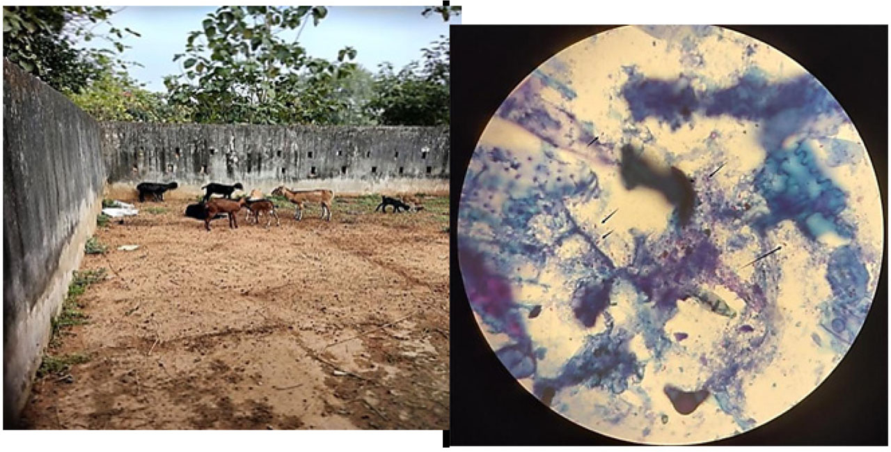
Figure 1.
Goats affected with PTB (left panel) and their faecal smear test (right panel). The left panel shows the isolated herd of goats affected with PTB in a study area of Odisha, India, and the right panel indicates a faecal smear showing acid-fast stained bacteria, as indicated by small black arrows.
Higher infection frequency has been reported in local animals compared to imported ones ( Shabana and Aljohani, 2020). Diagnostic approaches such as faecal smear, delayed-type hypersensitivity, agar gel immunodiffusion, PCR, and ELISA have shown variable detection rates (Barad et al., 2014 b; Thakur et al., 2019 b), indicating differences in sensitivity among methods.
Sex-specific prevalence
Female goats generally show higher PTB prevalence, possibly due to their longer lifespan. Hemalatha et al. (2013 a) reported higher prevalence in females than males. Similarly, Shabana and Aljohani (2020) observed greater infection frequency in females. In Odisha, prevalence was 39.16% in females and 34.67% in males (Mishra et al., 2024 a). However, further studies are required to establish definitive conclusions.
Global incidence
PTB prevalence in goats varies across countries and depends on diagnostic methods. The disease has been widely reported across Asia, Africa, the Arabian Peninsula, and Europe ( Islam et al., 2025). Herd-level prevalence in Europe, the USA, and Australia is in the range of 7–55%, around 40%, and 9–22%, respectively. Studies have reported widespread infection in countries such as the Netherlands ( Collins et al., 2001), Portugal (27% herd seropositivity; Mendes et al., 2004 a), Mexico (Estévez-Denaives et al., 2007 a; Claudia et al., 2016 a), Belgium (6%; Lilenbaum et al., 2007 b), and Poland (46.5%; Kaba et al., 2024). MAP has also been detected in zoo herbivores (Bruczyńska et al., 2023; Didkowska et al., 2021).
In some regions, such as Sudan, PCR-based detection shows higher prevalence than ELISA, indicating the importance of subclinical infections ( Tharwat et al., 2025). Molecular methods such as PCR provide more reliable detection in such cases ( Lee et al., 2025). A study in Denmark reported 70% herd-level positivity based on bulk milk testing. In France, herd-level prevalence was estimated at 55.2% (apparent) and 62.9% (true), while individual-level prevalence was 2.9% and 6.6%, respectively (Mercier et al., 2010 a). In Taiwan, very low prevalence rates were reported between 2011 and 2015 ( Lin et al., 2018), highlighting geographical variation.
India has the second-largest goat population globally, and PTB is endemic. Early reports include cases from Lahore ( Twort and Ingram, 1912; Sheather, 1918; Figure 1). Subsequent studies have reported prevalence across regions, including 8.7% in Maharashtra ( Srivastava and More, 1987 a), 31.9% in Uttar Pradesh, 23.3% in Punjab ( Singh et al., 2008), and around 29% in northern India. MAP seroprevalence of 23.1% has been reported in domestic animals (Singh et al., 2010 b), with regional variation in goats from Mathura (19.6%), Etawah (37.5%), Agra (40%), and Ajmer (21.9%). Higher prevalence (68.19%) has been reported in Odisha (Biswal et al., 2018 a). These findings indicate significant spatial variation in PTB prevalence across India and globally.
The incidence of MAP infection in sheep and goats varies worldwide; for example, mortality rates of up to 20% per year have been reported in certain Australian sheep herds ( Windsor, 2015). Similar economic losses, ranging from $200 to $250 annually in Latin America, have been reported due to reduced milk yield, culling, and related effects in cattle, goats, sheep, and other ruminants ( Espeschit et al., 2017). Barrero--Domínguez et al. (2019) reported high infection rates (87.5% seropositive herds) in Andalusian goat herds in Spain, leading to early culling, reduced milk production, and increased control costs. In Missouri, USA, MAP is endemic in Boer goats, with a herd-level prevalence of 54.7%, associated with impaired reproductive efficiency, increased mortality, and premature culling ( Pithua and Kollias, 2012).
A global meta-analysis in milk-producing goats estimated an overall prevalence of 8.24%, with ELISA-based detection ranging from 1.1% to 67.7% and PCR-based detection from 1.94% to 37.74% ( de Lacerda Roberto et al., 2021). Positive reports of MAP have also been documented in Austrian sheep and goats, with higher herd-level seropositivity in goats (11.1%) than in sheep (8.9%) ( Schrott et al., 2023). In northern Portugal, mid-sized sheep flocks showed a seroprevalence of 3.7% ( Quintas et al., 2021). Preventive measures such as vaccination, limiting contact between young and adult animals, improved hygiene, and controlled manure management have been suggested ( Correa-Valencia et al., 2021; Fernández et al., 2022; Schrott et al., 2023). Although recent studies indicate that vaccination may reduce clinical manifestations and induce immune responses (IFN-γ and antibodies), complete prevention has not been achieved ( Royo et al., 2025).
Caprine PTB is endemic in Odisha, India ( Biswal et al., 2020; Mishra et al., 2024 a). Biswal et al. (2020) detected subclinical PTB using 122 EDTA-mixed blood samples and 121 serum samples, with 19.01% and 70.25% positivity, respectively, by indirect ELISA. Genotyping using IS1311 PCR-REA indicated predominance of the ‘Indian Bison’ type strain. Overall seroprevalence in Odisha was reported as 38.75%, with subclinical cases (30.68%) more prevalent than clinical cases (8.06%) (Mishra et al., 2024 a).
Studies worldwide, including India, consistently indicate variable but often high prevalence of PTB (Mendes et al., 2004 b; Estévez-Denaives et al., 2007 a; Claudia et al., 2016 a; Lilenbaum et al., 2007 a; Mercier et al., 2010 a; Paliwal et al., 1984 a; Srivastava and More, 1987 a; Kumar et al., 1988 a; Singh et al., 2010 b; Rahman, 1985 a; Biswal et al., 2018 a). For example, 27% seropositivity was reported in Lisbon (Mendes et al., 2004 a), while in Mexico the disease was widespread among small ruminants (Estévez-Denaives et al., 2007 b; Claudia et al., 2016 b). ELISA-based screening showed 6% prevalence in Belgium (Lilenbaum et al., 2007 a), and herd-level prevalence in France reached 62.9%, with lower individual-level rates (Mercier et al., 2010 a). In India, prevalence varies by region, including 18% in Uttar Pradesh (Paliwal et al., 1984 b), 8.7% in Maharashtra ( Srivastava and More, 1987 a), and up to 23.1% overall in northern states, with higher rates in Agra (40%) and Etawah (37.5%) (Singh et al., 2010 b). In eastern India, particularly Odisha, a high prevalence of 68.19% has been reported using ELISA (Biswal et al., 2018 b).
Table 4.
Mode of transmission, pathogenesis and symptoms in animal
| Name of the animal | Mode of transmission | Pathogenesis | Symptoms detected | References |
|---|---|---|---|---|
| Goat (caprine species) | Ingestion of contaminated water, food, or milk from infected animals Intrauterine infection possible | Histopathology Ziehl-Neelsen staining Immunohistochemistry PCR (targeting IS900 sequence) Serological assays (e.g., PARACHEK® 2 Kit) | Subclinical stage: no visible symptoms Clinical stage: diarrhoea, severe weight loss, and poor body condition | Stefanova et al., 2024 b |
| Goat (caprine species) | Oral ingestion of contaminated faecal material, water, food, milk, or colostrum Potential intrauterine transmission | Enzyme-linked immunosorbent assay (ELISA), polymerase chain reaction (PCR) for MAP DNA detection, post-mortem necropsy and histopathological confirmation | Subclinical phase: MAP shedding in faeces without clinical symptoms Clinical phase: watery diarrhoea, weight loss, reduced milk production | Stefanova et al., 2024 a |
| Korean Black goats (Capra hircus aegagrus) | Transmission faecal–oral route, often occurring in ruminants, including goats, by ingesting contaminated feed or water | Chronic infectious enteric disease, leading to progressive intestinal inflammation and malabsorption | Weight loss, diarrhoea, reduced milk production, and poor body condition in infected animals | Lee et al., 2006 |
| Goat | Oral uptake through contaminated milk, water, feed, or the environment, ingestion of MAP shed in faeces or through contaminated tissues | MAP infects the host, primarily targeting the gastrointestinal tract, chronic granulomatous enteritis occurs, and in subclinical stages, bacteria proliferate intracellularly in macrophages, the pathogen can be shed intermittently in faeces. | Infected animals may not exhibit clinical signs initially, advanced cases show weight loss, diarrhoea, and reduced productivity | Jatav et al., 2022 |
| Cattle, sheep, goats | Oral ingestion of MAP through contaminated milk, water, feed, or environmental sources, inter-species transmission can occur, though less common | Chronic infectious granulomatous enteritis develops due to MAP infection, progression includes silent infection, subclinical stages, clinical disease, and advanced clinical disease stages. | Cattle: weight loss, diarrhoea, decreased milk production Sheep and goats: emaciation, anorexia, and severe disability | Borujeni et al., 2021 |
Clinical aspects of PTB in goats
Transmission mode of PTB
MAP is mainly transmitted through ingestion of feed or water contaminated with manure or faeces ( Lee et al., 2006; Borujeni et al., 2021; Jatav et al., 2022; Stefanova et al., 2024 a). Kids are commonly infected through consumption of contaminated colostrum and milk from infected dams. Transplacental transmission from an infected pregnant mother to the foetus has also been reported ( Park et al., 2017). Introduction of infected animals or carrier replacements into a herd is a major source of spread ( Barrett et al., 2011; Table 4). Wild rabbits may act as reservoir hosts, as reported in Scotland, where they excreted MAP in their faeces ( Greig, 1997).
In natural conditions, passive shedding may occur when MAP is detected in faeces without evidence of infection at necropsy, typically following oral ingestion without establishment of infection ( Whittington et al., 1998). In contrast, active infection involves multiplication of MAP in intestinal epithelial cells, followed by shedding through faeces and milk, often associated with clinical signs such as diarrhoea and weight loss. MAP can persist for long periods in the environment after being excreted in faeces, milk, and other secretions ( Eisenberg et al., 2010), making environmental contamination a major source of infection ( Ayele et al., 2001).
Clinical signs in PTB
PTB is a slowly progressive disease with a long incubation period, typically 2 or more years in small ruminants ( Kopecky and Larsen, 1975). The disease progresses through multiple stages. In the subclinical stage, animals do not show visible signs, and detection relies on identification of the organism through culture or histopathological examination. As the disease progresses to the clinical stage, animals exhibit characteristic signs such as progressive weight loss, diarrhoea, depression, and emaciation (Figure 2). Diagnosis at this stage is supported by faecal culture and increased serum antibody levels. In advanced stages, more severe manifestations such as persistent “pipe-stream” diarrhoea and inter-mandibular oedema may occur ( Coussens, 2004; Sohal et al., 2007, 2019; Mishra et al., 2024 b).
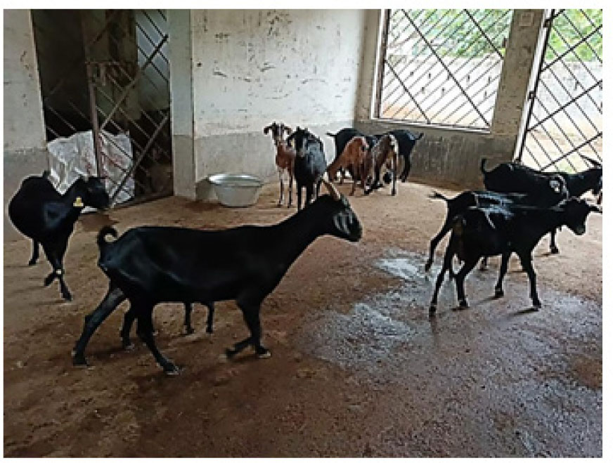
Figure 2.
MAP infected goats showing loss of body weight, emaciation.
The early stage of the disease manifests as progressive weight loss, decreased milk and meat yield, and premature culling, which may affect udder health and fertility. Infection may progress to a clinical form that includes chronic diarrhoea, emaciation, debilitation, and death ( Tiwari et al., 2006). Kruze et al. (2006) studied PTB in goats in Chile and noted characteristic clinical signs such as a dry and rough hair coat, emaciation despite a good appetite, and absence of diarrhoea. Hasonova and Pavlik (2006) reported progressive weight loss with or without diarrhoea, emaciation, and poor body condition. Production losses result from reduced milk yield, increased susceptibility to mastitis, high somatic cell counts in milk, and poor carcass quality ( Mohanty et al., 2022). Shulaw (1900) observed profuse watery diarrhoea in cattle, which is not a common feature in sheep and goats. Appetite is often maintained until advanced stages despite weight loss. Even in clinically affected animals, normal stools are common, although pasty or loose faeces may occasionally be observed. Eslami (2011) reported progressive weight loss as a key clinical sign.
Caprine paratuberculosis in goats
Goats affected with Johne’s disease may show pasty or loose faeces, loss of appetite, emaciation, dullness, and alopecia (Singh et al., 2013 b, c). MAP may persist in visceral organs such as the intestine for years without clinical signs ( Verma, 2013), making anatomical examination useful for detection ( Navarro et al., 2025). Under stress conditions such as malnutrition, pregnancy, lactation, environmental stress, or poor management, subclinical infection may progress to clinical disease, with continued shedding of acid-fast bacilli in milk and faeces ( Garg et al., 2015). Malone et al. (2013) reported reduced weight gain in infected goats compared to non-infected animals. Scherrer et al. (2019) observed clinical signs including moderate to marked emaciation, weight loss, and intermittent diarrhoea.
Intrauterine and transmammary transmission of MAP has been reported, with higher occurrence in clinically affected animals ( Lambeth et al., 2004). Milk and colostrum play important roles in transmission, as viable organisms are excreted in these fluids ( Raizman, 2007). Vaginal secretions may also act as a source of infection and can be used for screening after kidding ( Singh et al., 2007 d, f). Infection in young animals occurs through ingestion of contaminated colostrum, milk, or faecally contaminated materials. Environmental contamination is a major risk factor, particularly under conditions such as shared water sources, communal grazing, and use of surface water ( Angelidou et al., 2014; Florou et al., 2008; Pradhan et al., 2011).
Pathogenesis in PTB
After ingestion of contaminated milk or colostrum, MAP reaches the intestinal tract via the oral route ( Lambeth et al., 2004) and is translocated across the intestinal mucosa through M cells in Peyer’s patches. It then enters macrophages in the subepithelium, where it multiplies slowly. This intracellular multiplication induces a cell-mediated immune (CMI) response ( Henderson et al., 2000), while the humoral immune (HMI) response is minimal in early stages ( Milner et al., 1990), explaining the absence of detectable antibodies.
As infection progresses and MAP is released from macrophages, a humoral immune response develops, leading to detectable antibody production and forming the basis for ELISA-based diagnosis ( Milner et al., 1990). Based on immune response patterns, MAP infection may present as multi-bacillary (lepromatous) or paucibacillary (tuberculoid) forms. The multibacillary form, commonly reported in sheep, is associated with strong humoral responses and granulomatous enteritis ( Clarke and Little, 1996; Corpa et al., 2000; Tripathi et al., 2002). In contrast, the paucibacillary form, more typical in goats, involves a strong CMI response with lymphocytic infiltration and few detectable organisms in the lamina propria ( Clarke and Little, 1996; Pérez et al., 1996; Catton, 2002; Krüger et al., 2015 a, b).
Detection of MAP and PTB
Diagnostic approaches for PTB include faecal smear examination using Ziehl–Neelsen staining, histopathology, PCR targeting the IS900 sequence, ELISA, immunohistochemistry, and post-mortem confirmation (Stefanova et al., 2024 a, b; Borujeni et al., 2021). Although clinical signs may suggest the disease, definitive diagnosis relies on laboratory methods, including histopathological, biochemical, molecular, haematological, and pathological markers.
Gross pathological study
Gross lesions in sheep and goats may differ from those in cattle and can be difficult to detect ( Clarke et al., 1996; Clarke, 1997). Findings include distended lymphatic vessels on the jejunal serosa, corrugated intestinal mucosa, and enlarged oedematous mesenteric lymph nodes ( Kruze et al., 2006; Manjunatha et al., 2012; Singh et al., 2013 a, b). Multiple granulomas with calcified cores may be observed in lymph nodes. Lesions often involve thickening of jejunal Peyer’s patches, ulceration, and serositis (Krüger et al., 2015 a, b; Derakhshandeh et al., 2018). Granulomatous infiltration may be present in gut-associated lymphoid tissue and intestinal lymph nodes, along with arteritis in the intestinal submucosa ( Tripathi et al., 2002).
Additional findings include serous atrophy of fat and thickening of the terminal ileum, particularly at the ileocaecal junction, with occasional corrugations ( Derakhshandeh et al., 2018; Cheng et al., 2020). The ileum and caecum may show wall thickening, oedema, and mucosal haemorrhage. In some cases, minimal or no gross lesions are observed in other intestinal segments ( Chaturvedi et al., 2017). Severe cases may show loss of adipose tissue and bone marrow atrophy ( Scherrer et al., 2019). Enlarged, discoloured, and rigid lymph nodes with caseous lesions may also be present ( Zhao et al., 2021).
Recent tissue analyses indicate increased malondialdehyde (MDA) and caspase-3 levels, suggesting oxidative stress and apoptosis, particularly in intestinal mucosa, with comparatively higher anti-oxidant activity in mesenteric lymph nodes. These findings highlight the role of oxidative imbalance in disease progression and suggest MDA and caspase-3 as potential diagnostic and prognostic biomarkers for caprine PTB ( Ozturk et al., 2025).
Histopathological marker study
Histopathological examination is important for PTB diagnosis in goats and is based on evaluation of intestinal tissues, mesenteric lymph nodes, and Ziehl–Neelsen (ZN)-stained faecal smears in natural or experimental cases ( Corpa et al., 2000; Tafti and Rashidi, 2000). Lesions are generally classified into four categories. Focal lesions consist of small granulomata in the ileocaecal Peyer’s patches. Diffuse multibacillary lesions involve granulomatous enteritis across multiple intestinal regions. Diffuse lymphocytic lesions are characterised mainly by lymphocyte infiltration with some macrophages, while diffuse mixed lesions show infiltration by both lymphocytes and macrophages.
Histopathological findings include infiltration of the lamina propria of the jejunum and ileum with inflammatory cells, predominantly lymphocytes, along with eosinophilic hyaline material in epithelial cells. Similar lymphocytic infiltration may also occur in the caecum and colon ( Kruze et al., 2006). In affected kids, lesions are prominent in mesenteric lymph nodes, where mononuclear cells coexist with a few epithelioid cells. Ileal villi may show epithelial degeneration and transformation into goblet cells containing homogeneous material, while Peyer’s patches appear hypercellular with occasional epithelioid cells ( Kumar et al., 2007).
Lesion severity is graded (I–IV) based on the extent of MAP bacilli infiltration and the type of cellular infiltrate ( Hailat et al., 2010; Hemalatha et al., 2013 a; Zhao et al., 2021). Grades III and IV are considered positive, whereas Grades I and II are considered doubtful. Lesions may include small granulomas with few epithelioid cells or larger granulomas with extensive macrophage and epithelioid cell infiltration containing acid-fast bacilli ( Manjunatha et al., 2012).
Additional findings include granulomatous enteritis with macrophage infiltration into the lamina propria, sometimes with foamy cytoplasm, and chronic lymphoplasmacytic inflammation ( Scherrer et al., 2019). Proliferation of epithelioid cells may form nodules in mesenteric lymph nodes. Histopathological changes are broadly similar across goat breeds (Barad et al., 2014 b; Thakur et al., 2019 b). Other lesions may include degeneration and denudation of epithelial cells, lymphangitis, lymphangiectasia, and caseous necrosis with calcification in lymph nodes (Paliwal et al., 1984 a; Rajkumar, 1998; Derakhshandeh et al., 2018).
Acid-fast staining of faecal samples and microscopy
Direct faecal microscopy is a rapid method for detecting acid-fast bacilli (AFB) using ZN staining (Singh et al., 2013 a, b). However, sensitivity is low, and differentiation between pathogenic and non-pathogenic mycobacteria is difficult. Detection of clumps of AFB (three or more organisms) is considered indicative of infection, whereas single organisms are not diagnostic ( OIE, 2004). False-negative results may occur in low shedders (Lilenbaum et al., 2007 b).
Comparative studies have shown varying detection rates among methods. Thoresen et al. (1994) reported higher positivity with immunohistochemistry compared to acid-fast staining and culture. Shah et al. (2012) found faecal smear examination to be rapid and cost-effective under field conditions, although sensitivity varies. Advanced approaches such as high-resolution NMR spectroscopy combined with multivariate analysis have also been explored for improved detection ( Alonso et al., 2025).
Haematology and biochemical markers for PTB diagnosis
MAP is biochemically inert; therefore, biochemical tests are not directly suitable for its detection ( Gunnarsson and Fodstad, 1979). However, PTB-affected animals show alterations in haematological and biochemical parameters. Reduced levels of albumin and total protein have been reported in affected goats ( Kruze et al., 2007), along with conditions such as leukopenia and anaemia (Hemalatha et al., 2013 b).
Changes observed in affected animals include decreased total protein, albumin, and glucose, and increased levels of blood urea nitrogen (BUN), AST, ALT, ALP, GGT, and GLDH ( El-Deeb et al., 2014). Subclinical infection may also affect physiological and production parameters ( Jurkovich et al., 2016). Elevated globulin fractions and total protein have been reported in infected goats, with variable changes in albumin and albumin/globulin ratio ( Bonelli et al., 2017).
Other studies have reported increased levels of chloride, creatinine, triglycerides, and cholesterol ( Sharma et al., 2017). Haematobiochemical alterations such as reduced RBCs, PCV, total protein, globulin, and glucose have been suggested as supportive indicators ( Abdelaal et al., 2019). Oxidative stress markers also show variation, with reduced antioxidant levels and increased lipid peroxidation ( Balikci and Gurdogan, 2015).
Bacterial culture for PTB detection
Isolation of MAP by culture remains the gold standard for PTB diagnosis ( Butot et al., 2019). Media such as Herrold’s egg yolk medium (HEYM), Löwenstein–Jensen (LJ) medium, and modified Middlebrook 7H10 agar are used for cultivation. While HEYM and LJ media are suitable for bovine samples, small ruminant strains often grow better on LJ medium without sodium pyruvate ( Juste et al., 1991). Modified Middlebrook 7H10 agar is recommended to improve isolation efficiency ( Whittington et al., 2011).
MAP has been isolated from goat milk samples in India ( Singh and Vihan, 2004), and recent studies have reported molecular identification in goats from Himalayan regions ( Altaf et al., 2025) and surveillance in different countries ( Baghbadorani et al., 2025; Rasper-Hössinger et al., 2025). Faecal culture is considered sensitive, particularly as shedding may occur before detectable immune responses. Liquid culture systems have also been used for detection in faecal and tissue samples ( De-Grossi et al., 2019).
Diagnosis of PTB using molecular and immunological tools
1. PCR and ELISA
PTB diagnosis is based on clinical signs along with laboratory tests recommended by WOAH; however, detection of subclinical infection remains challenging due to the absence of visible symptoms. Diagnostic approaches include faecal smear examination using Ziehl–Neelsen staining, culture of faecal and tissue samples, PCR, serology, and necropsy. Among these, faecal culture can detect infection months before the onset of clinical signs, although it is time-consuming. ELISA is widely used as a cost-effective and rapid method for large-scale herd screening and for detecting antibodies against MAP in serum and milk. However, its sensitivity is low during early infection stages due to delayed humoral immune response (Karlik et al., 2014).
PCR-based molecular detection, particularly targeting the IS900 sequence, provides high specificity for identifying MAP DNA in faecal and tissue samples ( Mishra et al., 2007; Naser et al., 2014). Quantitative PCR (qPCR) allows estimation of bacterial load with greater sensitivity, although it requires specialised equipment. Limitations of PCR include the presence of inhibitors in faecal samples and potential environmental contamination ( Kralik and Beran, 2015). Sensitivity may also vary due to intermittent shedding and low bacterial load in early or subclinical stages ( Plain et al., 2014).
The IS900 element is the most commonly used genetic marker for MAP detection ( Collins et al., 1990; Douarre et al., 2010; Bhat et al., 2018; Hansen et al., 2019; Mishra et al., 2024 a). Although faecal culture remains the gold standard, it may require up to 16 weeks or more and has reduced sensitivity in subclinical cases ( Chaitanya et al., 2022). Pre-treatment of samples and specific growth requirements further increase cost and complexity ( Reddacliff et al., 2003; Randall and Collins, 1992).
For clinical cases, confirmation is typically achieved using faecal culture, histopathology, and PCR, whereas ELISA and direct faecal PCR are recommended for early or subclinical detection and herd-level screening ( Chaitanya et al., 2022). PCR-based methods, including real-time PCR, are widely used in surveillance programmes due to higher sensitivity compared to culture ( Bogli et al., 2005). Variants such as nested PCR have shown higher sensitivity than single or fluorescent PCR methods ( Englund et al., 2001; Fang et al., 2002).
Multiplex PCR assays targeting multiple sequences (e.g., 16S rRNA, IS900, and F57) enhance specificity ( Tasara et al., 2005). DNA-based subtyping techniques, including IS900 RFLP and IS1311 PCRREA, have been used for strain differentiation. PCR has been applied for detection of MAP in various sample types, including blood, faeces, and milk (Singh et al., 2010 a; Eslami et al., 2011; Millar et al., 1996; Mishra et al., 2024 a).
Advanced approaches include SNP-based assays for strain typing (Leão et al., 2016) and exosomal miRNA profiling as a potential non-invasive biomarker for PTB detection ( Bok et al., 2026). Loop-mediated isothermal amplification (LAMP) has also been reported as a rapid detection method with high sensitivity ( Wimalasena et al., 2025). Overall, ELISA is suitable for herd-level screening, while PCR-based methods are preferred for early detection and confirmation. A combined approach improves diagnostic accuracy and supports effective control of caprine PTB.
2. Detection of the immune responses
Commonly used methods for detecting immune responses against MAP include enzyme-linked immunosorbent assay (ELISA), complement fixation test (CFT), agar gel immunodiffusion (AGID), interferon-γ (IFN-γ) release assay, and delayed-type hypersensitivity (DTH). Immunological as-says such as ELISA detect antibodies in serum corresponding to the host response against MAP ( Yokomizo et al., 1983). It is the most widely used assay for quantifying anti-MAP antibodies in serum and milk (Sharma et al., 2008).
The sensitivity of ELISA is approximately 15% in livestock shedding low amounts of MAP in faeces, compared with up to 87% in animals with clinical disease ( Sweeney et al., 1995). ELISA and AGID are more sensitive for detecting multibacillary forms of infection but show reduced sensitivity in paucibacillary cases ( Clarke et al., 1996). Similarly, ELISA is more effective in detecting high shedders than low shedders; however, its sensitivity may decline over time in herds undergoing repeated testing, as high shedders are typically culled first ( Whitlock et al., 2000).
The sensitivity of ELISA in subclinical animals is lower than that of culture-based methods ( Kumar et al., 2007). Consequently, PCR is often preferred for early and rapid detection ( Abraham et al., 2014). Nevertheless, ELISA remains valuable for surveillance due to its ability to process large numbers of samples simultaneously ( Singh et al., 2019). Reported diagnostic sensitivities in Gaddi goats for ELISA, AGID, gross pathology, IS900/IS1311 tissue PCR, and Ziehl–Neelsen staining are 74.35%, 61.53%, 46.15%, 43.58%, and 35.89%, respectively (Thakur et al., 2019 a).
Alternative immunodiagnostic approaches include colloidal gold nanoparticle-based dipstick assays using MAP protoplasmic antigen ( Karthik et al., 2011). Although less sensitive than ELISA and PCR, these assays are simple and suitable for large-scale field screening. Recent studies have also reported improved ELISA specificity using MAP-derived polypeptides with minimal cross-reactivity in goats ( Moyano et al., 2026).
Zoonotic potential of PTB
Although PTB is not definitively classified as a zoonotic disease, Mycobacterium avium subsp. paratuberculosis has been detected in patients with Crohn’s disease (CD) ( Waddell et al., 2015). A report by the National Association for Colitis and Crohn’s Disease (UK) suggested a possible association between MAP and CD (NACC, 2003).
MAP has been detected in milk from infected animals, making dairy products a potential source of human exposure ( Sweeney et al., 1992; Miller et al., 2002; Favila-Humara et al., 2010; Singh et al., 2019). Notably, MAP may survive pasteurization under certain conditions ( Grant et al., 2002; Chaubey et al., 2016, 2017). The organism has also been identified in meat and environmental samples, indicating broader exposure risks. Consumption of contaminated or undercooked animal products, particularly intestinal tissues, may increase the likelihood of exposure ( Sechi et al., 2005; Mpenda and Buza, 2019). Additional potential transmission routes include contaminated ground beef from older dairy animals and breast milk from CD-affected women, suggesting possible neonatal exposure ( Rossiter and Henning, 2001; Naser et al., 2000).
Clinical manifestations of Crohn’s disease include abdominal pain, nausea, weight loss, night sweats, oral ulcers, and joint complications. In severe cases, diarrhoea with blood and pus, along with perianal inflammation and ulceration, may occur ( Hermon-Taylor and El-Zaatari, 2004). Epidemiological studies have reported MAP detection in animal handlers, with higher prevalence among individuals suffering from gastrointestinal disorders and those exposed to infected herds ( Shisodiya et al., 2009; Singh et al., 2011). Overall, while a causal relationship between MAP and Crohn’s disease remains inconclusive, accumulating evidence suggests a potential public health concern that warrants further investigation.
Prevention, control and future scope
There is no effective treatment available for PTB; therefore, prevention and control rely primarily on biosecurity, sanitation, and improved herd management practices. Regular screening of herds is essential, and manure-free areas should be designated for parturition, as faecal contamination is a major source of infection. Because MAP can be excreted in milk and colostrum, feeding pasteurised or heat-treated colostrum to kids is recommended. Heat treatment (e.g., 60°C for 60 min) has been shown to significantly reduce pathogen load, including MAP, E. coli, Salmonella enteritidis, and Listeria monocytogenes, without substantially affecting colostral IgG levels ( Godden et al., 2006; Kryzer et al., 2015).
Mixed farming systems, where goats are reared alongside cattle, may facilitate inter-species transmission of MAP. The bacterium has been isolated from goats, sheep, cattle, and humans, highlighting the need for greater attention to the socioeconomic and epidemiological implications of such practices. Critical gaps remain in understanding transmission dynamics, molecular epidemiology, host susceptibility, and the role of the gut microbiome in resistance to MAP infection.
Current control strategies include test-and-cull programmes, segregation of infected animals, and vaccination where available; however, their effectiveness is limited by practical and economic constraints. Research on pooled sampling for surveillance is limited, and cost-effective point-of-care diagnostic tools for goats are still lacking. The development of goat-specific vaccines with DIVA (Differentiating Infected from Vaccinated Animals) capability remains an unmet need.
Further research is required on the zoonotic potential of MAP, particularly in the context of the One Health approach. Comprehensive studies on the genetic diversity of MAP and its environmental persistence are essential for developing global control policies. Additionally, identification of reliable biomarkers for early and subclinical infection is critical. The role of wildlife reservoirs in disease transmission also requires detailed investigation. Future priorities should focus on improving herd hygiene, strengthening biosecurity, refining test- and-cull strategies, and advancing vaccine development. Integration of molecular diagnostics, genomic selection for disease resistance, and coordinated surveillance frameworks will be key to sustainable PTB control ( Tharwat et al., 2025).
Conclusion
MAP infection in goats leads to chronic enteric disease characterised by progressive weight loss, reduced productivity, and significant economic losses. This review consolidates current knowledge on the epidemiology, transmission, pathogenesis, and diagnosis of PTB, incorporating both conventional and advanced diagnostic approaches.
Despite progress in molecular and serological diagnostics, subclinical infections continue to hinder early detection and effective control. Strengthening biosecurity measures, implementing targeted test-and-cull strategies, and developing improved vaccines and diagnostic tools are essential for long-term management.
PTB remains under-recognised in many regions due to limitations in diagnostic capacity and awareness. Consequently, it persists as a “silent” disease with substantial impact on small ruminant production. Enhanced research efforts, improved surveillance, and increased stakeholder awareness are necessary to control disease spread and minimise its economic and public health implications.
Acknowledgements and declarations
Thanks to Honorable Vice Chancellor, Odisha University of Agriculture and Technology (OUAT) and Dean, College of Veterinary Science and Animal Husbandry, OUAT, Bhubaneswar, Odisha, India, for providing laboratory facilities. DBT Star College Scheme awarded to BRP (HRD-11011/33/2022-HRD-DBT) by Department of Biotechnology, Govt. of India is acknowledged. The authors declare that they have no competing interests. The authors confirm that the data supporting the findings of this study are available within the article. The AI tool was only used for the editing of the original text.
Notes
[2] Contributed by Authors contribution
Conceptualization, funding acquisition, project administration, resources, supervision, editing, validation, visualization by BRP and PKR. Investigation, data curation, writing – review and editing – PSM. Formal analysis, investigation, methodology, software, roles/writing – original draft by BPM, SSB, PD, JM, MKJ, and DKS.