
1. Introduction
Nontuberculous mycobacteria (NTM) are increasingly recognized as clinically relevant pathogens in both immunocompetent and immunocompromised individuals (Griffith et al. 2007; Adjemian et al. 2017; Ratnatunga et al. 2020; To et al. 2020). Over the past two decades, a steady global increase in the incidence of NTM pulmonary disease has been reported, particularly in patients with underlying structural lung diseases such as bronchiectasis and cystic fibrosis (CF), reflecting improved tuberculosis control, population aging, and enhanced diagnostic capabilities (Adjemian et al. 2017; Ratnatunga et al. 2020).
Among individuals at increased risk of NTM pulmonary disease, patients with chronic lung disorders constitute a particularly vulnerable population. Structural lung abnormalities, impaired mucociliary clearance, and chronic airway inflammation create favorable conditions for NTM colonization and persistence (Griffith et al. 2007; Martiniano et al. 2019). Cystic fibrosis predisposes patients to chronic respiratory infections due to impaired mucociliary clearance and structural lung damage (Martiniano et al. 2019; Zomer et al. 2023). Recurrent respiratory infections, prolonged antibiotic exposure, and frequent contact with healthcare systems further increase the risk of NTM acquisition in this population (Floto et al. 2016).
Within the broad spectrum of NTM, rapidly growing mycobacteria (RGM) have gained increasing clinical importance, particularly in CF care settings (Griffith et al. 2007). Among RGM, the M. abscessus complex (MABc) has emerged as one of the most clinically challenging NTM pathogens due to its intrinsic and acquired resistance to multiple antimicrobial agents, its capacity for long-term persistence in the airways, and its association with poor clinical outcomes (Floto et al. 2016; Victoria et al. 2021; Abdelaal et al. 2022). Epidemiological studies consistently show that M. abscessus is disproportionately represented among NTM isolated from CF respiratory samples and is associated with more aggressive disease and poorer clinical outcomes than other NTM species (Martiniano et al. 2019; Zomer et al. 2023).
The MABc comprises three closely related subspecies: M. abscessus subsp. abscessus, M. abscessus subsp. massiliense, and M. abscessus subsp. bolletii, which share substantial genomic similarity but differ in clinically relevant characteristics, including antimicrobial susceptibility profiles and treatment response (Victoria et al. 2021; Abdelaal et al. 2022). A defining feature of M. abscessus is its intrinsic resistance to multiple classes of antimicrobial agents, mediated by low cell envelope permeability, active efflux systems, antibiotic-modifying enzymes, and inducible resistance mechanisms, resulting in limited therapeutic options and frequent treatment failure (Egorova et al. 2021; Abdelaal et al. 2022). Consequently, treatment of M. abscessus pulmonary disease requires prolonged multidrug regimens that are often associated with significant toxicity and suboptimal outcomes, particularly in patients with complex comorbidities such as CF (Floto et al. 2016; Victoria et al. 2021).
Accurate laboratory diagnosis of M. abscessus infection remains challenging. Distinguishing transient colonization from clinically relevant disease requires integrating microbiological, radiological, and clinical criteria, as emphasized in international guidelines and laboratory recommendations (Griffith et al. 2007; Forbes et al. 2018). In CF patients, repeated isolation of NTM from respiratory samples is common, and careful interpretation is required to avoid both under- and overtreatment (Forbes et al. 2018). While culture-based methods remain essential, they are time-consuming and may delay species- and subspecies-level identification and the initiation of appropriate therapy.
Recent advances in diagnostic technologies have significantly improved laboratory approaches to NTM identification and characterization. Molecular methods enable rapid species identification and resistance prediction. At the same time, matrix-assisted laser desorption/ionization time-of-flight mass spectrometry (MALDI-TOF MS) has become an increasingly valuable tool for routine identification of rapidly growing mycobacteria once culture growth is obtained (Abdelaal et al. 2022; Kania et al. 2023). In parallel, emerging phenotypic typing methods, including Fourier-transform infrared (FT-IR) spectroscopy, have demonstrated potential for subspecies differentiation within the M. abscessus complex and for supporting epidemiological surveillance in routine laboratory settings (Curtoni et al. 2023).
Beyond diagnostic challenges, the epidemiology of M. abscessus has undergone substantial re-evaluation. Genome-based studies have demonstrated that M. abscessus populations are structured and include globally distributed lineages, with evidence consistent with patient-to-patient transmission in CF care environments, challenging the traditional paradigm of exclusive environmental acquisition (Bryant et al. 2016; Bronson et al. 2021). These findings underscore the importance of infection control measures and robust epidemiological surveillance in specialized CF centers.
Taken together, M. abscessus represents a paradigmatic example of an emerging pathogen at the intersection of environmental exposure, host susceptibility, and microbial adaptability. In patients with cystic fibrosis, infection with M. abscessus poses substantial diagnostic and therapeutic challenges and remains a major unmet clinical need.
Therefore, this study aimed to perform a longitudinal microbiological characterization of sequential M. abscessus subsp. abscessus isolates obtained during chronic pulmonary infection in a cystic fibrosis patient, with particular emphasis on antimicrobial resistance evolution, biofilm formation, and phenotypic relatedness assessed by FT-IR spectroscopy.
2. Materials and methods
2.1. Patient and case description
The study included four sequential clinical isolates of M. abscessus subsp. abscessus recovered from bronchoalveolar lavage (BAL) samples obtained from the same female patient between 2017 and 2025. The patient was 24 years old at the time of the first documented isolation of M. abscessus in 2017, which occurred following an episode of severe pneumonia complicated by pleural empyema requiring surgical intervention. Cystic fibrosis was subsequently confirmed in 2018 by positive sweat chloride testing and CFTR gene analysis revealing the pathogenic F508del variant.
The four isolates analyzed in this study were recovered in 2017, 2018, 2024, and 2025, respectively. During the follow-up period, the patient experienced recurrent respiratory exacerbations and repeated recovery of M. abscessus from respiratory specimens despite prolonged multidrug treatment.
Available medical records documented repeated exposure to amikacin and macrolide-based treatment, together with other antimicrobial agents, including imipenem, linezolid, clofazimine, doxycycline, tigecycline, moxifloxacin, and trimethoprim–sulfamethoxazole, administered according to the clinical course and treatment tolerance. In 2021, treatment was intensified with amikacin, imipenem, and tigecycline, followed by a continuation regimen including moxifloxacin and trimethoprim–sulfamethoxazole. Inhaled amikacin was subsequently introduced but was used intermittently because of limited bronchial tolerance. Despite these treatment regimens, M. abscessus continued to be recovered from respiratory samples. Throughout the follow-up period, the patient experienced recurrent respiratory exacerbations, progressive bronchiectasis, and persistent microbiological positivity. The repeated recovery of M. abscessus subsp. abscessus between 2017 and 2025 documented the organism’s long-term microbiological persistence in this patient over an eight-year observation period.
The retrospective nature of the study limited detailed epidemiological information on possible exposure to other individuals with M. abscessus infection and healthcare contacts.
2.2. Microbiological analyses
2.2.1. Clinical specimens, culture, and phenotypic identification
Respiratory specimens, including bronchoalveolar lavage fluid (BALF), were collected repeatedly during the disease course in accordance with international recommendations for diagnosing non-tuberculous mycobacterial (NTM) pulmonary disease (Griffith et al. 2007; Forbes et al. 2018).
All specimens were processed on the day of collection. Digestion and decontamination were performed using the N-acetyl-L-cysteine–sodium hydroxide (NALC–NaOH) method. Owing to frequent polymicrobial contamination of respiratory specimens obtained from patients with cystic fibrosis (CF), an additional decontamination step using 5% oxalic acid was applied when required (Forbes et al. 2018).
Acid-fast bacilli (AFB) smear microscopy was performed using Auramine O fluorescent staining. Positive smears were confirmed by Ziehl–Neelsen staining and examined under oil immersion at × 1000 magnification (Figure 1).
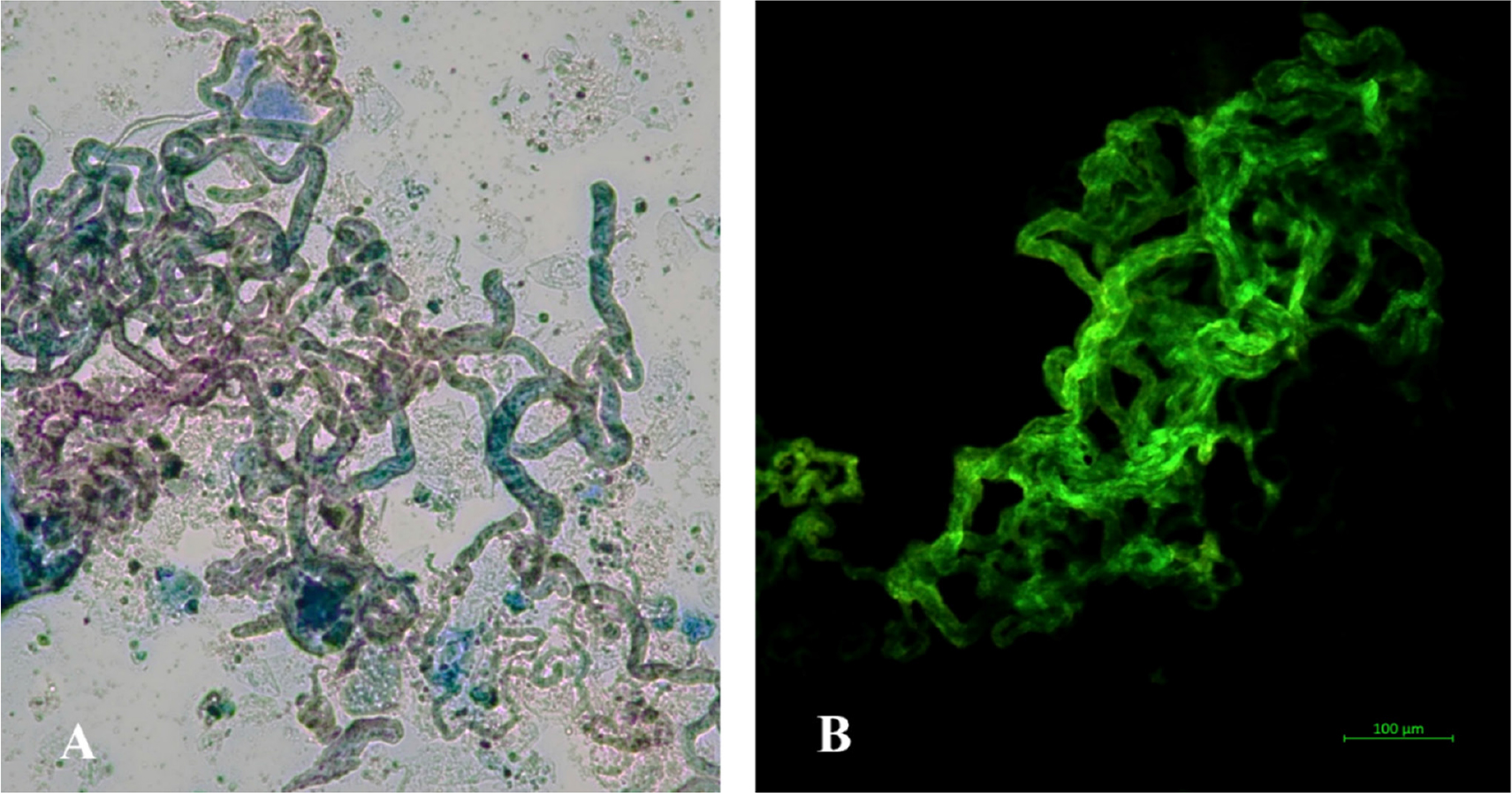
Figure 1.
M. abscessus subs. abscessus visualized by Ziehl–Neelsen staining (A) and auramine fluorescence staining (B). Magnification ×400.
Following decontamination, specimens were inoculated onto Löwenstein–Jensen (LJ) solid medium and into the BACTEC MGIT 960 liquid culture system (Becton Dickinson, Sparks, MD, USA). Cultures were incubated at 37°C and monitored according to the manufacturer’s instructions. Visible growth was observed within 5–10 days, consistent with the growth characteristics of rapidly growing mycobacteria (Griffith et al. 2007).
2.2.2. Molecular identification and resistance-associated determinants
Initial identification of non-tuberculous mycobacteria (NTM) was performed using the GenoType CM direct ver. 1.0 line probe assay (LPA) (Hain Lifescience GmbH, Nehren, Germany). DNA extraction was carried out using the GenoLyse ver. 2.0 kit in accordance with the manufacturer’s instructions. (GenoType CM-direct VER 1.0 IFU-295-01, Hain Lifescience GmbH, Nehren, Germany). The assay enabled identification of the isolates as members of the MABc, but did not permit subspecies differentiation.
Subspecies identification within the M. abscessus complex and molecular detection of resistance-associated mutations were performed using the GenoType NTM-DR ver. 1.0 assay (GenoType NTM-DR VER 1.0 – Instructions for Use. IFU-297-03. Hain Lifescience GmbH, Nehren, Germany). This assay enabled subspecies-level identification within the MABc and detection of resistance-associated variants in erm(41), rrl, and rrs, associated with inducible macrolide resistance, acquired macrolide resistance, and aminoglycoside resistance, respectively. Assay validity was assessed using the internal control zones included on each test strip, according to the manufacturer’s instructions. Each test run included a negative control containing molecular biology-grade water instead of DNA to monitor potential contamination.
2.2.3. Identification of Mycobacterial Isolates by MALDI-TOF MS
Mycobacterial isolates were identified using matrix-assisted laser desorption/ionization time-of-flight mass spectrometry (MALDI-TOF MS) with the MALDI Biotyper system. Isolates were cultured on Löwenstein–Jensen medium prior to analysis. Sample preparation was performed using the MBT Mycobacteria Kit according to the manufacturer’s instructions.
Briefly, approximately 1 μL of bacterial biomass was transferred into a Suspension Vial containing 300 μL of Washing Solution. Subsequently, 900 μL of Inactivation Reagent was added, followed by vigorous vortexing and incubation at room temperature for 30 min to ensure bacterial inactivation. After centrifugation and supernatant removal, the pellet was extracted with 100 μL acetonitrile and 100 μL 70% aqueous formic acid. The mixture was vortexed thoroughly, centrifuged again, and 1 μL of the supernatant was spotted onto a MALDI target plate. After air-drying, each spot was overlaid with 1 μL of Bruker HCCA matrix solution.
Mycobacterial isolates were identified using the MALDI-TOF Biotyper system with the MBT wMycobacteria Module integrated into MBT Compass software. Samples were analyzed using the “Mycobacteria” sample type with an acquisition method optimized for Mycobacterium spp., and the obtained spectra were automatically compared with the MBT Mycobacteria Library. Mass spectra acquisition and species identification were performed according to the manufacturer’s recommendations and score-based criteria implemented in the MBT Compass software (MBT Mycobacteria Module User Manual. Revision A. Bremen, Germany: Bruker Daltonics GmbH & Co. KG.). Instrument calibration and performance verification were performed using the Bruker Bacterial Test Standard (BTS), based on an extract of Escherichia coli DH5α, according to the manufacturer’s recommendations. Mycobacterium fortuitum subsp. fortuitum ATCC 6841 was used as an identification control and was processed in parallel with the clinical isolates using the same extraction procedure. The peak-picking mass range for Mycobacteria samples was 3000–15,000 Da, with log(score) thresholds of 1.6 for low-confidence and 2.0 for high-confidence identification. All isolates were analyzed in triplicate.
2.2.4. Antimicrobial susceptibility testing
Antimicrobial susceptibility testing (AST) was performed by broth microdilution using Sensititre RAPMYCO panels (Thermo Fisher Scientific, Cleveland, OH, USA) in accordance with Clinical and Laboratory Standards Institute (CLSI) recommendations for rapidly growing mycobacteria (CLSI M24, CLSI M24S). Staphylococcus aureus ATCC 29213, Enterococcus faecalis ATCC 29212, and Pseudomonas aeruginosa ATCC 27853 were included as quality-control strains. Minimum inhibitory concentrations (MICs) were determined after 3–5 days of incubation. For clarithromycin, incubation was extended to 14 days to assess inducible macrolide resistance. MICs of bedaquiline and delamanid were determined separately by broth microdilution. Susceptibility categories for agents with available CLSI breakpoints were interpreted according to CLSI M24S.
2.2.5. Biofilm formation assay
Biofilm-forming capacity was assessed using a crystal violet microtiter plate assay. Bacterial suspensions were inoculated into sterile 96-well polystyrene plates containing brain–heart infusion broth (BHI), incubated at 30 °C for 5 days under static conditions.
Following incubation, wells were gently washed with phosphate-buffered saline (PBS) to remove non-adherent cells. Adherent biomass was stained with 0.1% crystal violet for 15 min, rinsed with distilled water, and air-dried. The retained dye was subsequently solubilized using 96% ethanol containing acetic acid, and optical density was measured at 595 nm (OD595).
Uninoculated BHI broth subjected to the same incubation, washing, staining, and solubilization procedure was used as the negative control. Biofilm-forming capacity was evaluated relative to the negative control. The classification thresholds were applied according to the previously published criteria described by Lira et al. (2025): isolates were classified as non-biofilm producers (OD ≤ ODc), weak biofilm producers (ODc < OD ≤ 2×ODc), moderate biofilm producers (2×ODc < OD ≤ 4×ODc), or strong biofilm producers (OD > 4×ODc), according to previously published criteria (Lira et al. 2025).
2.2.6. FT-IR spectroscopy and strain similarity analysis
Spectral similarity among MABc isolates was evaluated using Fourier-transform infrared (FT-IR) spectroscopy with the IR Biotyper system (Bruker Daltonics, Bremen, Germany). Sample preparation, spectral acquisition, and data analysis were performed according to the manufacturer’s recommendations. Briefly, fresh bacterial biomass from solid culture was transferred into microcentrifuge tubes containing 50 µL of 70% ethanol and thoroughly homogenized. Next, 50 µL of sterile distilled water was added, and the suspension was mixed before further processing according to the standard IR Biotyper extraction protocol. Aliquots of the prepared suspension were spotted onto a silicon target plate and allowed to dry at room temperature. Each clinical isolate was cultured in parallel on three separate LJ media. From each culture replicate, four technical FT-IR measurements were obtained, resulting in 12 spectra per clinical isolate. The clinical isolate was the experimental unit; the three parallel cultures were culture replicates, and the four spectra from each culture were technical replicates. Replicate spectra were not considered independent biological observations. Infrared spectra were acquired using the IR Biotyper instrument and processed using the dedicated software package (IR Biotyper Client Software, Bruker Daltonics). Spectral comparison was performed following automated quality control and normalization. Principal component analysis (PCA) and hierarchical clustering using the unweighted pair group method with arithmetic mean (UPGMA) were applied to assess spectral similarity among isolates (Bronson et al. 2021; Curtoni et al. 2023).
3. Results
3.1. Clinical course and microbiological confirmation of chronic infection
Repeated recovery of M. abscessus subsp. abscessus from bronchoalveolar lavage samples collected over eight years, together with progressive radiological abnormalities and recurrent respiratory exacerbations, supported the diagnosis of chronic pulmonary disease. The first isolate was recovered in 2017 following severe pneumonia complicated by pleural empyema. Subsequent cultures remained persistently positive despite prolonged multidrug therapy.
High-resolution computed tomography (HRCT) demonstrated progression of parenchymal infiltrative lesions in the upper lung fields compared with previous imaging studies, with new consolidations involving the lingula and right upper lobe. Repeated bronchoscopic procedures revealed abundant thick purulent bronchial secretions.
Despite previous multidrug therapy, microbiological positivity persisted, and treatment response remained limited. Partial radiological regression was observed after subsequent therapy; however, residual bronchiolitis-type changes and disseminated nodular lesions remained present.
Taken together, repeated microbiological confirmation, persistent smear positivity, progressive radiological abnormalities, and the need for prolonged multidrug treatment supported the diagnosis of chronic clinically significant pulmonary disease caused by M. abscessus rather than transient colonization.
3.2. Species/subspecies identification and molecular resistance determinants
MALDI-TOF MS identified all isolates as members of the M. abscessus complex (MABc). Due to the limited discriminatory capacity of MALDI-TOF MS for subspecies differentiation within the MABc, further molecular characterization was performed using the GenoType NTM-DR assay. Molecular analysis confirmed all isolates as M. abscessus subsp. abscessus (Table 1).
Table 1.
Molecular characterization of M. abscessus subsp. abscessus isolates using the GenoType NTM-DR assay.
| Isolate | Species identification | erm(41) T28 | rrl mutation | rrs mutation | Macrolide resistance | Aminoglycoside resistance |
|---|---|---|---|---|---|---|
| 128/2017 | M. abscessus subsp. abscessus | Present | Not detected | Not detected | Yes | No |
| 126/2018 | M. abscessus subsp. abscessus | Present | Not detected | Not detected | Yes | No |
| 184/2024 | M. abscessus subsp. abscessus | Present | A2058C* | A1408G** | Yes | Yes |
| 199/2025 | M. abscessus subsp. abscessus | Present | A2058C* | A1408G** | Yes | Yes |
Genotypic resistance analysis revealed the erm(41) T28 sequence variant in all isolates, consistent with inducible macrolide resistance. In addition, mutations in the rrl (A2058C) and rrs (A1408G) genes associated with aminoglycoside resistance were detected in isolates 184/2024 and 199/2025 (Table 1).
3.3. Antimicrobial susceptibility profiles
Antimicrobial susceptibility testing demonstrated dynamic changes in the resistance profile of sequential M. abscessus subsp. abscessus isolates over the course of chronic infection (Table 2). The early isolates recovered in 2017 and 2018 remained susceptible to amikacin, whereas the isolates obtained in 2024 and 2025 exhibited high-level amikacin resistance (MIC ≥64 µg/mL). Clarithromycin MICs increased during follow-up, with high-level resistance in later isolates (MIC ≥16 µg/mL), consistent with molecular detection of resistance-associated mutations (Table 1).
Table 2.
Minimum inhibitory concentrations (MICs) and susceptibility categories of sequential M. abscessus subsp. abscessus isolates.
| Antibiotic/Tested strains | MIC [µg/mL] | |||
|---|---|---|---|---|
| 128/2017 | 126/2018 | 184/2024 | 199/2025 | |
| LZD | 4 (S) | 4 (S) | 4 (S) | 4 (S) |
| AMK | 4 (S) | 4 (S) | ≥64 (R) | ≥64 (R) |
| CLR | 1 (S) | ≥16 (R) | ≥16 (R) | ≥16 (R) |
| SXT | 8/152 (R) | ≥8/152 (R) | ≥8/152 (R) | ≥8/152 (R) |
| CIP | ≥4 (R) | ≥4 (R) | ≥4 (R) | ≥4 (R) |
| MXF | ≥8 (R) | ≥8 (R) | ≥8 (R) | ≥8 (R) |
| IPM | ≥64 (R) | ≥64 (R) | ≥64 (R) | ≥64 (R) |
| FOX | ≥128 (R) | ≥128 (R) | ≥128 (R) | ≥128 (R) |
| TOB | ≥16 (R) | ≥16 (R) | ≥16 (R) | ≥16 (R) |
| DOX | ≥16 (R) | ≥16 (R) | ≥16 (R) | ≥16 (R) |
| TGC | 0.5 | 0.5 | 0.5 | 0.5 |
| BDQ | 0.5 | 0.5 | 0.5 | 0.5 |
| DLM | ≥16 | ≥16 | ≥16 | ≥16 |
Resistance to ciprofloxacin, cefoxitin, imipenem, tobramycin, and trimethoprim–sulfamethoxazole was observed throughout the entire study period. Linezolid remained active against all isolates, with stable MIC values of 4 µg/mL, whereas tigecycline and bedaquiline showed consistently low MIC values during follow-up. Delamanid MICs remained ≥16 µg/mL in all isolates. However, CLSI recommendations currently do not provide clinical susceptibility categories for these agents (Table 2).
3.4. Biofilm-forming capacity
All analyzed MAB isolates formed biofilms in vitro. Early isolates recovered in 2017 and 2018 exhibited a rough colony morphotype on Middlebrook 7H10 agar and were classified as moderate biofilm formers based on crystal violet assay results.
In contrast, later isolates obtained in 2024 and 2025 showed a mixed morphotype and increased biofilm-forming capacity, and were classified as strong biofilm formers (Figure 2).
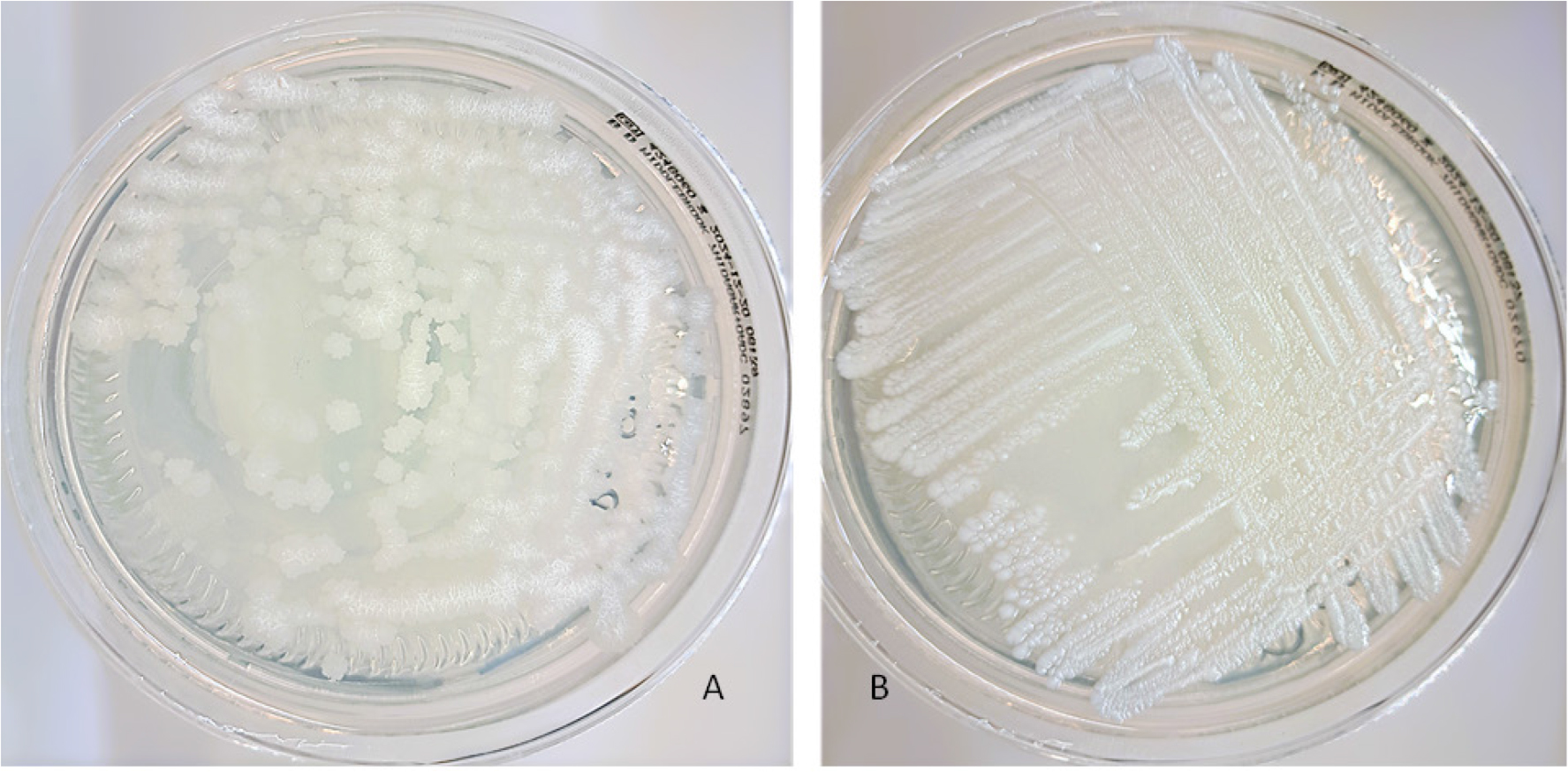
Figure 2.
Representative rough and smooth colony morphotypes of Mycobacterium abscessus subsp.abscessus on Middlebrook 7H10 agar. Rough colonies (A) show a dry, irregular, and wrinkled appearance, whereas smooth colonies (B) are moist, shiny, and have regular margins.
Detailed morphotype characteristics and quantitative biofilm assay results are presented in Table 3.
Table 3.
Morphotype characterization and biofilm-forming ability of M. abscessus isolates assessed using Middlebrook 7H10 agar, and Crystal Violet staining methods.
| Strains No. | Species | Morphotype7H10 agar | Biofilm forming | ||
|---|---|---|---|---|---|
| Crystal violet staining method | |||||
| OD595 | OD595/OD595 NC Ratio * | Characterization | |||
| 128/2017 | M. abscessus subsp. abscessus | Rough | 4.89 | 3.74 | moderate biofilm former |
| 126/2018 | M. abscessus subsp. abscessus | Rough | 3.78 | 3.15 | moderate biofilm former |
| 184/2024 | M. abscessus subsp. abscessus | Mixed | 5.98 | 4.63 | strong biofilm former |
| 199/2025 | M. abscessus subsp. abscessus | Mixed | 5.98 | 4.96 | strong biofilm former |
3.5. FT-IR-based similarity of sequential isolates
Analysis of spectral similarity among sequential isolates was assessed by Fourier-transform infrared (FT-IR) spectroscopy using IR Biotyper technology, based on FT-IR spectra obtained from the 1300–800 cm−1 (fingerprint region: carbohydrates and phosphates), 3200–2800 cm−1 (lipids; aliphatic chains), and 1500–1400 cm−1 (amide II proteins and lipid CH2/CH3 deformation) spectral ranges.
Two additional closely related M. abscessus isolates, unrelated to the patient and cultured on Löwenstein–Jensen medium for 6 days, were included as comparative controls.
Hierarchical clustering analysis using Euclidean distance and UPGMA demonstrated high similarity among all patient-derived isolates collected over multiple years. The dendrogram analysis showed a cophenetic correlation coefficient (CCC) of 0.981, indicating that the dendrogram well represented the pairwise distance relationships in the original distance matrix. PCA and LDA analyses demonstrated partial temporal separation of the isolates while maintaining overall spectral similarity. The additional closely related control isolates also exhibited detectable spectral variability despite their known relatedness (Figure 3, Figure 4).
Overall, FT-IR analysis demonstrated high spectral similarity among sequential patient isolates collected over the eight-year observation period.
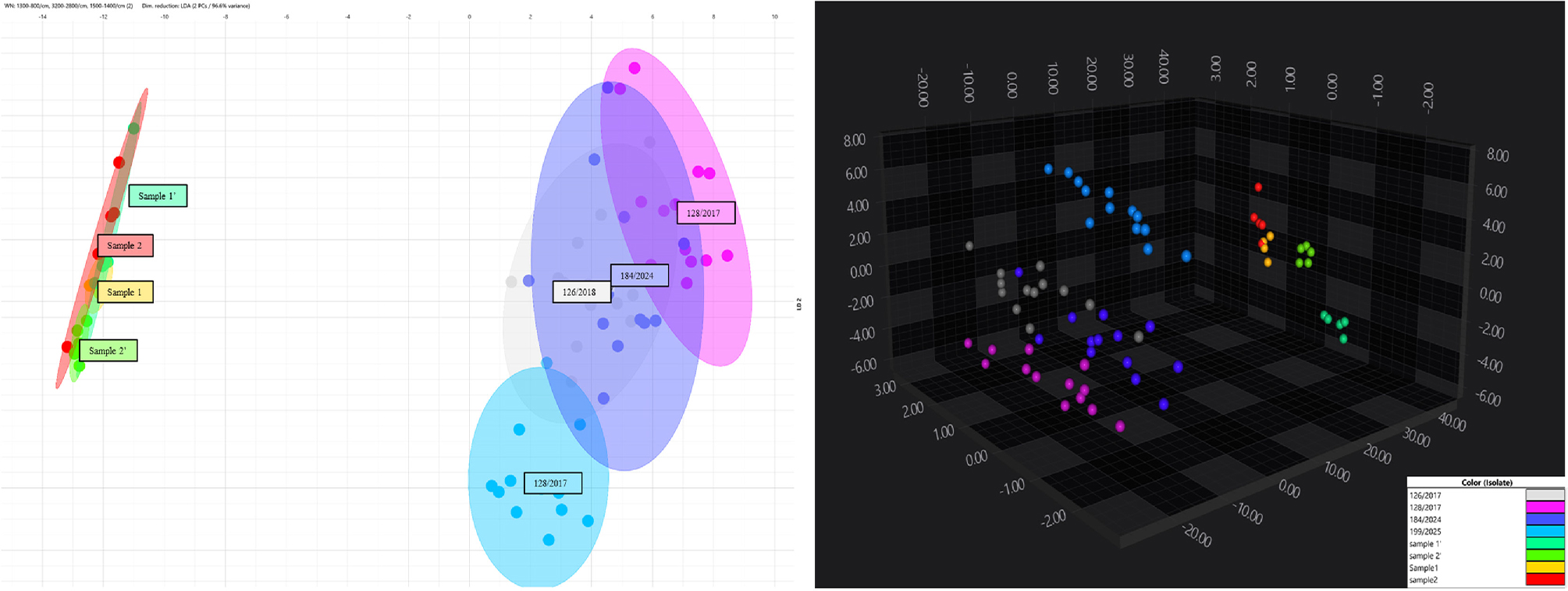
Figure 3.
Two-dimensional and three-dimensional FT-IR spectral profiles of the analyzed isolates.
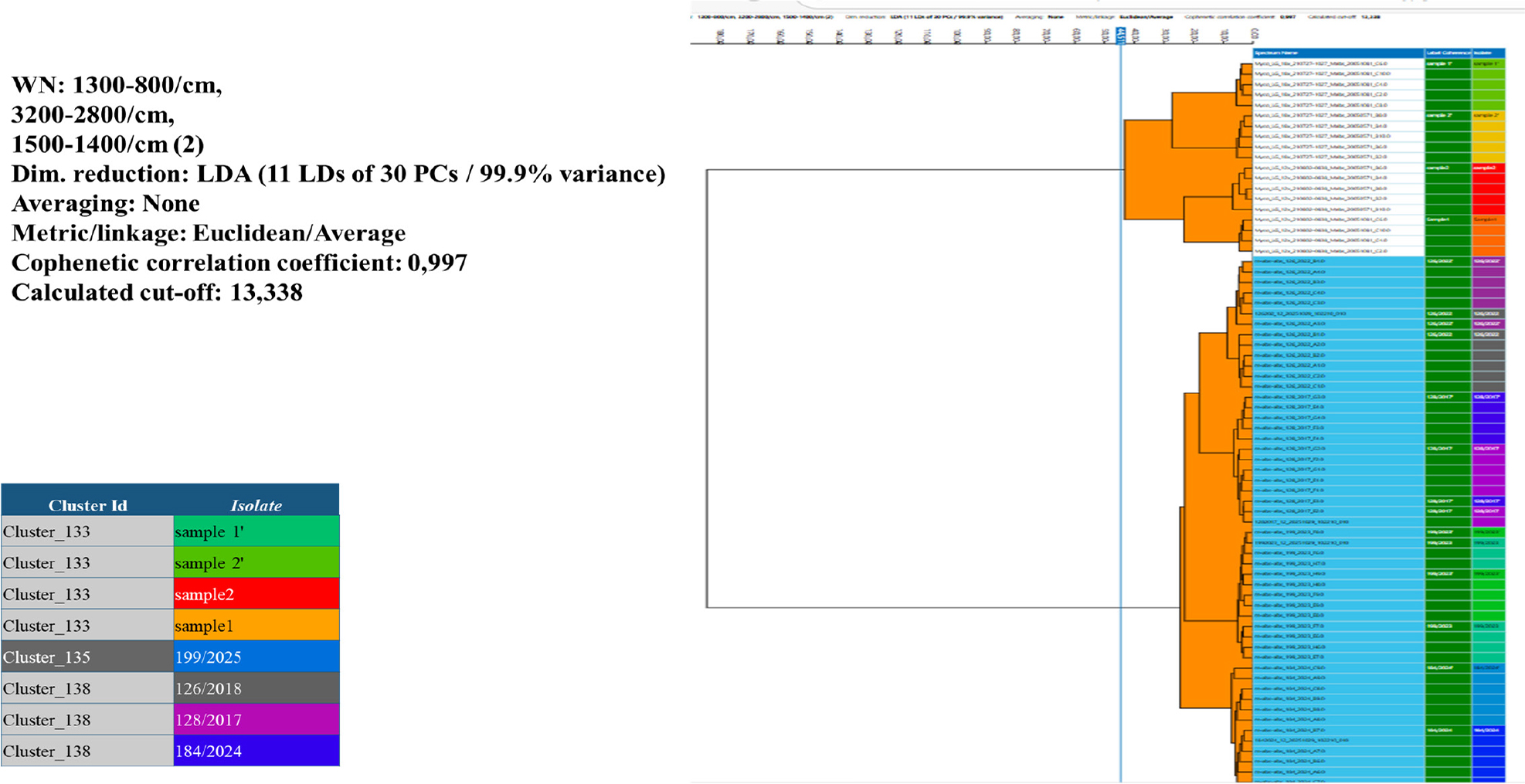
Figure 4.
Dendrogram showing the degree of phenotypic similarity among M. abscessus isolates collected between 2017 and 2025, generated based on FT-IR spectra using the IR Biotyper system.
4. Discussion
All four M. abscessus subsp. abscessus isolates analyzed in this study were recovered from the same cystic fibrosis patient over eight years, providing a unique opportunity to investigate the long-term microbiological evolution of chronic infection. Despite prolonged antimicrobial therapy and repeated clinical interventions, the organism persisted throughout the observation period, indicating a remarkable capacity for adaptation and survival within the respiratory tract.
Pulmonary infection caused by M. abscessus represents one of the most challenging infectious complications in patients with cystic fibrosis (CF). Compared with other non-tuberculous mycobacteria, M. abscessus is associated with a more rapid decline in lung function, frequent pulmonary exacerbations, and lower treatment success rates (Floto et al. 2016; Martiniano et al. 2019).
In the present case, repeated isolation of M. abscessus together with progressive radiological abnormalities and severe airflow obstruction supported the diagnosis of clinically relevant chronic pulmonary disease rather than transient colonization. This observation is consistent with previous CF cohort studies demonstrating the adverse prognostic impact of persistent M. abscessus infection (Zomer et al. 2023).
Accurate diagnosis of M. abscessus infection in CF patients requires integrating microbiological, radiological, and clinical data. Repeated recovery of NTM from respiratory specimens is common in this population, but only a subset of patients fulfills the criteria for active disease (Griffith et al. 2007; Forbes et al. 2018).
In the present study, line probe assays enabled rapid identification of the M. abscessus complex. At the same time, GenoType NTM-DR provided clinically relevant subspecies differentiation and resistance-associated determinants. MALDI-TOF MS served as an additional culture-based confirmatory tool. Furthermore, FT-IR spectroscopy enabled rapid assessment of spectral similarity among sequential isolates and may serve as a practical phenotypic tool complementary to more resource-intensive genomic methods.
Management of M. abscessus pulmonary disease remains difficult because of intrinsic antimicrobial resistance, inducible macrolide resistance mediated by erm(41), prolonged treatment duration, and frequent drug toxicity (Victoria et al. 2021; Abdelaal et al. 2022).
One of the most notable findings of the present study was the progressive acquisition of antimicrobial resistance during long-term persistence of M. abscessus. While early isolates remained susceptible to amikacin, clarithromycin resistance was already observed in the 2018 isolate, and the later isolates recovered after several years of treatment exhibited high-level resistance accompanied by the emergence of rrs and rrl resistance-associated mutations.
This observation illustrates the remarkable adaptive capacity of M. abscessus under prolonged antimicrobial pressure and highlights the importance of repeated susceptibility testing during chronic infection. In the presented case, treatment modification was required because of recurrent pancreatitis, illustrating the narrow therapeutic window frequently encountered in CF patients with multiple comorbidities. In addition, the biofilm-forming capacity of the isolates may have contributed to persistence despite prolonged therapy. Interestingly, acquiring additional resistance determinants was accompanied by increased biofilm-forming capacity. Early isolates were classified as moderate biofilm producers, whereas later isolates exhibited strong biofilm formation. Biofilm development is a major survival strategy of M. abscessus and may significantly reduce antibiotic penetration and antimicrobial efficacy (Lira et al. 2025; Ferrell et al. 2024). The observed transition from a rough morphotype to a mixed morphotype may further reflect ongoing adaptation during chronic infection. Morphotype switching has been associated with alterations in cell envelope composition, virulence, and biofilm architecture, potentially facilitating long-term persistence within the CF airway environment (Lira et al. 2025; Ferrell et al. 2024).
A major clinical challenge in chronic M. abscessus infection is distinguishing long-term persistence from repeated reinfection. In the present study, FT-IR analysis demonstrated high similarity among the spectral profiles of sequential isolates collected over the eight-year observation period. This finding indicates a high degree of phenotypic similarity among the isolates and is compatible with the long-term persistence of a phenotypically similar population over time. However, FT-IR assesses phenotypic rather than nucleotide-level similarity and therefore cannot establish genetic clonality or definitively exclude reinfection with a genetically distinct strain. Confirming clonality and assessing within-host microevolution would require whole-genome sequencing.
Notably, the high FT-IR spectral similarity was observed despite longitudinal changes in antimicrobial susceptibility, resistance determinants, biofilm-forming capacity, and colony morphotype. These observations suggest that isolates with broadly similar phenotypic spectral profiles may nevertheless exhibit substantial variation in clinically relevant characteristics during prolonged infection. Given the single-patient design and the limited number of sequential isolates, interpret these changes as case-specific observations rather than evidence of a defined evolutionary trajectory.
Taken together, the present findings suggest that chronic M. abscessus infection in this patient was characterized not only by prolonged microbiological persistence but also by continuous phenotypic adaptation. The emergence of additional resistance determinants, increased biofilm production, and morphotype alterations observed during follow-up may reflect progressive adaptation during chronic infection and may contribute to treatment failure and adaptive changes.
Although whole-genome sequencing remains the reference approach for investigating genetic relatedness and within-host evolution, FT-IR spectroscopy may provide complementary phenotypic information on the similarity of sequential isolates. Further studies incorporating larger longitudinal cohorts and genomic analyses are needed to determine the clinical and epidemiological utility of this approach.
5. Conclusion
To sum up, sequential M. abscessus subsp. abscessus isolates recovered from a cystic fibrosis patient over eight years demonstrated highly similar FT-IR spectral profiles, consistent with the long-term persistence of phenotypically similar isolates. However, FT-IR does not allow definitive conclusions regarding genetic clonality or the exclusion of reinfection.
Despite this overall phenotypic similarity, the isolates exhibited temporal changes during chronic infection, including the emergence of resistance-associated rrs and rrl mutations, increased MICs for amikacin and clarithromycin, and enhanced biofilm-forming capacity. These findings may reflect adaptation during prolonged infection; however, confirmation of clonality and within-host evolution would require whole-genome sequencing. The combined use of molecular diagnostics, antimicrobial susceptibility testing, biofilm assessment, and FT-IR spectroscopy provided complementary information on longitudinal microbiological changes, highlighting the potential value of integrated microbiological monitoring in managing chronic M. abscessus pulmonary disease in patients with cystic fibrosis.
6. Limitations
This study has several limitations. First, it presents longitudinal observations from a single patient and a limited number of isolates, which restricts the generalizability of the findings. Second, molecular resistance analysis was confined to resistance determinants included in the GenoType NTM-DR assay and did not involve whole-genome sequencing (WGS). Consequently, additional genetic mechanisms associated with antimicrobial resistance and bacterial adaptation may have remained undetected.
Furthermore, although FT-IR spectroscopy demonstrated a high degree of similarity among the spectral profiles of sequential isolates, this method assesses physicochemical and phenotypic characteristics rather than direct genetic relatedness. Therefore, the FT-IR findings cannot establish clonality or definitively distinguish long-term persistence from reinfection. Definitive assessment of clonality and within-host evolution would require whole-genome sequencing.
Notes
[5] Ethic approval
All the experimental protocols were approved by the Bioethics Committee at the Regional Medical Chamber in Krakow (L.dz.OIL/KBL/2/2025) and the Committee for Ethics of Scientific Research at the Jagiellonian University Collegium Medicum in Krakow (Approval No. 118.0043.1.20.2025).
The research project was partially funded by Jagiellonian University Medical College’s statutory funds (N42/DBS/000499).
[6] Data availability statement
The data that support the findings of this study are available from the corresponding author upon reasonable request.
[7] Authors contribution statement
K. Kania conceptualized the study, performed microbiological and molecular analyses, interpreted the data, and drafted the manuscript.
K. Wójcik contributed to microbiological and molecular analyses and data interpretation.
M. Grzesiak performed FT-IR Biotyper analyses and contributed to data interpretation.
A. Szczepański contributed to manuscript editing and reference management.
I. Gross-Sondej was involved in the long-term clinical care of the patient, contributed to the interpretation of the clinical course of chronic pulmonary M. abscessus infection in cystic fibrosis, critically reviewed the manuscript for its clinical content, and approved the final version of the manuscript.
K. Sładek contributed to the clinical characterization of the patient, supported the interpretation of the clinical course and treatment history, critically reviewed the manuscript for its clinical content, and approved the final version of the manuscript.
K. Klesiewicz supervised the study, coordinated the research activities, contributed to data interpretation, and critically revised the manuscript. All authors read and approved the final manuscript. All authors contributed to data analysis, critically revised the manuscript, and approved the final version of the manuscript.