
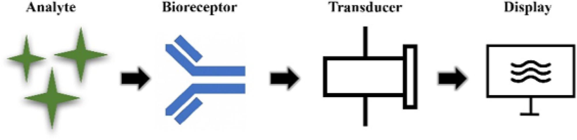
Fig. 1:
General working principle of a biosensor showing the interaction among the target analyte, the biological recognition element, the transducer, and the signal processing unit.
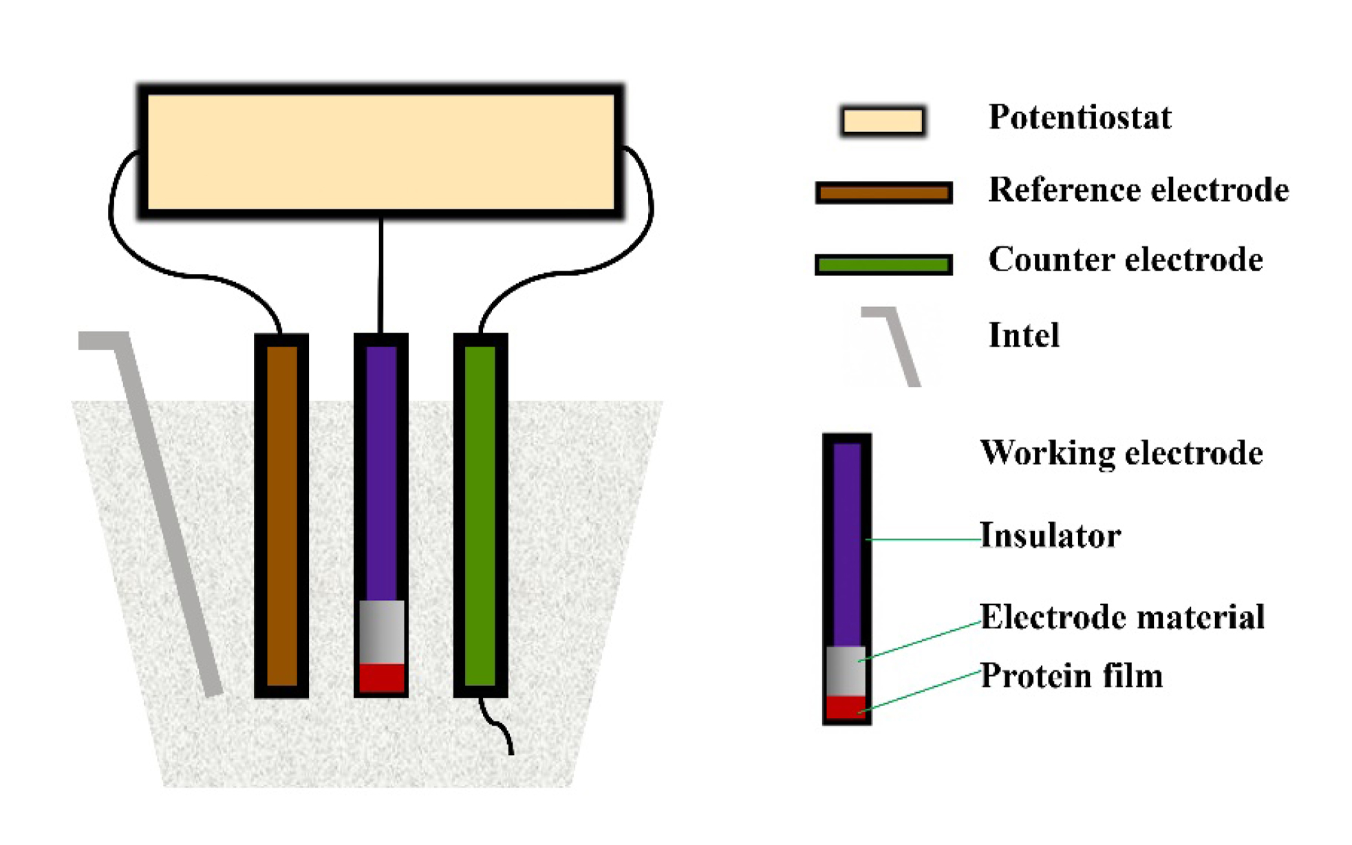
Figure 2.
Basic structure of an electrochemical biosensor cell, illustrating the working, reference, and counter electrodes that convert biological recognition events into measurable electrical signals.
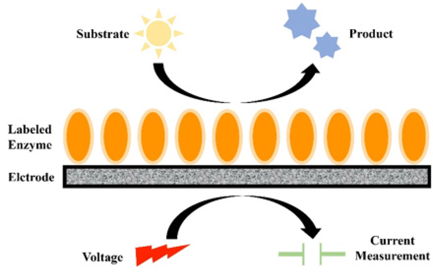
Fig. 3:
Working principle of an amperometric biosensor. The biological interaction at the sensing surface produces an oxidation or reduction current proportional to the concentration of the target analyte.
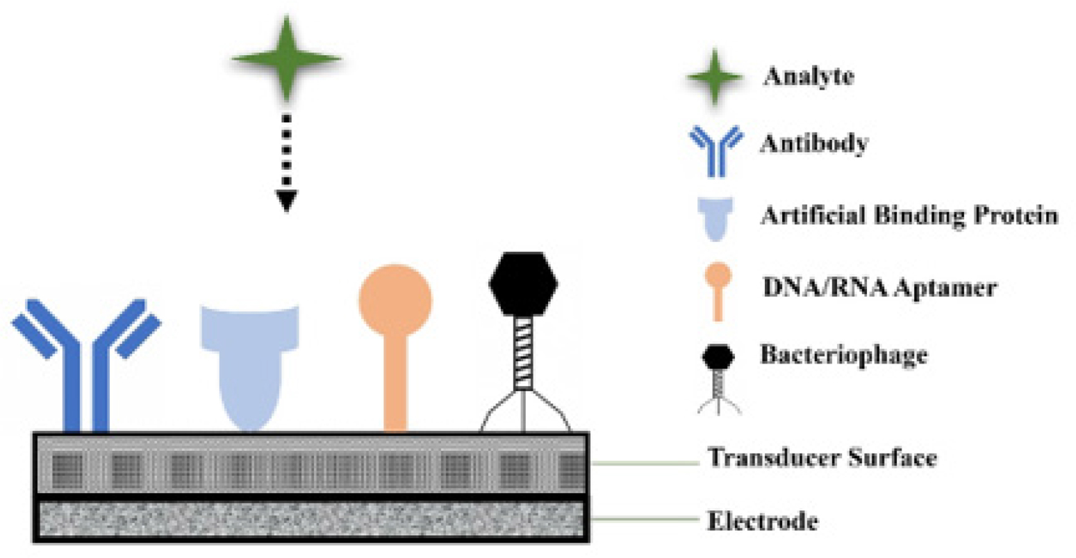
Fig. 4:
Schematic representation of an impedimetric biosensor for bacterial detection, showing immobilized bioreceptors on the electrode surface and the resulting change in charge-transfer resistance upon target binding.
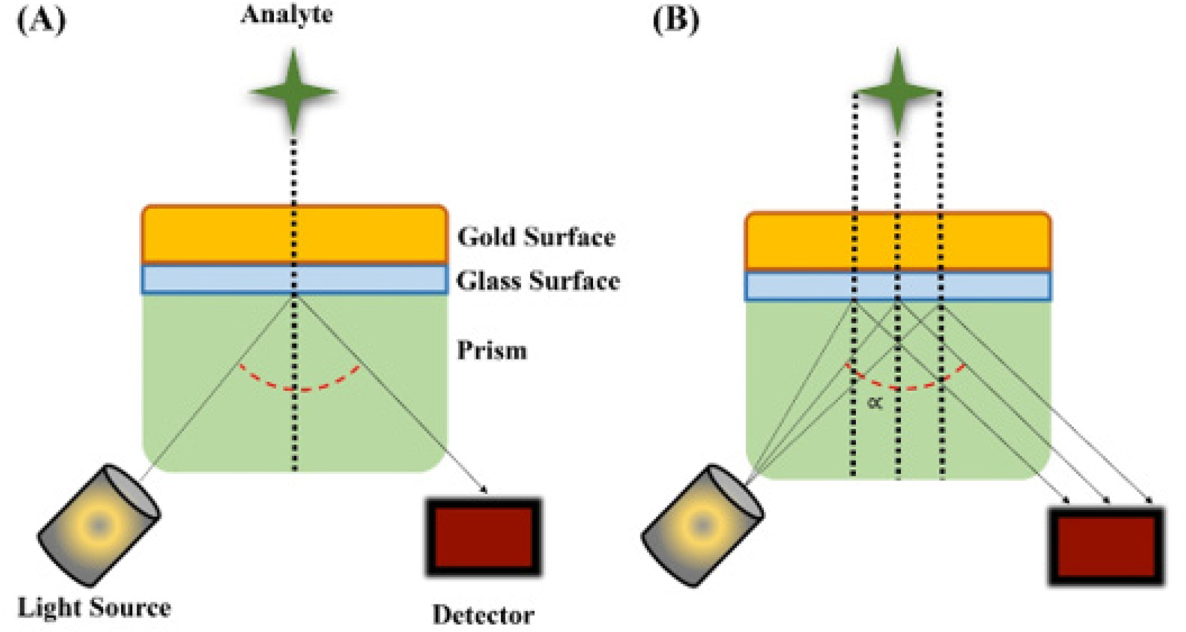
Fig. 5:
Surface plasmon resonance (SPR)-based biosensor. (A) Changes in the angle of refracted light after analyte binding. (B) Generation of an evanescent field at the thin gold surface, enabling label-free, real-time detection.
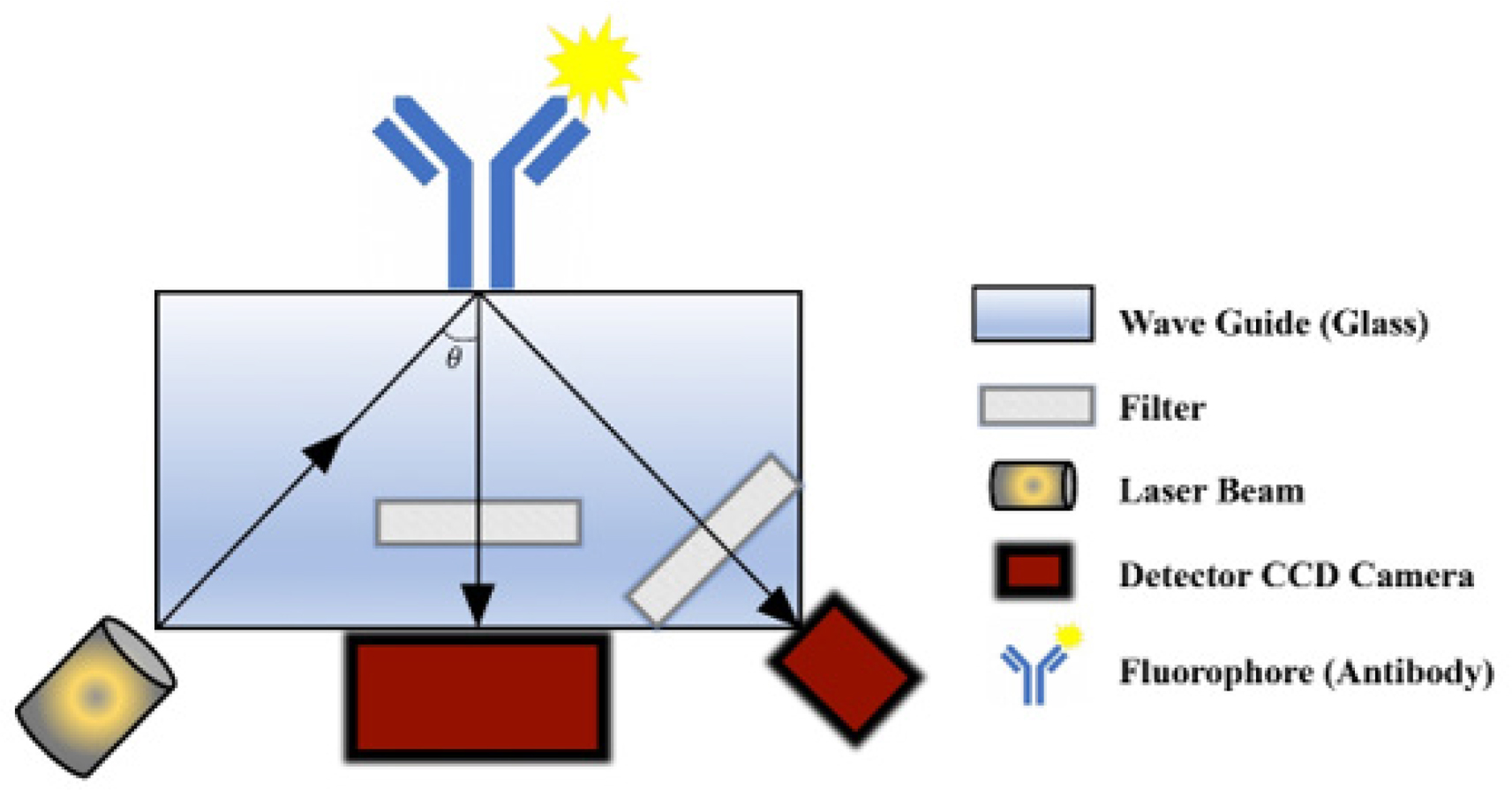
Fig. 6:
Total internal reflection fluorescence (TIRF) array biosensor system showing the optical, fluidic, waveguide, and detection components used for pathogen detection by fluorescence.
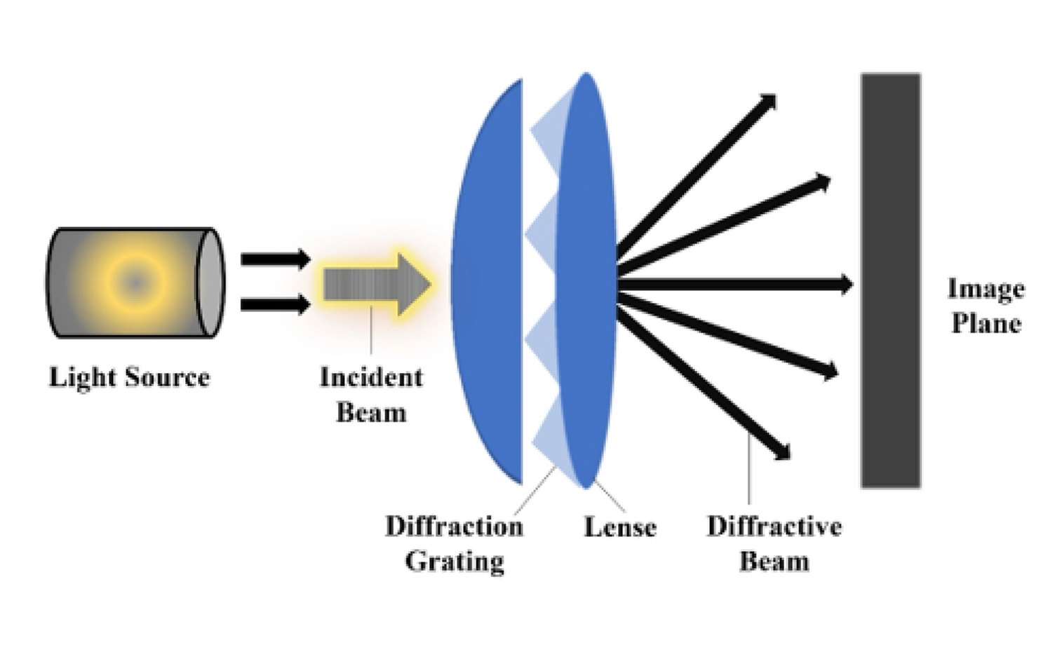
Fig. 7:
Principle of a diffractive-optic biosensor. Binding of the target analyte to immobilized capture molecules increases diffraction intensity, enabling label-free detection.
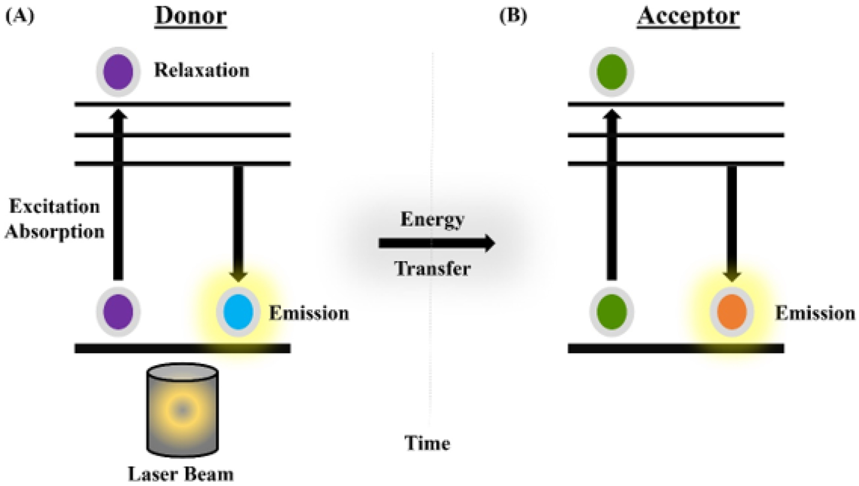
Fig. 8:
Fluorescence resonance energy transfer (FRET) event. (A) Excitation of the donor molecule by a laser beam. (B) Donor and acceptor photon-emission signals over time. (C) Use of donor-acceptor energy transfer to estimate intramolecular distance.
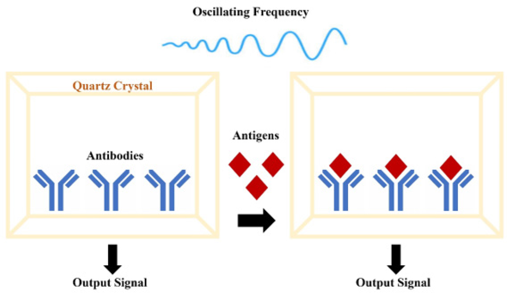
Figure 9.
Quartz crystal microbalance (QCM)-based mass-sensitive biosensor. Antibodies immobilized on the crystal surface specifically capture target antigens, increasing sensor mass and shifting the oscillation frequency, which is converted into an electrical signal.
Table 1.
Comparative summary of biosensor platforms reported for Campylobacter spp. detection
| Biosensor platform | Target analyte / recognition element | LOD | Sample type | Advantages | Disadvantages / limitations | References |
|---|---|---|---|---|---|---|
| Amperometric biosensor | Campylobacter spp. cells; antibodies immobilized on bilayer lipid membrane or magnetic beads | One cell/sample to 2 × 10⁴ CFU/ml; reported ranges up to 107 CFU/ml | PBS, ground turkey meat, washing water, chicken carcass rinse | Rapid, portable, compatible with food matrices, low-cost transducers | Signal may depend on antibody quality, sample preparation, and matrix effects | Ivnitski et al. 2000; Che et al. 2001; Viswanathan et al. 2013 |
| Impedimetric biosensor | C. jejuni cells; monoclonal anti-C. jejuni antibody on Fe3O4 nanoparticle-modified electrode | 103 CFU/ml | Patient stool / PBS buffer | Label-free, relatively rapid, suitable for miniaturization | Electrode fouling and non-specific binding may affect reproducibility | Huang et al. 2010 |
| Conductometric / capacitance-based biosensor | C. jejuni growth/ metabolism; conductance or capacitance change | Detection times reported for 1 to 104 CFU/ml | Optimal broth medium / injured cells | Useful for monitoring viable cells and metabolic activity | Longer detection time than direct molecular biosensors; requires growth conditions | Cappelier et al. 2000; Line and Pearson, 2003 |
| SPR biosensor | C. jejuni cells; polyclonal or monoclonal antibodies on gold sensor chip | 1.1 × 10⁵ CFU/ml to 8 × 10⁶ CFU/ml; some systems tested 102 to 109 CFU/ml | Pure culture, meat rinse, apple juice, PBS | Label-free, real-time, multiplex channel formats possible | Higher instrumentation cost; surface regeneration and matrix interference need optimization | Yang et al. 2013; Masdor et al. 2014; Wang and Salazar, 2015 |
| TIRF / array biosensor | C. jejuni and C. coli; fluorescent antibody-based sandwich immunoassay | 5 × 10² to 4 × 10³ CFU/ml depending on matrix | Ground turkey, milk, yogurt, carcass wash | High sensitivity, real-time fluorescence detection, suitable for multiplex arrays | Requires fluorescent labels, optical alignment, and specialized instrumentation | Gnanaprakasa et al. 2011 |
| Diffractive optics / BARDOT | C. jejuni DNA or colony light-scattering pattern | 5 nM DNA for dotLab; BARDOT classification accuracy around 85% | PBST buffer, agar colonies, fresh and spiked chicken samples | Label-free options, non-destructive colony screening, high-throughput potential | Requires pattern analysis and validation with naturally contaminated samples | Gnanaprakasa et al. 2011; He et al. 2014 |
| FRET biosensor | Campylobacter DNA; fluorescent ssDNA probe and gold nanoparticles | 0.8 to 7 × 10⁴ nM | Laboratory DNA samples | High molecular specificity and useful for nucleic acid detection | Requires labeled probes and controlled assay conditions | Darbha et al. 2008 |
| Mass-sensitive / QCM biosensor | C. jejuni cells; lectins or antibody-based capture molecules on quartz crystal | Not consistently reported; applied for C. jejuni detection in experimental systems | Bacterial suspensions / assay buffer | Label-free, real-time mass-change monitoring | Surface immobilization, cross-reactivity, and matrix robustness require optimization | Safina et al. 2008; Yakovleva et al. 2011; Masdor et al. 2016 |