
1. Introduction
Zoonotic Campylobacter spp. are globally associated with bacterial diarrhea (Njoga et al. 2025). The Gram-negative Campylobacter species are microaerophilic, curved or spiral-shaped bacteria that possess a single, polar, unsheathed flagellum at one or both ends. C. jejuni is a common foodborne pathogen that causes infectious diarrhea worldwide (Abulreesh et al. 2017). In humans, infection predominantly occurs through handling and ingestion of contaminated poultry meat (uncooked or raw), tap water, and raw milk. Infection may present with abdominal pain, vomiting, mild, self-limiting gastroenteritis with fever (1–3 days), and bloody, watery diarrhea (3–7 days) (Kim et al. 2021; Graham et al. 2024). Campylobacter-associated infections may lead to Guillain-Barre syndrome (GBS), a rare, life-threatening autoimmune condition that frequently results in long-term sequelae such as irritable bowel syndrome and reactive arthritis (Abulreesh et al. 2017). Therefore, Campylobacter detection is crucial for food safety and public health. Traditional culture-based methods are considered the “gold standard” for Campylobacter spp. detection and diagnosis. However, these techniques are time-consuming (5–7 days), and in heavily contaminated samples, overgrowth of competing microflora can produce false-negative results by preventing Campylobacter spp. from reaching detectable levels (Abulreesh et al. 2005). C. jejuni can cause serious outbreaks, particularly in older adults and infants. Therefore, rapid detection is important for curbing the spread of the pathogen and preventing a detrimental outbreak (Huang et al. 2021). Multiple Campylobacter spp. detection methods are available and are classified as conventional cultural methods [plating/culturing, microscopic and biochemical confirmation, and enumeration] and modern immunological and molecular methods [testing kits and PCR/hybridization] (Neyaz et al. 2024). However, these techniques can be expensive and time-consuming, with laborious sample preparation, and may generate incomplete or misleading data. In this regard, biosensors may offer significant advantages over traditional approaches (Oyarzabal and Battie, 2012). To our knowledge, the use of biosensors for Campylobacter spp. detection is limited and scattered. Therefore, this review provides an overview of different types of biosensors to improve understanding and detection of Campylobacter species.
2. Classification of Biosensors
Biosensors are analytical instruments that integrate a biological recognition component with a signal transducer to identify a specific target (Ali et al. 2017). Biosensors for Campylobacter spp. detection can be categorized as electrochemical, optical, or mass-sensitive devices based on the transduction mechanism employed (Yang et al. 2013; Yasmin et al. 2016). Figure 1 summarizes the basic concept of biosensor operation.
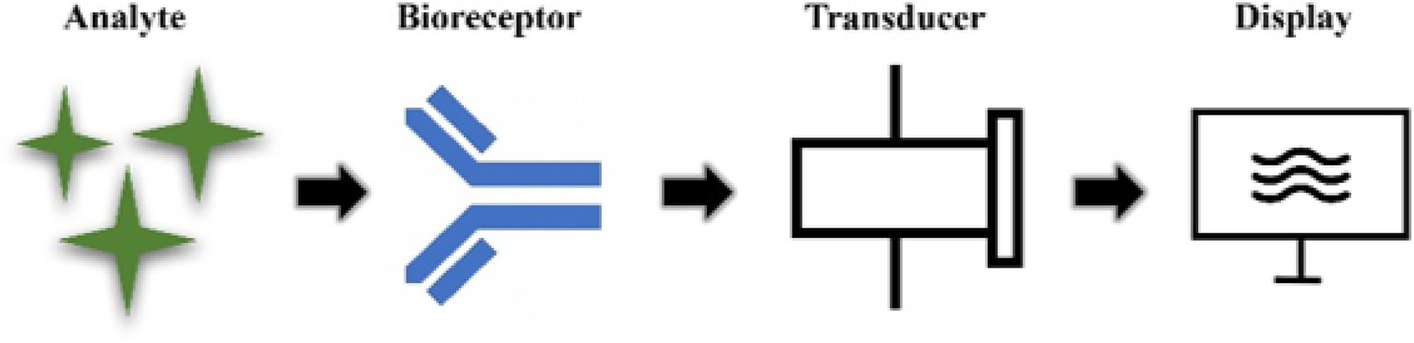
Fig. 1:
General working principle of a biosensor showing the interaction among the target analyte, the biological recognition element, the transducer, and the signal processing unit.
2.1. Electrochemical biosensors
Electrochemical biosensors are extensively researched platforms for detecting Campylobacter spp. because of their rapid analysis, high sensitivity, portability, and compatibility with diverse food matrices (Yang et al. 2013; Aleksic and Kapetanovic 2013; Sumitha and Xavier 2023). These biosensors convert biological recognition events into quantifiable electrical signals (Fig. 2) and can be categorized as amperometric, impedimetric, and conductometric systems. Numerous electrochemical biosensors have been developed for the identification of Campylobacter spp. in food, environmental, and clinical specimens.
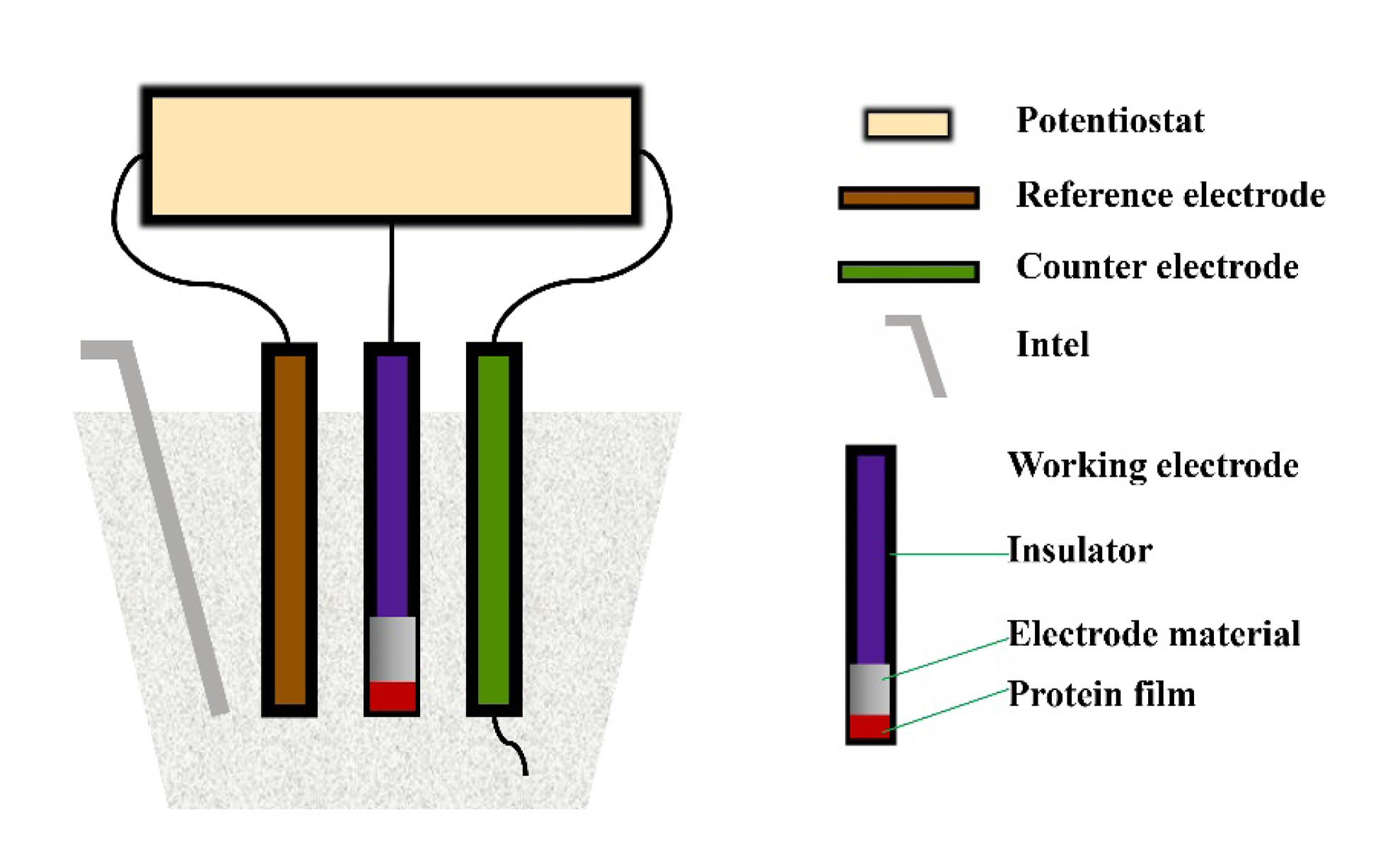
Figure 2.
Basic structure of an electrochemical biosensor cell, illustrating the working, reference, and counter electrodes that convert biological recognition events into measurable electrical signals.
2.1.1. Amperometric biosensors
Amperometric biosensors measure current changes generated during biological recognition events and have been widely used for pathogen detection (Fig. 3). A biosensor design used electron transfer through a bilayer lipid membrane (BLM) to detect Campylobacter spp. It involved immersing a stainless-steel (SS) electrode in an egg phosphatidylcholine-based squalene-decane solution (1:1) to form a BLM. Soaking the electrode for 5 hours immobilized Campylobacter spp.-specific antibodies. The dielectric BLM blocked electron transfer to the electrode. However, antibody conformational perturbation upon bacterial binding opened a channel for ion permeation through the BLM, yielding a current peak in the amperometric biosensor. The current peaks were of the same size irrespective of the cell concentration, indicating channel opening or closing, whereas the concentration determined the current peak’s width. The current peaks corresponded to a flow of approximately 1010 ions/s, suggesting an LOD of one cell/sample (Ivnitski et al. 2000).
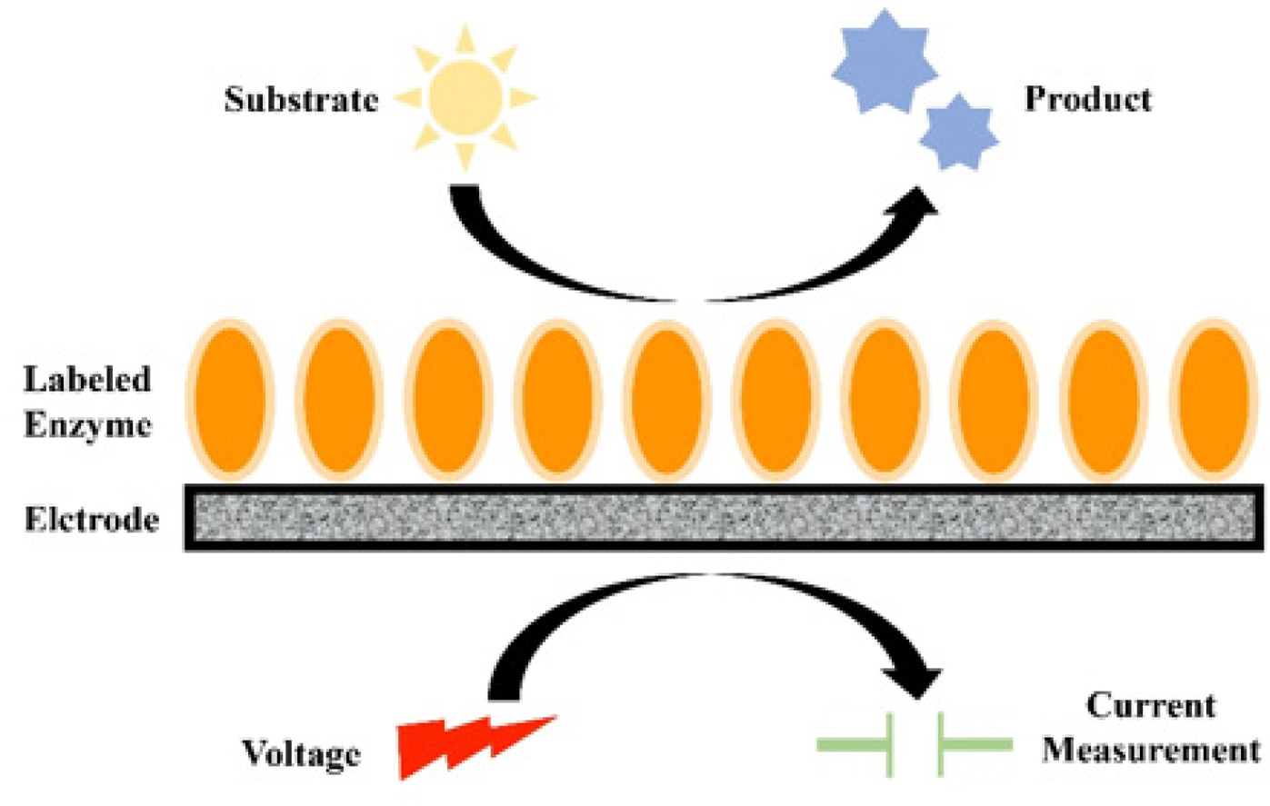
Fig. 3:
Working principle of an amperometric biosensor. The biological interaction at the sensing surface produces an oxidation or reduction current proportional to the concentration of the target analyte.
Another design used magnetic beads to confine the bacteria. Initially, streptavidin was used to modify the beads, followed by immobilization of biotinylated rabbit anti-C. jejuni via an avidin-biotin interaction and phosphatase labeling. The bead-bacteria conjugates were formed by mixing and shaking the beads with bacteria for an hour. The immunomagnetic separation method compared eight bead types of varying sizes and modifications to achieve optimal cell capture. After removing the supernatant, the conjugates were incubated with phenyl phosphate for 1 hour to produce phenol. A tyrosinase-modified carbon-paste electrode detected the phenol, and the current signal was recorded. This design was used to measure C. jejuni in ground turkey meat, washing water, chicken carcass, and PBS. The study revealed similar bacterial results in the poultry samples and PBS. A logarithmic concentration was used for linear fitting. The linear detection range remained 102–107 CFU/ml, with a fitting variation (R2) of 0.94, whereas the limit of detection was noted as 2×10⁴ CFU/ml (Che et al. 2001). Viswanathan et al. (2013) employed an antibody-bacteria-antibody strategy. Nanocrystals were used to design amperometric biosensors for detecting Campylobacter spp., including C. jejuni. Carboxylic acid-functionalized multi-walled carbon nanotubes (MWCNT) were used to modify a screen-printed electrode (SPE), followed by immobilization of Campylobacter spp.-specific antibodies. They observed a limit of detection (LOD) range of 1×10³ to 5×10⁵ CFU/ml, whereas 4×10² CFU/ml was noted in a sigmoidal current response.
Amperometric biosensors have demonstrated superior analytical performance for detecting Campylobacter spp., with reported LODs ranging from a single Campylobacter cell per sample to 2 × 10⁴ CFU/ml, depending on the sensing technique. These biosensors have been effectively evaluated in phosphate-buffered saline (PBS), poultry rinse water, raw poultry meat, ground turkey meat, and several other food matrices. Their rapid response, portability, and compatibility with complex food samples make amperometric biosensors among the most promising electrochemical platforms for routine Campylobacter spp. surveillance.
2.1.2. Impedimetric biosensors
Impedimetric biosensors detect changes in electrical impedance caused by target binding and have shown promise for rapid detection of Campylobacter spp. (Fig. 4). Several impedance-based biosensors have been reported in recent years. Fe3O4 nanoparticles were used to develop an impedimetric biosensor for detecting C. jejuni in patient stool, with a glassy carbon electrode serving as the working electrode. The electrode was coated with an O-Carboxymethylchitosan (OCMCS)-modified Fe3O4 nanoparticle suspension, followed by immobilization via immersion in the anti-Campylobacter jejuni monoclonal antibody 2D12. Non-specific binding was blocked by applying BSA. Bacteria-containing PBS (pH 7) buffer was used to incubate the electrodes for 40 min at 37.5 °C, and the limit of detection was reported as 103 CFU/ml (Huang et al. 2010).
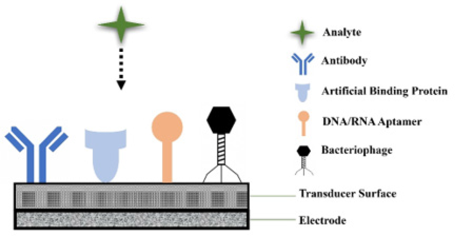
Fig. 4:
Schematic representation of an impedimetric biosensor for bacterial detection, showing immobilized bioreceptors on the electrode surface and the resulting change in charge-transfer resistance upon target binding.
Impedimetric biosensors offer several advantages over amperometric biosensors, including label-free detection, streamlined assay design, and reduced reagent use. They directly detect changes in electrical impedance caused by bacterial adhesion, without requiring electro-active labels or enzyme substrates. However, impedimetric sensors are typically more susceptible to electrode fouling, non-specific adsorption, and fluctuations arising from complex food matrices, which can compromise reproducibility. Despite these constraints, their rapid response and simple sensor architecture make them appealing options for point-of-care and food safety applications for Campylobacter spp. detection.
2.1.3. Conductometric biosensors
Conductometric biosensors measure changes in electrical conductivity caused by microbial growth or metabolic activity. Despite fewer conductometric platforms reported for Campylobacter spp. detection than for amperometric or impedimetric systems, they have shown promise for monitoring bacterial growth and viability in culture media.
Monitoring changes in the medium’s conductance could reveal Campylobacter growth (Cappelier et al. 2000). A capacitance-based detection technique has been reported to monitor C. jejuni growth, thereby facilitating analysis of injured C. jejuni cells in an optimal broth medium. This method involved monitoring bacterial metabolism in addition to analyzing capacitance changes in the solution. The detection times (DTs) at minimum capacitance were linked to the initial cell concentrations, which were 17, 21, 25, 29, and 35 h for 104, 103, 102, 10, and 1 CFU/ml, respectively (Line and Pearson 2003).
Although only a relatively small number of conductimetric biosensors have been specifically developed for Campylobacter spp. (Wang et al. 2023; Vizzini et al. 2026), recent advances in electrochemical sensing, microfluidic platforms, and portable biosensor technologies offer renewed possibilities for conductimetric detection (Wang et al. 2023; Madadelahi et al. 2025). Improvements in electrode materials, nanostructured conductive polymers, and integrated lab-on-a-chip systems are expected to increase sensitivity, reduce detection time, and enable real-time monitoring of Campylobacter spp. in food processing environments (Vizzini et al. 2026). Future studies should aim to transfer these technological advances into conductimetric platforms optimized for Campylobacter spp. detection.
2.2. Optical biosensors
Optical biosensors provide rapid, highly sensitive detection by measuring optical signal changes generated by target recognition and include SPR, TIRF, diffractive optics, and FRET-based systems (Yang et al. 2013; Singh et al. 2023).
2.2.1. Surface plasmon resonance (SPR) biosensors
SPR biosensors enable real-time, label-free detection based on changes in refractive index at the sensor surface (Fig. 5) and have been successfully applied to Campylobacter spp. detection (Poltronieri et al. 2014; Al Mahmud et al. 2023). The SpreetaTM dual-channel SPR sensor is a commercial SPR biosensor for the real-time detection of Campylobacter species. It involves immobilizing the rabbit polyclonal IgG antibody on the sensor surface. In a study, this sensor efficiently detected C. jejuni (102 to 109 CFU/ml) in meat rinse and pure culture samples (Wang and Salazar 2015). Yang et al. (2013) reviewed the development of an eight-channel SPR sensor for C. jejuni detection in apple juice (pH 3.7 and 7.4) and buffer (pH 7.4). Glass chips served as substrates in this biosensor, with gold (Au) and chromium (Cr) deposited by vacuum evaporation. A self-assembled monolayer (SAM) of Biotinylated alkanethiol: Oligo (ethylene glycol) (BAT: OEG) was used to functionalize the sensor. Then, biotinylated polyclonal rabbit antibody (PAb) and streptavidin were added. All eight channels were functionalized with different antibodies, enabling simultaneous multi-analyte detection with a detection limit of 1.1×10⁵ CFU/ml in apple juice and PBS. Moreover, SPR biosensors can be optimized to investigate bacterial interactions for the development of antimicrobials against pathogenic bacteria. Masdor et al. (2014) developed an SPR biosensor platform for C. jejuni detection through in-house cultivation of C. jejuni strains. A covalent attachment technique was used to raise polyclonal antibodies against C. jejuni, which were then used to functionalize SPR sensor chips, followed by induction of gold (Au) chips for direct C. jejuni detection within a limit of 8×10⁶ CFU/ml. The SPR-based sandwich biosensor was also compared with ELISA and proved to be an excellent alternative for C. jejuni detection.
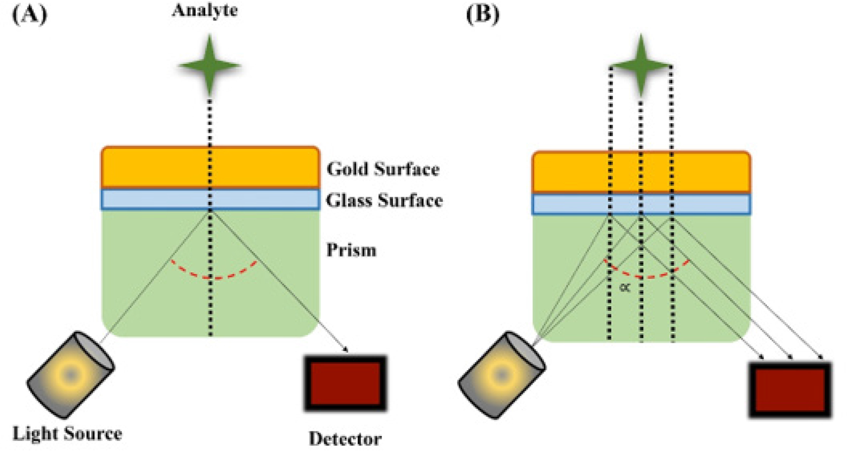
Fig. 5:
Surface plasmon resonance (SPR)-based biosensor. (A) Changes in the angle of refracted light after analyte binding. (B) Generation of an evanescent field at the thin gold surface, enabling label-free, real-time detection.
2.2.2. Total internal reflection fluorescence (TIRF) (Array biosensor)
TIRF biosensors employ fluorescence-based detection and offer high sensitivity for pathogen identification in complex samples (Fig. 6) (Colson et al. 2023). Gnanaprakasa et al. (2011) conducted a sandwich immunoassay to test a sensor’s capability to detect C. coli and C. jejuni in buffer and in various foods, including lettuce leaf, turkey, milk, and carcass wash. EZ-link biotin-LC-HNS was used to biotinylate monoclonal rabbit antibodies against C. jejuni, and a Cy5 bifunctional dye was used for labeling. Poly(dimethyl) siloxane (PDMS) flow cells were used to pattern these antibodies on the glass waveguide. The limit of detection for C. jejuni was 5 × 10², 4 × 10³, 5 × 10², 5 × 10², and 2 × 10³ CFU/ml in ground turkey ham, ground turkey sausage, milk, and yogurt, respectively.
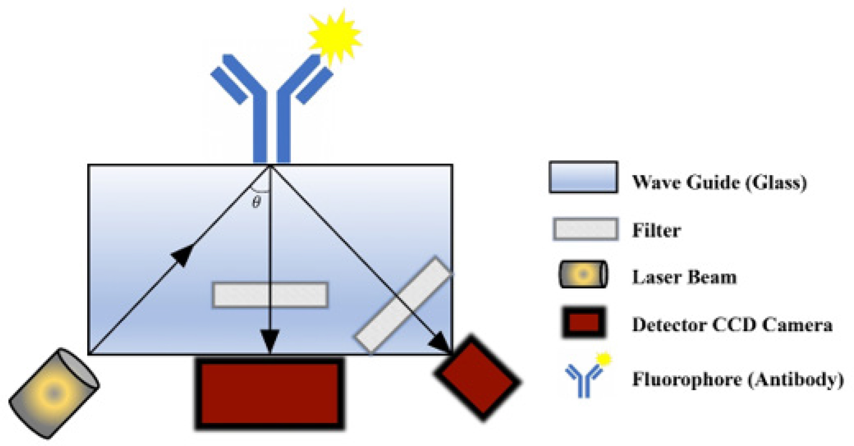
Fig. 6:
Total internal reflection fluorescence (TIRF) array biosensor system showing the optical, fluidic, waveguide, and detection components used for pathogen detection by fluorescence.
2.2.3. Diffractive optics
Diffractive optical biosensors detect target binding through changes in diffraction patterns (Fig. 7) (McGovern et al. 2023) and have been applied to the identification of Campylobacter spp. Gnanaprakasa et al. (2011) employed the dotLab system to detect unique DNA segments of C. jejuni and compared the results with those from the Spreeta SPR biosensor. The results revealed that unique DNA segments of the hippuricase gene were expressed only in C. jejuni, demonstrating specificity for this pathogen. The LOD remained at 5 nM for dotLab and 2.5 nM for SPR in PBST buffer (PBS & Tween). In another investigation, a novel biosensor-based detection method, BARDOT (Bacterial Rapid Detection using Optical scattering Technology), was developed for high-throughput screening of Campylobacter colonies on an agar plate without disruption. During the study, principal component analysis (PCA) and image pattern characterization were performed on 6909 bacterial colonies. The data demonstrated 85% detection accuracy and showed significantly different light-scattering patterns for C. coli and C. jejuni compared with Listeria monocytogenes, Salmonella spp., and Escherichia coli O157:H7. The BARDOT biosensor is recommended for detection of Campylobacter spp. in naturally contaminated fresh chicken meat and spiked ground chicken (He et al. 2014).
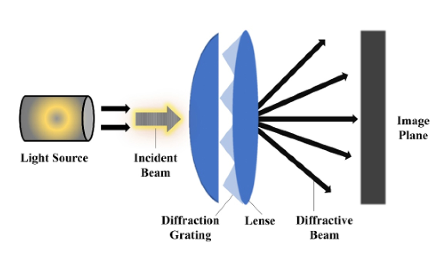
Fig. 7:
Principle of a diffractive-optic biosensor. Binding of the target analyte to immobilized capture molecules increases diffraction intensity, enabling label-free detection.
2.2.4. Fluorescence resonance energy transfer (FRET)
FRET biosensors use energy transfer between fluorescent molecules to detect specific nucleic acid targets (Fig. 8) and have been explored for Campylobacter spp. DNA detection. Darbha et al. (2008) developed a gold nanoparticle-based miniaturized FRET probe for Campylobacter DNA screening. They reported that nanoparticle-based FRET can monitor biological events over a 10 nm distance limit. The reduction of HAuCl4 was used to synthesize gold nanoparticles for the probe, whereas homocysteine (Hcys) and mercapto propionic acid (MPA) were used for surface modification. A fluorescent staining dye (Rhodamine B) aqueous solution tagged with Campylobacter-associated single-stranded DNA (ss-DNA, 5’GTAACTTC-CATTTCTTTTGG3’) was used to immerse the gold nanoparticles. This biosensor had a linear detection range of 0.8 – 7×10⁴ nM.
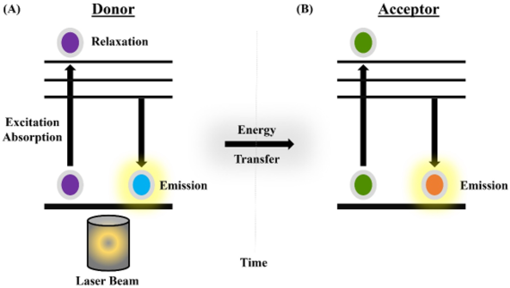
Fig. 8:
Fluorescence resonance energy transfer (FRET) event. (A) Excitation of the donor molecule by a laser beam. (B) Donor and acceptor photon-emission signals over time. (C) Use of donor-acceptor energy transfer to estimate intramolecular distance.
2.3. Mass-sensitive biosensors
Mass-sensitive biosensors detect changes in sensor mass after target binding (Fig. 9) (Sharma et al. 2013; Singh et al. 2024) and have been investigated for Campylobacter detection using quartz crystal microbalance (QCM)-based platforms. A lectin-based QCM biosensor has been successfully used for C. jejuni detection. Lectins are non-immune glycoproteins from animals and plants. They specifically bind various carbohydrates and offer greater stability than antibodies (Safina et al. 2008; Yakovleva et al. 2011). Masdor et al. (2016) developed a QCM-based immunosensor for C. jejuni detection. Commercial mouse monoclonal and rabbit polyclonal antibodies were used to construct direct sandwich assays with gold nanoparticles (AuNPs) for C. jejuni detection. A limit of detection (LOD) of 150 CFU/mL was observed in solution. The QCM sensor exhibited excellent specificity and sensitivity for Campylobacter spp. detection. The data also revealed low cross-reactivity with Listeria monocytogenes (3%), Salmonella Typhimurium (7%), and Escherichia coli (0).
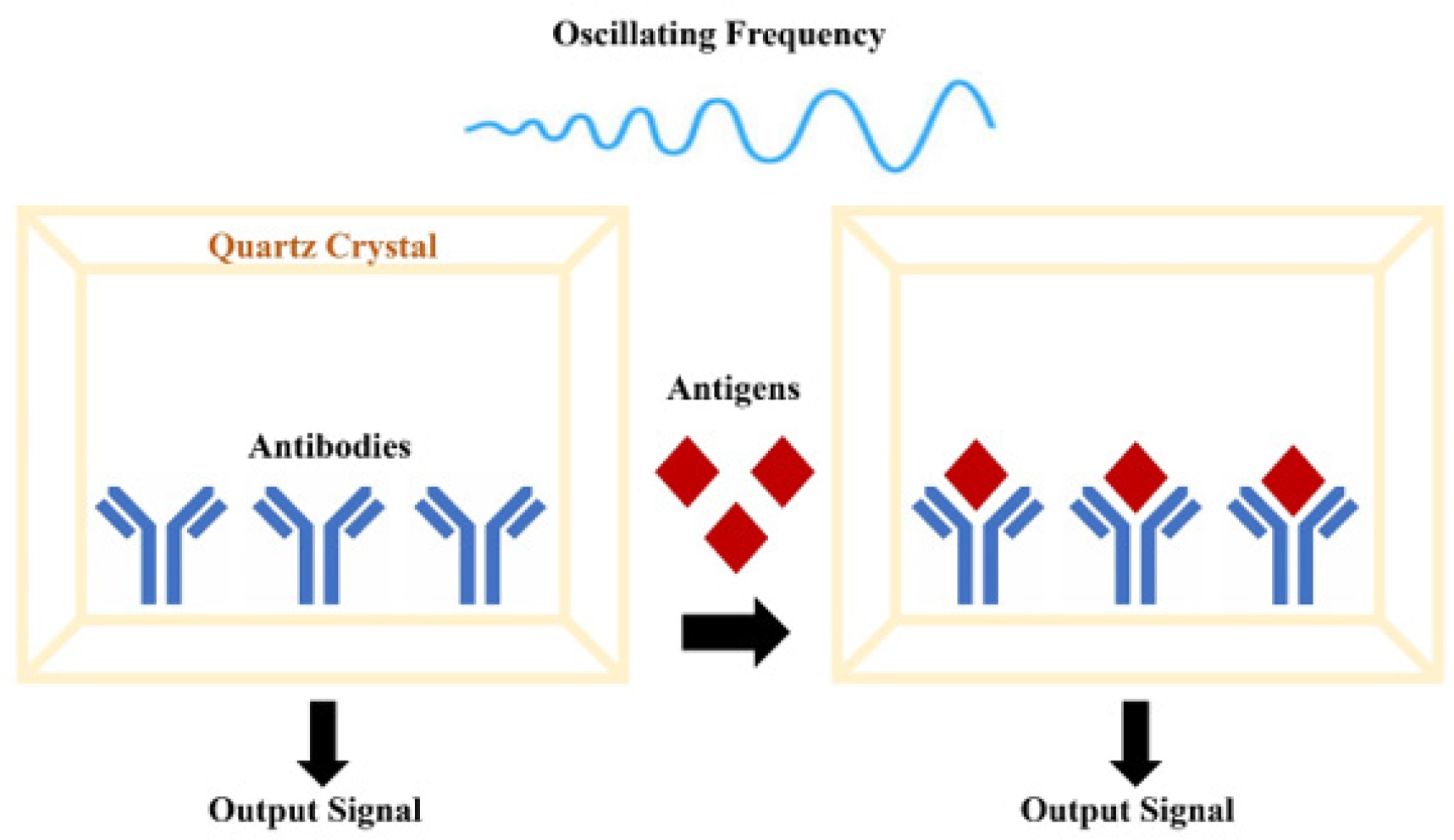
Figure 9.
Quartz crystal microbalance (QCM)-based mass-sensitive biosensor. Antibodies immobilized on the crystal surface specifically capture target antigens, increasing sensor mass and shifting the oscillation frequency, which is converted into an electrical signal.
Table 1.
Comparative summary of biosensor platforms reported for Campylobacter spp. detection
| Biosensor platform | Target analyte / recognition element | LOD | Sample type | Advantages | Disadvantages / limitations | References |
|---|---|---|---|---|---|---|
| Amperometric biosensor | Campylobacter spp. cells; antibodies immobilized on bilayer lipid membrane or magnetic beads | One cell/sample to 2 × 10⁴ CFU/ml; reported ranges up to 107 CFU/ml | PBS, ground turkey meat, washing water, chicken carcass rinse | Rapid, portable, compatible with food matrices, low-cost transducers | Signal may depend on antibody quality, sample preparation, and matrix effects | Ivnitski et al. 2000; Che et al. 2001; Viswanathan et al. 2013 |
| Impedimetric biosensor | C. jejuni cells; monoclonal anti-C. jejuni antibody on Fe3O4 nanoparticle-modified electrode | 103 CFU/ml | Patient stool / PBS buffer | Label-free, relatively rapid, suitable for miniaturization | Electrode fouling and non-specific binding may affect reproducibility | Huang et al. 2010 |
| Conductometric / capacitance-based biosensor | C. jejuni growth/ metabolism; conductance or capacitance change | Detection times reported for 1 to 104 CFU/ml | Optimal broth medium / injured cells | Useful for monitoring viable cells and metabolic activity | Longer detection time than direct molecular biosensors; requires growth conditions | Cappelier et al. 2000; Line and Pearson, 2003 |
| SPR biosensor | C. jejuni cells; polyclonal or monoclonal antibodies on gold sensor chip | 1.1 × 10⁵ CFU/ml to 8 × 10⁶ CFU/ml; some systems tested 102 to 109 CFU/ml | Pure culture, meat rinse, apple juice, PBS | Label-free, real-time, multiplex channel formats possible | Higher instrumentation cost; surface regeneration and matrix interference need optimization | Yang et al. 2013; Masdor et al. 2014; Wang and Salazar, 2015 |
| TIRF / array biosensor | C. jejuni and C. coli; fluorescent antibody-based sandwich immunoassay | 5 × 10² to 4 × 10³ CFU/ml depending on matrix | Ground turkey, milk, yogurt, carcass wash | High sensitivity, real-time fluorescence detection, suitable for multiplex arrays | Requires fluorescent labels, optical alignment, and specialized instrumentation | Gnanaprakasa et al. 2011 |
| Diffractive optics / BARDOT | C. jejuni DNA or colony light-scattering pattern | 5 nM DNA for dotLab; BARDOT classification accuracy around 85% | PBST buffer, agar colonies, fresh and spiked chicken samples | Label-free options, non-destructive colony screening, high-throughput potential | Requires pattern analysis and validation with naturally contaminated samples | Gnanaprakasa et al. 2011; He et al. 2014 |
| FRET biosensor | Campylobacter DNA; fluorescent ssDNA probe and gold nanoparticles | 0.8 to 7 × 10⁴ nM | Laboratory DNA samples | High molecular specificity and useful for nucleic acid detection | Requires labeled probes and controlled assay conditions | Darbha et al. 2008 |
| Mass-sensitive / QCM biosensor | C. jejuni cells; lectins or antibody-based capture molecules on quartz crystal | Not consistently reported; applied for C. jejuni detection in experimental systems | Bacterial suspensions / assay buffer | Label-free, real-time mass-change monitoring | Surface immobilization, cross-reactivity, and matrix robustness require optimization | Safina et al. 2008; Yakovleva et al. 2011; Masdor et al. 2016 |
3. Challenges
C. jejuni is a global foodborne human pathogen. Traditionally, cultural and molecular methods are used to analyze food, blood, urine, and saliva samples for pathogen detection. The fastidious nature of C. jejuni complicates the development of optimal methods and culture media, as it requires capnophilic (3 to 5% CO2) and microaerophilic (5% O2) conditions for growth. Sensitivity to low temperatures, desiccation, ambient oxygen, and antibiotic resistance are additional constraints in developing optimal media (Neyaz et al. 2024). Differentiation of Campylobacter species by conventional techniques is another limitation, as they exhibit nearly identical biochemical characteristics. Bacteria can enter a viable but non-culturable (VBNC) state under unfavorable conditions (Abulreesh et al. 2006). Moreover, cultural techniques have limited specificity and sensitivity and require trained personnel and specialized equipment (Neyaz et al. 2024). Therefore, these methods cannot perform real-time pathogen detection. Immunological methods (ELISAs) are often employed to detect specific surface epitopes. The need for specialized equipment and technical staff makes these methods time-consuming and costly (Silva et al. 2011; Oyarzabal and Battie 2012). Molecular techniques (PCR amplification) offer relatively rapid Campylobacter identification without essentially requiring bacterial culture. However, PCR-based approaches require genetic probes that match the target sequence. Mismatched pairing can yield false positives, and genetically mutated strains may also escape detection. Conventional PCR also lacks quantification capability. By contrast, rapid real-time PCR analysis can be accomplished within hours, but it requires specific reagents and equipment, which raises the cost.
4. Future perspective
Rapid bacterial identification is critical for clinical diagnosis, treatment, and outcomes. Point-of-care screening could yield immediate results, enabling appropriate precautionary measures and barrier nursing to reduce cross-infection risks. Rapid and accurate identification of food-borne pathogens in contaminated products is essential to prevent further disease. Therefore, sensitive, rapid, and cost-effective methods for detecting pathogens are in high demand for point-of-care testing (POCT) and field use. Biosensor innovation has addressed this issue to some extent. Multiple types of biosensors have been developed and can be applied in the laboratory, in the field, or at the point of care. However, biosensor development faces limitations, including reliance on synthetic cell-surface receptors and biological agents (enzymes, antibodies, and nucleic acids) (Ahmed et al. 2014; Vigneshvar et al. 2016; Zeng et al. 2016).
Antibodies are common bioreceptors in biosensor research. Their purification, production, and stability in biosensor design are challenging because of short shelf life and a gradual decline in binding efficiency. Recent advancements include the development of protein adhirons and mimetic antibodies, which are smaller and can be easily mass-produced and customized in bacterial systems. Challenges include (i) difficulties in achieving sensitivity and specificity in complex samples (food, blood, and feces) and (ii) difficulties in reducing cost and size for SPR, cantilever-based, and QCM sensors. Therefore, novel manufacturing processes should be adopted to improve biosensor reliability. Currently, almost two hundred companies work on bioelectronics and biosensor systems for blood glucose monitoring. Therefore, the efforts of academicians, researchers, and industries should be synchronized to commercialize laboratory-based biosensors. Selecting reproducible, cost-effective, and stable base materials is the first important step toward commercial biosensors. A broad range of base materials has been investigated; however, the individual compatibility of sensor systems should be carefully assessed. Single-chip multiplexing offers a broad range of simultaneous detection that can lower detection time and cost. These advancements require large-scale optimization, which may face funding limitations (Ahmed et al. 2014; Vigneshvar et al. 2016; Zeng et al. 2016).
Recent advances in biosensor technology are enabling rapid detection of Campylobacter spp. The combination of artificial intelligence (AI), CRISPR-based diagnostics, and lab-on-a-chip (LOC) technologies is expected to improve the sensitivity and functionality of biosensor platforms. AI can aid in interpreting complex sensor data and enable rapid analysis of food and environmental specimens, while CRISPR-Cas technology provides precise detection of Campylobacter spp. through sequence-specific nucleic acid identification. Furthermore, LOC devices integrated with electrochemical or optical biosensors offer compact, portable platforms that enable multiplex detection using minimal sample volumes. As these technologies advance, they are expected to facilitate expedited, precise, and field-deployable detection of Campylobacter spp., thereby enhancing food safety monitoring and public health surveillance (Huang et al. 2021; Colson et al. 2023; Wang et al. 2023; Madadelahi et al. 2025).
5. Conclusion
Biosensors demonstrate significant potential for rapid detection and identification of Campylobacter spp. and offer distinct advantages over traditional detection and identification approaches, particularly in detection speed and sensitivity. Despite the advancements achieved in the last 20 years, additional efforts are required to enhance sensor resilience, streamline their use with complex specimens, and enable routine use beyond laboratory settings. As technological advancements persist, biosensors are expected to assume a more significant role in food safety monitoring and the early detection of Campylobacter spp., thereby enhancing public health protection.
Notes
Acknowledgement
We would like to thank Dr. Marwah Sobhan Neyaz for her assistance in figure preparation