
1. Introduction
Klebsiella pneumoniae bacilli are part of the natural gut microbiota in humans, they also colonize the nasopharynx and skin in many healthy people, usually without causing symptoms. These bacteria can also act as opportunistic pathogens. They cause serious infections: pneumonia, urinary tract infections, septic infections, intra–organ abscesses, and other diseases (Ochońska et al. 2024).
Resistance of K. pneumoniae to antibiotics and other antimicrobials is one of the greatest challenges for modern medicine, spanning epidemiological surveillance, microbiological diagnostics, and infection therapy. Multidrug–resistant (MDR) and highly virulent strains of K. pneumoniae (hvKp) are among the most important bacterial pathogens responsible for health-care–associated infections (HAIs). These microorganisms, in particular, cause dangerous nosocomial infections and are increasingly etiological factors in severe community–acquired infections (van Duin et al. 2015; van Duin and Paterson 2020).
2. Drug resistance in K. pneumoniae
For K. pneumoniae, natural resistance is characteristic, as is its high capacity to acquire resistance through various genetic processes (Lee et al. 2016; Navon–Venezia et al. 2017). Acquired drug resistance in K. pneumoniae to antibiotics and/or other antimicrobial agents arises from spontaneous mutations in the bacterial genome or from the acquisition of genetic information through horizontal gene transfer (HGT). HGT mechanisms significantly affect the spread of resistance among K. pneumoniae strains and across other bacterial genera and species, including phylogenetically unrelated ones (Fig. 1) (Lee et al. 2016; Navon–Venezia et al. 2017). HGT occurs via mobile genetic elements (MGEs), which can constitute up to 40% of the total genetic information in a given strain (Dziewit and Bartosik 2011). In addition to plasmids and bacteriophage genomes, MGEs include transposable elements (insertion sequences and transposons), DNA–integrating elements such as integrative and conjugative elements (ICEs) and integrative and mobilizable elements (IMEs), and introns (Dziewit and Bartosik 2011). MGEs are among the most recombinogenic agents, as they encode various recombinases whose activity can alter the structure of the microorganism's genetic material (Dziewit and Bartosik 2011). Exposure to antimicrobial substances from different classes can lead to the emergence of cross–resistance and the selection of genes that carry them (Catalano et al. 2022).
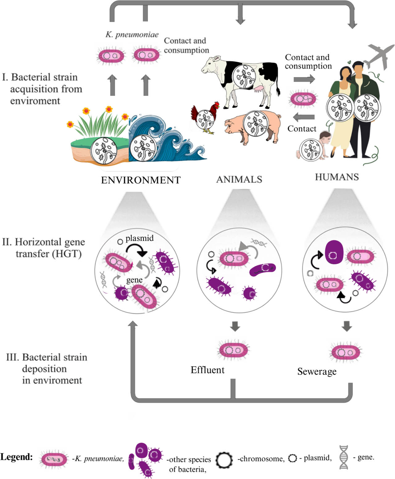
Fig. 1.
Diagram illustrating horizontal gene transfer (HGT) of resistance to antibiotics and/or other antimicrobial agents (ARGs) among K. pneumoniae strains. Own graphic design based on (Meek et al., 2015; Wyres et al., 2018 and Catalano et al., 2022).
The most common causes of the emergence and development of antibiotic resistance in K. pneumoniae strains are the overuse or inappropriate use of antimicrobials, not only in medicine but also in other areas of life (e.g., veterinary medicine, agriculture, fisheries, and horticulture) (Meek et al. 2015). The spread of resistance to antimicrobials, including antibiotics, is associated with the transfer of antibiotic resistance genes (ARGs) to other bacteria, through direct contact – i.e., from colonized animals to humans (especially from farm animals) – and through indirect contact (an environment contaminated with resistant strains) (Meek et al. 2015; Wyres et al. 2018). Indirect carriers of antibiotic–resistant bacteria or their ARGs can be: aerosols, raw materials and food of animal origin, feces, animal manure, soil, farmlands and crops, water resources, sewage, and other environmental pollutants. Hospitals, livestock farms, veterinary clinics, food processing plants, and food warehouses should also be considered concentration sites for antibiotic–resistant bacteria and their ARGs. The spread of drug–resistant strains is also facilitated by the growing phenomenon of travel and long–distance movement of people (Wyres et al. 2018).
2.1. Mechanisms of resistance in K. pneumoniae
K. pneumoniae has developed three major mechanisms of resistance to antibiotics and/or chemotherapeutics, i.e., enzyme resistance, receptor resistance, and transport resistance (Galani et al. 2022). This resistance can arise from inactivation, altered binding to the target site, or increased efflux of the drug. In addition, this resistance can be potentiated by the production of various beta-lactamases, mainly extended–spectrum beta-lactamases (ESBLs), or by biofilm formation (Fig. 2) (Galani et al. 2022).
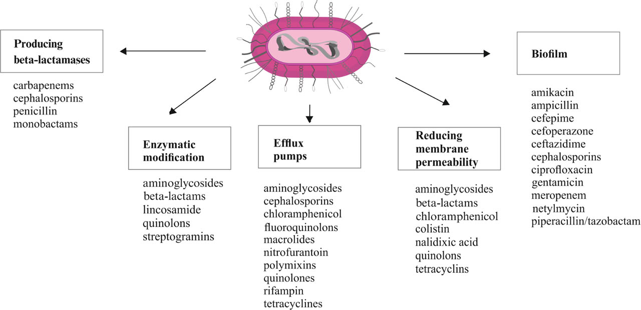
Fig. 2.
Diagram showing the resistance mechanisms of K. pneumoniae, along with the classes of antibiotics and/or chemotherapeutics to which each mechanism applies. Own graphic design based on (Hughes et al., 2020; Karami-Zarandi et al., 2022; Dzierżanowska-Fangrat, 2023).
2.1.1. Enzymatic resistance
Enzymatic resistance is the best understood, most common, and most extensively studied mechanism of resistance to beta-lactam antibiotics in K. pneumoniae strains. This type of resistance is associated with the production of extracellular beta-lactamases that degrade antibiotics in this class (Nordmann and Poirel 2014; Calbo and Garau 2015; Caltagirone et al., 2017; Chong et al., 2018).
A fundamental framework for beta-lactamase subdivisions, including all known classes, groups, and subgroups of enzymes, was established long before beta-lactam antibiotics were introduced into therapeutics (Paterson et al. 2003; Chong et al. 2018; Galani et al. 2022). The current vast diversity of beta-lactamases results from the spread of resistance genes among bacterial strains through conjugation, transduction, and transformation (Galani et al., 2022). As with other ARGs, a variety of MGEs play a key role in the mobilization and HGT of beta-lactamase genes. Moreover, over evolutionary time, additional mutations arise, and homologous recombination drives differentiation of these enzymes, driven by the massive use of antibiotics and/or chemotherapeutics (Meek et al. 2015; Wyres et al. 2018).
Currently, there are two main classification systems for beta-lactamases, i.e., the structural and functional systems (Table I) (Galani et al. 2022). The structural system proposed by Ambler is based on the analysis of the amino acid sequences of beta-lactamases and classifies the enzymes into four classes designated A through D, based on evolutionary affinity (Ambler 1991; Galani et al. 2022). Classes A, C, and D include serine beta-lactamases, while class B enzymes are metallo-beta-lactamases (Galani et al., 2022). All of these beta-lactamases from classes A, C, and D are described in more detail later in this article.
Table I.
Classification of preferred substrates for beta-lactam antibiotics, beta-lactamase inhibitors (BLIs), and bacterial beta-lactamases. Own graphic design based on (Livermore 1995; Bush and Jacoby 2010; Mączyńska 2015; Bush 2018).
| Molecular class according to Ambler | Bush-Jacoby-Mederios functional group | Preferred substrates in the group of beta-lactam antibiotics | Inhibited by beta-lactamase inhibitors (BLIs): | Bacterial beta-lactamases - representative enzymes | ||
|---|---|---|---|---|---|---|
| AV | CA or TZB | EDTA | ||||
| A | 2a | P | + | + | − | PC1 |
| 2b | P, Cp | + | + | − | TEM-1, TEM-2, SHV-1 | |
| 2be | P, Cp, E, Mb | + | + | − | ESBL (e.g. CTX-M), TEM, SHV, K1 (OXY) in K. oxytoca | |
| 2br | P | + | − | − | TEM-30, SHV-10 | |
| 2c | P, Cbp | + | ± | − | CARB-1, PSE-1, PSE-3, PSE-4 | |
| 2e | Cp | + | + | − | Inducible cephalosporinases Proteus vulgaris | |
| 2f | P, Cp, Cb, E, Mb | + | ± | − | KPC | |
| B | 3a | P, Cp, E, Cb | + | − | + | IMP-1, VIM-1, NDM-1, L1 |
| 3b | Cb | − | − | + | CphA | |
| C | 1 | Cp | + | − | − | AmpC (e.g. MIR-1), CMY |
| 1e | Cp, E | + | − | − | GC1 | |
| D | 2d | P, Clox | + | ± | − | OXA-1, OXA-10 (PSE-2) |
| 2de | P, Cb | + | − | − | OXA-23, OXA-48 | |
| 2df | P, E, Mb | + | ± | − | OXA-11, OXA-15 | |
[i] Abbreviations: AV, avibactam; CA, clavulanic acid; Cb, carbapenem; Cbp, carboxypenicillins; Cp, cephalosporin; Clox, cloxacillin; E, extended-spectrum cephalosporin; EDTA, ethylenediaminetetraacetic acid; Mb, monobactam; P, penicillin; TZB, tazobactam; (+), hydrolyzed; (−), unhydrolyzed; (±), barely hydrolyzed.
The second classification system, developed by Bush and Jacob, groups beta-lactamases based on the rates of hydrolysis of beta-lactam antibiotics and on an assessment of these enzymes' susceptibility to classic beta-lactamase inhibitors (clavulanic acid, sulbactam, and tazobactam) and other compounds, including aztreonam, cloxacillin, EDTA, and NaCl (Table I) (Bush and Jacoby 2010; Galani et al. 2022). However, aztreonam and cloxacillin are not classic beta-lactamase inhibitors; rather, they are compounds that can be inactivated by beta-lactamases or that inhibit these enzymes through mechanisms distinct from those of classic inhibitors (clavulanic acid, sulbactam, tazobactam). Aztreonam is a synthetic antibiotic in the monobactam class, with a structure that differs significantly from other beta-lactam antibiotics. It is highly resistant to hydrolysis by most beta-lactamases produced by Gram-negative bacteria. However, it is hydrolyzed by many enzymes, including: extended-spectrum plasmid-mediated beta-lactamases (ESBLs) of class A (mutated forms of classical enzymes such as TEM and SHV-1) and class D enzymes (e.g., OXA-1); chromosomal AmpC cephalosporinase produced by mutants of certain Enterobacterales species in the genera Enterobacter, Serratia, Citrobacter, and indole-positive Proteus, as well as non-fermenting rods of the genus Pseudomonas; and plasmid-mediated enzymes derived from AmpC-type beta-lactamases produced by Klebsiella spp. and Escherichia spp. (Dzierżanowska 2024). The presence of a characteristic monocyclic beta-lactam ring makes this antibiotic naturally resistant to class B beta-lactamases (MBLs). This resistance is due to spatial separation from the nucleophilic zinc ion present in the active site of these enzymes (Musawa et al. 2024). Because ESBL and AmpC beta-lactamases are widely co-produced in MBL-positive pathogens, aztreonam monotherapy has proven clinically ineffective. For strains that co-produce MBLs, ESBLs, and AmpCs, combination therapy or the use of the latest molecules has become the standard of care (Musawa et al. 2024; Khattab et al. 2025). Cloxacillin is an isoxazolyl penicillin resistant to staphylococcal beta-lactamases (penicillinases). Cloxacillin acts as a “suicide substrate,” binding the beta-lactamase active site and forming a stable acyl-cloxacillin-enzyme complex that prevents the enzyme from degrading other beta-lactam antibiotics in the vicinity (Kiener et al. 1980). EDTA (ethylenediaminetetraacetic acid) inhibits metallo-beta-lactamases (MBLs), enzymes classified as class B by Ambler, by chelating (binding) zinc ions (Zn2+), which are essential for their catalytic activity. MBLs (e.g., NDM, VIM, IMP) require one or two zinc ions in their active site to cleave the beta-lactam ring of an antibiotic. MBLs deprived of zinc become inactive, restoring bacterial susceptibility to carbapenems and other beta-lactam antibiotics (Bahr et al. 2021). There is no scientific evidence that sodium chloride (NaCl) directly binds to or inactivates beta-lactamase enzymes, the target of classic beta-lactamase inhibitors. However, high salt concentrations (high ionic strength) may indirectly affect enzyme function through several mechanisms: a) changes in protein structure – high salt concentrations can affect the conformational stability of proteins, including enzymes, which in some cases can lead to changes in their active site and a reduction in catalytic activity (Sinha and Khare 2014); b) effects on bacteria–resistance mechanisms, such as the production of beta-lactamases, are often linked to the life processes of bacteria. Changes in the osmotic environment (e.g., high salinity) can affect bacterial metabolism and, consequently, the production or secretion of these enzymes (Chambers and Hackbarth 1987); c) inhibition of autolysis (self-destruction) – beta-lactam antibiotics inhibit bacterial cell wall synthesis, which activates the bacteria's own “suicide” enzymes (autolysins). NaCl blocks the action of autolysins, preventing the bacterium from “falling apart” even though its cell wall is damaged (Chambers and Hackbarth 1987; Koch et al. 2001); d) effect on overall survival – rather than neutralizing the drug, NaCl alters the physiology of the bacteria (e.g., by increasing osmotic pressure or strengthening the cell wall) (Chambers and Hackbarth 1987; Esumeh et al. 2017). Accordingly, some of the listed substances can be used in phenotypic screening tests to detect enzymes such as ESBL–type beta-lactamases or carbapenemases (KPC, MBL, OXA-48), even though they are not inhibitors sensu stricto (Chambers and Hackbarth 1987; Koch et al. 2001; Sinha and Khare 2014; Esumeh et al. 2017). Bush and Jacoby's system classifies beta-lactamases into four major functional groups, numbered 1 to 4 (Paterson et al. 2003; Bush and Jacoby 2010).
Group 1 includes chromosomal AmpC cephalosporinases, which are completely inhibited by cloxacillin but respond poorly to clavulanic acid. Cephalosporins, penicillins, and monobactams (aztreonam) are preferred substrates of AmpC cephalosporinases (Livermore 1995; Nordmann and Poirel 2014). Group 2, the most structurally and functionally diverse, includes 12 subgroups (2a, 2b, 2be, 2br, 2ber, 2c, 2ce, 2e, 2f, 2d, 2de, 2df) of enzymes that include both penicillinases and narrow–spectrum cephalosporinases, such as: LEN-1 (from K. pneumoniae strain), OKP (other K. pneumoniae beta-lactamases), and SHV (sulfhydryl variable) (Livermore 1995; Haeggman et al. 2004; Nordmann and Poirel 2014; Arakawa 2020). Group 2 also includes broad-spectrum beta-lactamases (BSBL), also known as secondary beta-lactamases, subsequently extended–spectrum beta-lactamases (EBSL), and beta-lactamases with an extremely broad substrate spectrum covering virtually all beta-lactam antibiotics (Nordmann and Poirel 2014). The latter include carbapenemases of the KPC type (Klebsiella pneumoniae carbapenemases), acquired beta-lactamases hydrolyzing class D carbapenems (subgroup 2df), or CHDL enzymes (carbapenem-hydrolysing class D beta-lactamases), including OXA-48 (oxacilinase-48). The activity of enzymes of the OXA group, including OXA-48, is not inhibited by clavulanic acid, sulbactam, and tazobactam, with the exception of certain narrow–spectrum OXA enzymes (e.g., OXA-1), which may exhibit partial susceptibility to classical beta-lactamase inhibitors (Pitout et al. 2019). Group 3 includes metallo-beta-lactamases (MBLs) represented by imipenemase (IMP)-type beta-lactamases, Verona integron-encoded metallo-beta-lactamases (VIMs), and New Delhi metallo-beta-lactamase (NDM-1) (Livermore 1995; Nordmann and Poirel 2014). MBLs hydrolyze penicillins, cephalosporins, and carbapenems. These enzymes are encoded on class 1 and 3 integrons, are inhibited by EDTA, and are not inhibited by beta-lactamase inhibitors, distinguishing them from the activity of group 2 enzymes. Group 4 includes several long–identified penicillinases, while these poorly understood enzymes were omitted from the latest version of the functional classification system (Livermore 1995).
In K. pneumoniae strains, alongside the evolution of enzymatic resistance to beta-lactam antibiotics, the acquisition of enzymatic resistance to aminoglycoside antibiotics also occurred (Butler et al. 2021). Compared with beta-lactams, aminoglycosides have a much narrower spectrum of activity and are less likely to induce resistance in bacteria. The primary mechanism of bacterial resistance to this class of antibiotics is the production of plasmid-encoded enzymes. These enzymes modify aminoglycoside antibiotics by blocking the functionally active amine and hydroxyl groups (Butler et al. 2021). Among the most common aminoglycoside-modifying enzymes (AMEs) are: amino-glycoside N-acetyltransferases (AAC), which modify amino groups using acetyl–coenzyme A as the acetyl donor; O-phosphotransferases (AMEs), in turn, aminoglycoside phosphotransferases (APH), which modify hydroxyl groups using adenosine-5′-triphosphate (ATP); and O-nucleotidyltransferases (ANT), which modify hydroxyl groups using ATP (Butler et al. 2021). Natural aminoglycosides such as gentamicin, kanamycin, streptomycin, and tobramycin are readily modified (Butler et al. 2021). Aminoglycoside resistance may also result from reduced affinity of the 30S ribosomal subunit for the antibiotic, caused by mutations in bacterial DNA, 16S rRNA, and ribosomal proteins, as well as from reduced antibiotic uptake into the bacterial cell due to activation of efflux pumps that actively remove the drug from the cell (Butler et al. 2021).
2.1.1.1. Species-specific beta-lactamases
In K. pneumoniae, species–specific class A beta-lactamases were present even before the era of widespread antibiotic use. These enzymes include broad-spectrum beta-lactamases (LEN-1, OKP or SHV) that primarily hydrolyze penicillins (ampicillin, amoxicillin, carbenicillin, and ticarcillin) (Haeggman et al. 2004; Fevre et al. 2005; Arakawa 2020). These enzymes are chromosomally encoded and are constitutively expressed at low levels. Species-specific beta-lactamases do not hydrolyze cephalosporins and are sensitive to beta-lactamase inhibitors (Livermore 1995; Haeggman et al. 2004; Bush 2018).
2.1.1.2. Broad-spectrum beta-lactamases (BSBLs)
The emergence of broad-spectrum beta-lactamases (BSBLs), also known as secondary beta-lactamases, results from mutations and the transfer of genes for new beta-lactamases with a broader substrate hydrolysis spectrum onto a plasmid. BSBLs are usually plasmid-encoded, and the genes encoding them are often located within transposons (Haeggman et al. 2004; Dzierżanowska et al. 2010). In Enterobacteriaceae, the most common BSBLs are TEM-1, TEM-2, and SHV-1 (Dzierżanowska et al. 2010). TEM-1 was the first enzyme, discovered in 1965 in a strain of Escherichia coli isolated from a patient–resident of Athens, Greece, named Temoneira, after whom it was given its shortened name (Fig. 3). The TEM-1 enzyme hydrolyzes aminopenicillins most strongly, carboxypenicillins and first-generation cephalosporins to a lesser extent, does not degrade broad-spectrum cephalosporins, carbapenems, and monobactams, and is inhibited by clavulanic acid. TEM-2, like TEM-1, is a beta-lactamase with a broad substrate spectrum and exhibits high hemolytic activity. SHV-1 is another secondary beta–lactamase first described in 1972 in K. pneumoniae, also referred to as PIT-2 after its discoverer (Pitton 1972). Currently, SHV-1 is commonly found in this bacterial species (Haeggman et al. 2004; Dzierżanowska et al. 2010).
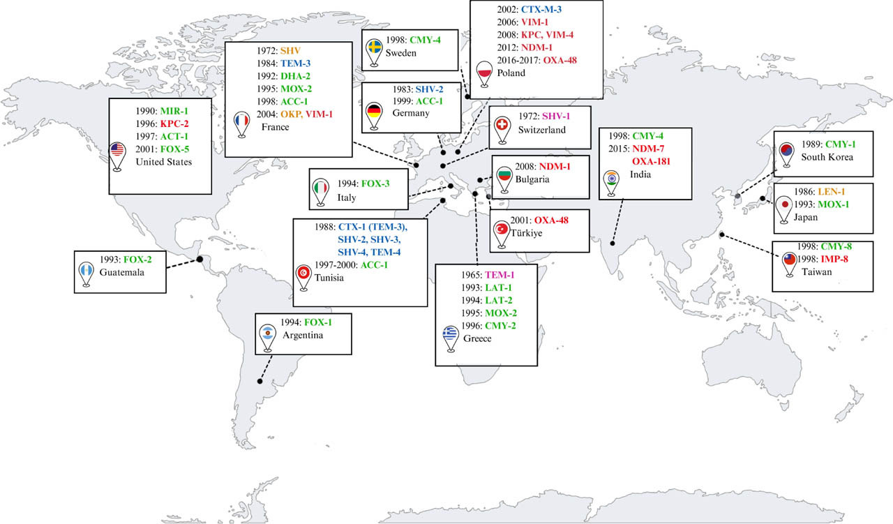
Fig. 3.
World map showing the spread and origin of K. pneumoniae strains producing: species-specific beta-lactamases (marked in orange); selected BSBL-type beta-lactamases (marked in purple); selected ESBL-type beta-lactamases (marked in blue); selected plasmid-encoded AmpC beta-lactamases (marked in green); and selected carbapenemases KPC, NDM, and OXA-48 (marked in red). Own graphic design based on (Jacoby 2009; Dzierżanowska et al. 2010; Mączyńska et al. 2015; Castanheira et al. 2021).
2.1.1.3. Extended–spectrum beta-lactamases (ESBLs)
Extended–spectrum beta-lactamases (ESBLs) were first detected in K. pneumoniae in 1983 in Europe (France, Germany) and then in the United States in 1989 (Bush 2018). These enzymes arose from point mutations in the genes responsible for the synthesis of BSBLs. ESBLs hydrolyze nearly all penicillins (except temocillin), cephalosporins (except cephamycins), and monobactams (aztreonam). ESBLs do not hydrolyze carbapenems. The activity of most of these beta-lactamases is inhibited by clavulanic acid, sulbactam, and tazobactam. Currently, among the numerous known ESBL variants, the most common are beta-lactamases from the TEM, SHV, CTX-M, and OXA families, described in more detail below. Lesser–known endemic enzymes include those from the BEL, BES, GES, PER, SFO, TLA, and VEB families, the description of which is beyond the scope of this publication (Bush 2018; Castanheira et al. 2021).
Beta-lactamases of the TEM family are common ESBL enzymes in K. pneumoniae. The first described enzyme from this family, CTX-1 (TEM-3), was detected in this bacterial species in 1984 (Philippon et al. 1989). Evolution has produced a large diversity of TEM-type beta-lactamase variants worldwide, with about 243 known (Bradford 2001; Bush 2018; Castanheira et al. 2021). In Poland, the scientific team of Gniadkowski et al. (1998) traced two evolutionary paths of the TEM family enzymes, i.e., the first path leading from the secondary beta-lactamase TEM-1 to the ESBL type TEM-93 and the second evolutionary path leading through different variants of ESBL types: TEM-25, TEM-29 and TEM-85, and, successively, through TEM–48, TEM-49 and TEM-47 (Gniadkowski et al. 1998). Moreover, in the studies of the group of Gniadkowski et al. (1998), a new variant TEM-68 resulting from a point mutation of TEM-47 was described (Gniadkowski et al. 1998). After 2000, few strains of K. pneumoniae producing TEM-30, TEM-32 and TEM-37 were detected (Gniadkowski et al. 1998).
SHV family beta-lactamases that contain a sulfhydryl group in the active site are also among the ESBL enzymes frequently produced by K. pneumoniae (Tsang and KlebNETGSP AMR Genotype-Phenotype Group et al. 2024). After the first SHV-2 enzyme was detected in Klebsiella ozenae, subsequent Klebsiella strains producing diverse, heterogeneous SHV variants have spread worldwide (Gniadkowski et al. 1998; Paterson et al. 2003; Dzierżanowska et al. 2010; Bush 2018; Castanheira et al. 2021). Currently, 228 variants of these widespread (cosmopolitan) enzymes are known. In Poland, the most frequently detected SHV variants are SHV-2, SHV-5, and SHV-12 (Baraniak et al. 2005; Liakopoulos et al. 2016; Castanheira et al. 2021).
Enzymes from the CTX-M family (cefotaximase from Munich) are considered the dominant group of ESBLs, replacing TEM and SHV. The first CTX-M-type beta-lactamases were detected in the 20th century, appearing simultaneously in several locations around the world, including France, Japan, Germany, and Italy (Castanheira et al. 2021). These initial reports were followed by the widespread dissemination of CTX-M in several countries. The worldwide expansion of isolates carrying these ESBLs was later termed the “CTX-M pandemic” (Castanheira et al. 2021). CTX-M enzymes originate from plasmid–transferred and mutated chromosomal genes of Klebsiella oxytoca OXY (K1) beta-lactamases. CTX-M enzymes exhibit strong hydrolytic activity toward cefotaxime, while they hydrolyze ceftazidime less effectively. Currently, CTX-M enzymes are the most common type of ESBL, with the dominant variants worldwide being CTX-M-15, CTX-M-14, and CTX-M-27 (Castanheira et al. 2021). In Poland, the most common variants of CTX-M enzymes are CTX-M-3 and CTX-M-15 (Baraniak et al. 2005).
OXA-type beta-lactamases constitute the majority of class D beta-lactamases in the Ambler classification and Bush–Jacoby–Medeiros functional group 2d (Yoon and Jeong 2021). This broad group of enzymes shows substantial variability in substrate profiles and amino acid sequences. These enzymes hydrolyze oxacillin. Several variants of OXA–type beta-lactamases have been reported that hydrolyze cephalosporins, cephems, and/or monobactams. According to Yoon and Jeong 2021, a total of 965 OXA-type beta-lactamases were detected in the beta-lactamase database (BLDB) used in this work (Yoon and Jeong 2021). Most extended–spectrum oxacillinases are derived from OXA-2 and OXA-10. OXA-2 derivatives include: OXA-15, OXA-32, OXA-34, OXA-36, OXA-53, OXA-141, OXA-161, OXA-210, and OXA-226. OXA-10 derivatives include OXA-11, OXA-13, OXA-14, OXA-16, OXA-17, OXA-19, and OXA-28 (Castanheira et al. 2021). One of the most common is OXA-1. Importantly, this group of enzymes gives rise to CHDL-class D beta-lactamases (e.g., OXA-48) that hydrolyze carbapenems, as described below (Pitout et al. 2019).
2.1.1.4. AmpC cephalosporinases
AmpC cephalosporinases are a distinct group of beta–lactamases that have evolved along a distinct evolutionary path (Tamma et al. 2019; Rodríguez–Guerrero et al. 2022). Chromosomal enzymes (chromo-some–mediated AmpC beta-lactamase, cAmpC) are encoded by evolutionarily “old” ampC genes located in the chromosomes of many species of Gram-negative bacilli, including Acinetobacter spp., Aeromonas spp., Citrobacter freundii, Enterobacter spp., Hafnia alvei, Morganella morganii, Proteus vulgaris, Providencia stuartii, Pseudomonas aeruginosa, Serratia marcescens, and Yersinia enterocolitica (Jacoby et al. 2009; Meini et al. 2019; Tamma et al. 2019). These bacteria gave rise to several families of acquired AmpCs, such as ACT, MIR, CMY-2, DHA, ACC, MOX, FOX, and LAT. The most common beta-lactamases from the CMY-2 family are derived from the C. freundii AmpC (Papanicolaou et al. 1990; Bauernfeind et al. 1999; Meini et al. 2019; Tamma et al. 2019; Rodríguez-Guerrero et al. 2022). Variants of these enzymes have been identified, among others, in Greece (LAT-1, LAT-2, MOX-2, CMY-2), France (DHA-2, MOX-2, ACC-1), the United States (ACT-1, FOX-5, MIR-1), South Korea (CMY-1), India and Sweden (CMY-4), Taiwan (CMY-8), Germany and Tunisia (ACC-1), Japan (MOX-1), Argentina (FOX-1), Guatemala (FOX-2), and Italy (FOX-3) (Fig. 3) (Jacoby et al. 2009; Dzierżanowska et al. 2010; Mączyńska et al. 2015; Castanheira et al. 2021). In K. pneumoniae and other bacterial species (e.g., K. oxytoca, Proteus mirabilis, Salmonella enterica) that normally do not produce cAmpC or produce it in negligible amounts (e.g., E. coli), plasmid-mediated AmpC beta-lactamase (pAmpC) can be found (Jacoby et al. 2009; Meini et al. 2019; Tamma et al. 2019).
The production of the AmpC enzyme depends on the ampD, ampG, and ampR gene system. AmpC production can be either induced or constitutive, resulting in different resistance phenotypes. Constitutive expression of the ampC gene means that AmpC enzymes are produced constantly at a steady level, whereas inductive expression involves production of the AmpC enzyme only after an inducer (e.g., an antibiotic) appears in the environment (Jacoby 2009; Meini et al. 2019; Tamma et al. 2019). The level of AmpC enzyme production may differ between the two expression types, reflecting variations in the rate of gene transcription that depend on the nucleotide sequence of the promoter, i.e., the site where expression begins. The speed of the process depends primarily on whether the antibiotic is a so-called weak or strong inducer. Antibiotics identified as strong inducers of the AmpC production pathway include aminopenicillins, amoxicillin/clavulanic acid, amoxicillin, narrow-spectrum (i.e., first-generation) cephalosporins, and cephamycins (Jacoby 2009; Meini et al. 2019; Tamma et al. 2019). Mutation of the regulatory gene (e.g., ampD) or deletion of the ampR gene causes permanent unblocking of expression, resulting in constant, very high-level beta-lactamase production regardless of the presence of the inducer (Jacoby 2009; Meini et al. 2019; Tamma et al. 2019). This phenomenon, referred to as AmpC derepression, enables bacteria to hydrolyze a wide range of substrates, i.e., oxyimino-beta-lactams (penicillins, cephalosporins, and monobactams) and 7-alpha-methoxycephalosporins (cefoxitin and cefotetan). At the same time, AmpC enzymes are not inhibited by commonly known inhibitors (clavulanate, sulbactam, and tazobactam). For infections caused by AmpC cephalosporinase-producing Enterobacterales, cefepime or carbapenems are the primary treatment options. The choice of regimen depends on the severity and location of the infection, with carbapenems the drugs of choice for the most serious infections (e.g., bacteremia, sepsis, pneumonia, intra-abdominal infections). Cefepime is a recommended, safe alternative to carbapenems, especially when the antibiogram indicates susceptibility to cefepime. Treating selected infections with cefepime is an important component of antimicrobial stewardship strategies aimed at reducing the overuse of carbapenems, thereby minimizing the risk of carbapenem-resistant organisms (the so-called “carbapenem-sparing” approach) (Tebano et al. 2024; Slain et al. 2025). For urinary tract infections caused by AmpC cephalosporinase-producing Enterobacterales, current treatment options include nitrofurantoin, fosfomycin, pivmecillinam, fluoroquinolones, cefepime, piperacillin with tazobactam, and carbapenems (Bader et al. 2020; Tebano et al. 2024; Slain et al. 2025).
Strains producing cAmpC in an inducible manner usually appear susceptible to third-generation cephalosporins, which are weak inducers, but can readily generate constitutively expressing mutants resistant to these drugs (which are good substrates), resulting in treatment failure. pAmpC is usually constitutively expressed. pAmpC production is common in community–acquired infections, while cAmpC producers are mainly involved in healthcare–associated infections (HAIs) (Meini et al. 2019).
K. pneumoniae belongs to the bacilli that lack the natural chromosomal AmpC mechanism (with the exception of Klebsiella mobilis). Currently, chromosomal ampC genes, both inducible and derepressed, have been transferred to plasmids and spread in the environment, especially in hospitals (Papanicolaou et al. 1990; Jacoby 2009; Meini et al. 2019; Tamma et al. 2019; Rodríguez–Guerrero et al. 2022). The elements ISEcp1, ISCR1, and IS26 are responsible for the transfer of natural ampC genes. This process has been accelerated by the widespread use of antibiotics from the 7-alpha-methoxycephalosporin group (cefoxitin and cefotetan) and combinations containing beta-lactamase inhibitors (amoxicillin with clavulanic acid, ampicillin with sulbactam, or piperacillin with tazobactam) (Rodríguez–Guerrero et al. 2022). Currently, outbreaks caused by K. pneumoniae strains with an acquired, derepressed AmpC mechanism, carrying plasmids encoding AmpC beta-lactamase genes of molecular class C, have been reported (Rodríguez–Guerrero et al. 2022). Moreover, in K. pneumoniae strains with reduced envelope permeability, AmpC enzymes may cause resistance to carbapenems, as observed in epidemic outbreaks registered, among others, in the United States and in numerous European countries: Spain, the Netherlands, Sweden, and the United Kingdom (Papanicolaou et al. 1990; Rodríguez–Guerrero et al. 2022).
K. pneumoniae remains a major producer of acquired AmpC cephalosporinase variants, first observed in 1988 (Jacoby 2009). Unlike their induced precursors, most acquired cephalosporinases are produced constitutively because the ampC gene is often mobilized without the accompanying regulatory gene ampR on the chromosome. Acquired AmpC confers resistance to penicillins, cephalosporins (except fourth–generation cephalosporins), monobactams, and beta-lactam-inhibitor combinations. As with other beta-lactamases, the level of resistance in strains depends on the level of enzyme expression. In clinical terms, acquired AmpC-producing K. pneumoniae strains are as dangerous as ESBL (+) strains, but epidemiologically they pose little threat due to their much lower incidence (Papanicolaou et al. 1990; Jacoby 2019; Meini et al. 2019; Tamma et al. 2019; Rodríguez–Guerrero et al. 2022).
2.1.1.5. Carbapenemases of classes A, B and D in the Ambler classification
2.1.1.5.1. Class A: KPC type carbapenemases
Among the known families of carbapenemases classified as molecular class A are: NMC-A/IMI, SME, KPC, SFC, BKC, FRI, and GES (Sawa et al. 2020; Ding et al. 2023). The chromosome is the primary location of genes encoding enzymes such as NMC-A/IMI, SME, and SFC, whereas genes encoding enzymes such as KPC, BKC, FRI, and GES are located on plasmids (Sawa et al. 2020; Ding et al. 2023). These genes can be located on various genetic elements, including simple or complex transposons, integrons, and phage-like genomic islands. Among class A carbapenemases, most enzymes are detected predominantly or partially in Enterobacterales as acquired beta-lactamases (Sawa et al. 2020; Ding et al. 2023).
KPCs (Klebsiella pneumoniae carbapenemases), one of the three most common types of carbapenemases in Enterobacterales, are important enzymes from the clinical and epidemiological point of view (Fig. 3) (Baraniak et al. 2011; Bush and Bradford 2019; Sawa et al. 2020). The first description of K. pneumoniae producing a carbapenemase KPC appeared in 1996 in North Carolina, USA. Since then, K. pneumoniae strains with this resistance phenotype have spread to many places around the world (Szewczyk 2019; Ochońska et al. 2021; Parra–Sellera et al. 2024). K. pneumoniae remains the dominant producer of KPCs, which constitute the largest category of class A carbapenemases in terms of the number of types and variants, while serine carbapenemases within this class constitute a significant minority (Bush and Bradford 2019; Sawa et al. 2020).
The production of KPC carbapenemases is determined by the gene blaKPC, which has a highly conserved nucleotide sequence. To date, several blaKPC haplo-types have been described, encoding KPC variants that differ by single amino acids (Ding et al. 2023). Currently, there are over 150 variants of the blaKPC gene, including most of the new variants detected over the last 3 years (Ding et al. 2023). Globally, the most commonly observed gene variants are blaKPC, and the two oldest variants remain, i.e., KPC-2 and KPC-3 (Ojdana et al. 2015; Machulska et al. 2017; Ochońska et al. 2021). In most K. pneumoniae strains producing KPC-type carbapenemases, blaKPC genes are located within specific elements of Tn4401, which belongs to the family of simple transposons Tn3 (Ding et al. 2023). Tn4401 fragments carrying blaKPC genes are located mainly on plasmids, among which the key role is played by specific molecules with two replicons, IncFIIK and IncFIBK (pKpQIL type), but also other plasmids: IncA/C, IncI2, IncR, IncX, and ColE1 (Ding et al. 2023).
KPC-type carbapenemases have the broadest substrate spectrum (Baraniak et al. 2011; Bush and Bradford 2019; Sawa et al. 2020). These enzymes effectively hydrolyze virtually all classical beta-lactam antibiotics, i.e., penicillins, 1st–4th generation cephalosporins, monobactams, and carbapenems (Codjoe et al. 2018). Among these groups, cefoxitin and ceftazidime remain poor substrates for KPC. This does not translate into susceptibility of KPC (+) strains to these antibiotics; moreover, in the common KPC-3 variant, mutations are known that increase KPC activity toward ceftazi-dime (Mehta et al. 2015). Cefiderocol, a new fifth-generation cephalosporin, has very good in vitro activity against KPC (+) strains, although, according to current reports, clinical experience with this drug is limited (McCreay et al. 2021; Syed et al. 2021). In the treatment of infections caused by KPC (+) strains, non-beta-lactam beta-lactamase inhibitors (avibactam, relebactam, and vaborbactam) in the following combinations are effective: ceftazidime with avibactam, imipenem with relebactam, and meropenem with vaborbactam (Bush et al. 2019).
On a global scale, the spread of KPC-type carbapenemases occurs through clonal expansion, i.e., the transmission of microorganisms containing blaKPC genes. Baraniak et al. (2017) presented an example of clonal spread of K. pneumoniae ST258 strains belonging to the CG258 clonal group, which includes closely related genotypes ST437 and ST512 (Baraniak et al. 2017). Hyperepidemic CG258 clones of K. pneumoniae are responsible for nationwide epidemics in Brazil, China, the Netherlands, Israel, Canada, Korea, Norway, Sweden, Italy, and many other countries (Pitout et al. 2015; Dautzenberg et al. 2016; Baraniak et al. 2017; Liu et al. 2022). Over the last 10 years, in Poland and in other countries, including France and Italy, significant changes have been noted in K. pneumoniae KPC (+) populations, consisting of the replacement of CG258 clones by other clones (emerging clones) with outstanding expansiveness, such as ST101, ST147, and ST307 (Loconsole et al. 2020; Peirano et al. 2020; Ochońska et al. 2021). Such clones, unlike CG258, are not closely related to KPC, but their variants producing ESBL-type beta-lactamases and/or other carbapenemases are often observed (Peirano et al. 2020). International CG258 K. pneumoniae clones are very often characterized by a heteroresistance phenotype, meaning that strains are simultaneously resistant to carbapenems and to antibiotics and/or chemotherapeutics from other therapeutic groups, mainly aminoglycosides, fluoroquinolones, tetracyclines, sulfonamides, and chloramphenicol (Liu et al. 2022; Krul and Dalla Costa 2024).
2.1.1.5.2. Class B: carbapenemases type MBL/NDM
Molecular class B includes metallo-beta-lactamases (MBLs) (Bush et al. 2019). Natural MBLs occur in many environmental bacteria of limited clinical importance, such as Aeromonas hydrophila, Bacillus cereus, Bacteroides fragilis, Chryseobacterium indologenes, Elisabethkingia meningoseptica, Myroides odoratimimus, and Stenotrophomonas maltophilia (Boyd et al. 2020). MBLs constitute the most distinct evolutionary and structural lineage. Functionally, these enzymes are distinguished from other carbapenemase classes by their dependence on zinc ions and their natural ability to hydrolyze carbapenems (Codjoe et al. 2018). The substrate spectrum of most MBL enzymes is broad and includes penicillins and cephalosporins from the first to the fourth generation. MBLs do not hydrolyze monobactams (aztreonam) and are not inhibited by beta-lactam inhibitors (clavulanate, sulbactam, and tazobactam). Similarly to KPC (+) strains, cefiderocol shows in vitro activity against MBL (+) strains. Ceftazidime with avibactam, ceftolozane with tazobactam, imipenem with relebactam, and meropenem with vaborbactam remain inactive against strains producing MBLs (Bush et al. 2019; Syed et al. 2021; Shortridge et al. 2022). In the treatment of infections caused by strains with this resistance phenotype, attempts are being made to use substances that demonstrate the ability to inhibit MBL enzymes, such as diazabicyclooctanes (zidebactam and nacubactam), cyclic boronic acids (taniborbactam), or indole-2-carboxylates (InCs) (Bush et al. 2019; Yahav et al. 2020; Brem and Schofield 2022).
Among the 20 families of acquired MBL-type carbapenemases commonly found in K. pneumoniae enterobacteria, the most clinically and epidemiologically significant are the NDM (New Delhi metallo-beta-lactamases) enzymes (Boyd et al. 2020; Ochońska et al. 2021; Biedrzycka et al. 2022). About 65 variants of NDM-type enzymes are known, with NDM-1 and NDM-5 predominating in pathogenic microorganism populations (Reference Gene Catalog - Pathogen Detection, https://www.ncbi.nlm.nih.gov/pathogens/refgene/). The production of NDM-type carbapenemases is encoded by the blaNDM gene, which, together with the bleomycin resistance gene (bleMBL), forms an operon that is part of the complex transposon Tn125 (Fiett et al. 2014). In Enterobacterales bacilli, larger or smaller fragments of Tn125 elements are observed, and the blaNDM genes are located on plasmids of many groups: IncC, IncF, IncH, IncM, IncN, IncR and IncX, and their various subgroups. This localization indicates high transposition and recombination activity of Tn125 and/or other “secondary” elements containing Tn125 fragments (Fiett et al. 2014).
The diversity of plasmids carrying blaNDM genes means that, both globally and in individual countries, the distributions of K. pneumoniae MBL (+) clones (genotypes) are more diverse than those of K. pneumoniae KPC (+) strains (Parra Selerra et al. 2024). Uncontrolled spread of a given K. pneumoniae clone (genotype) can lead to its dominance in a given region (Fiett et al. 2014; Loconsole et al. 2020; Peirano et al. 2020; Krul and Dalla Costa 2024).
The Indian subcontinent is the primary reservoir of microorganisms producing NDM-type enzymes, which are widely distributed in the environment and have been spreading worldwide since 2006 among hospital pathogens (Bose et al. 2022). The endemic state of NDM has been reported in the Persian Gulf countries, North African countries, and the southwestern Balkans (Subramanian et al. 2016). Since 2008, K. pneumoniae NDM (+) strains have been systematically detected across Europe (Baraniak et al. 2016; Baraniak et al. 2019; Parra Sellera et al. 2024).
2.1.1.5.3. Class D: carbapenemases type OXA–48
Class D is the most diverse category of beta–lactamases in terms of evolution and structure. Enzymes in this group most effectively hydrolyze isoxazolyl penicillins (oxacillin and cloxacillin), which is why they are called oxacillinases. CHDL carbapenemases (CHDL) are a minority among oxacillinases and mainly comprise serine beta-lactamases from the OXA family, produced naturally by bacilli of the genera Acinetobacter, Aeromonas, and Shewanella (Bush et al. 2019; Yoon and Jeong 2021). Most OXA enzymes are unable to hydrolyze carbapenems and are classified as ESBL enzymes. New variants that have emerged during evolution and exhibit an extended substrate-hydrolysis spectrum show weak hydrolytic activity toward carbapenems (Bush et al. 2019; Yoon and Jeong 2021).
In K. pneumoniae, the plasmid variant of OXA-47 located in class 1 integrons does not show hydrolytic activity against ceftazidime and imipenem, whereas the acquired CHDL (OXA-48 variant) already shows strong carbapenemase properties (Nordmann–Poirel 2014; Mączyńska 2015). The OXA-48 mechanism was first detected in a strain of K. pneumoniae in Turkey in 2001. After this discovery, observational studies showed a high prevalence of OXA-48-type carbapenemase in the Arabian Peninsula, the Middle East, North Africa, and Europe (Pitout et al. 2019). The activity of OXA-48-type oxacillins, similar to other CHDLs, is highly specific among carbapenemases, as it covers only penicillins, first-generation cephalosporins, and carbapenems, while the efficiency of carbapenem hydrolysis is relatively low. Resistance to temocillin is characteristic of OXA-48-producing strains. These enzymes are not sensitive to EDTA and clavulanic acid (Nordmann–Poirel 2014; Mączyńska 2015; Bush et al. 2019; Yoon and Jeong 2021). CHDLs do not inactivate third- and fourth-generation cephalosporins, monobactams, and cefiderocol. OXA-48 and other CHDLs are not inhibited by clavulanate, sulbactam, and tazobactam, which is why antibiotic combinations containing them are not used. The preferred first-line treatment for severe infections caused by OXA-48-producing bacterial strains is typically a combination of beta-lactam antibiotics and beta-lactamase inhibitors (e.g., ceftazidime with avibactam) (Pitout et al. 2019). In cases of non-response, resistance, or mixed infections (e.g., those additionally producing MBLs), cefiderocol demonstrates high efficacy (Tarski et al. 2024). In the absence of newer drugs or due to specific clinical circumstances, the use of older antibiotics in combination (e.g., colistin, tigecycline, aminoglycosides) is considered, always based on an individual antibiotic susceptibility test (Stewart et al. 2018). It is worth noting that OXA-48 microorganisms (as in other CPE) commonly harbor ESBLs, which means that these two beta-lactamases together hydrolyze all classical beta-lactams (Nordmann–Poirel 2014; Mączyńska 2015; Bush et al. 2019; Yoon and Jeong 2021).
The formation of the group of OXA-48 derivatives, i.e., OXA-181, OXA-204, OXA-232, OXA-163, OXA-244, and OXA-245, was associated with substitution or deletion of single amino acids (Pitout et al. 2019). blaOXA-48-type genes occur in various mobile genetic elements, most often in complex transposons of the Tn1999 type. These genes can also be located within the ISEcp1 transposition modules defined as Tn2016-type elements (Izdebski et al. 2018). These transposons are most often located on plasmids, but their diversity is lower than that of the blaKPC, blaNDM, and blaVIM genes. Tn1999-type elements usually lie on plasmids from the IncL group. The evolutionary line pOXA-48a is characterized by an exceptionally high potential for conjugative transfer of blaOXA-48 genes and their derivatives in Enterobacterales populations at the scale of single hospitals, countries, and the world. Tn2013 modules with the blaOXA-181 gene or its variants often correlate with plasmids from the IncX3 group, but also with IncM, IncT, and ColE (Izdebski et al. 2018).
K. pneumoniae is one of the most commonly reported species of redundant carbapenemase–producing (RCP) bacteria. These bacteria possess dual or multiple carbapenemases in various combinations, are widely distributed worldwide, and their incidence is increasing over time (Yuan et al. 2024). In the study by Yuan et al. (2024), it was shown that K. pneumoniae carrying certain carbapenemase combinations, i.e., NDM + OXA (56.76%) and KPC + VIM (50.00%), are associated with high mortality. In patients with RCP strains isolated from the bloodstream and respiratory tract, the mortality rates are 58.70% and 69.23%, respectively. Plasmid analysis of RCP strains suggests that they may acquire additional antibiotic resistance phenotypes and virulence factors (Yuan et al. 2024).
2.1.2. Receptor resistance
Receptor resistance in K. pneumoniae concerns beta–lactam antibiotics, aminoglycosides and fluoroquinolones. This type of resistance involves modifying the antibiotic's target binding site, called the receptor, where it binds to the bacterial cell. The effect of this process is various conformational changes, depending on the type of antibiotic. In the case of beta-lactam antibiotics, the mechanism of receptor resistance is associated with permeability barriers or with a decrease in affinity for penicillin-binding proteins (PBPs) belonging to the genera PBP-3, PBP-4 and PBP-5. This mechanism is recorded in 60% of Enterobacteriaceae (Dzierżanowska–Fangrat 2023). Receptor mechanisms of resistance to aminoglycosides may result from changes in ribosomal proteins (mainly streptomycin) or, less frequently, from mutations occurring in ribosomal RNA (rRNA). In the epidemiological aspect, the most important mechanism determining the resistance of K. pneumoniae to most aminoglycosides used in therapy (amikacin, gentamicin, kanamycin, netilmicin and tobramycin) is the production and expression of plasmid-encoded methyltransferases that methylate the 16S rRNA subunit: ArmA (aminoglycoside resistance methyltransferase), RmtA (resistance-associated methyltransferase A), RmtB (resistance-associated methyltransferase B), RmtC (resistance-associated methyltransferase C), RmtD (resistance-associated methyltransferase D), RmtG (resistance-associated methyltransferase G) (Ishizaki et al. 2018, Yang et al. 2022).
For fluoroquinolone antibiotics, the primary mechanism of resistance is mutations in the quinolone resistance-determining region (QRDR). The most common mutations occur in the genes responsible for the synthesis of gyrase (gyrA) or topoisomerase IV (parC). The first enzyme introduces negative supercoils into the DNA helix, and the second enzyme separates DNA strands necessary for the replication process (Dzierżanowska–Fangrat 2023). Mutations in the gyr gene lead to a change in a single amino acid in the enzyme. Resistance associated with topoisomerase IV results from stepwise mutations, through which bacterial strains gradually acquire higher levels of resistance (Dzierżanowska–Fangrat 2023).
2.1.3. Transport resistance
Transport resistance may result from mutations in genes encoding outer membrane proteins (OMPs). OMPs are anchored in the bacterial cell membrane and constitute the first barrier encountered by antibacterial agents. The ability to penetrate the cell membrane is necessary for inhibiting bacterial division or killing bacteria (Rocker 2020). In K. pneumoniae, two major porin proteins (OmpK35 and OmpK36), present in large numbers of copies in the membrane, play a key role in antibiotic transport. Resistant strains may be characterized by a deficiency of porins in the cell membrane, in which case their functions are taken over by minor proteins (LamB, OmpK37/26 and PhoE), the expression of which in the membrane depends on the needs of the cell. These proteins compensate for the deficiency of non–specific porins by allowing the free flow of nutrients while blocking the flow of antibiotics (Rocker 2020).
The second possible variant of transport resistance used by K. pneumoniae bacilli is the pumping of antibiotics out of the cell via the efflux mechanism. The transport of antibiotics is determined by the presence of transport proteins in the cell membrane. Depending on structure, the number of transmembrane sequences, substrate specificity, and mechanism of action, five families of pumps are distinguished: MFS (major facilitator superfamily), SMR (small multidrug resistance family), MATE (multidrug and toxic compound extrusion family), ABC (ATP–binding cassette superfamily), and RND (resistance–nodulation–cell–division family) (Huang et al., 2022). Due to the complex structure of the cell envelopes in Gram-negative bacteria, transport proteins form a tripartite structure composed of a protein of the inner cytoplasmic membrane, a protein of the outer membrane, and a protein of the periplasmic space.
In K. pneumoniae, there is an AcrAB-TolC pump previously described in E. coli (Huang et al. 2022). This pump belongs to the RND family of pumps that use a proton gradient across the cell membrane to trigger the movement of substrate particles (Huang et al. 2022). The AcrAB-TolC pump is responsible not only for the removal of fatty acid salts from the bacterial cell, but also for the ejection of antibiotics, including aminoglycosides, beta-lactams, fluoroquinolones, glycocyclines, and tetracyclines. As a result of derepression of the acrAB gene, the cell membranes of resistant K. pneumoniae strains are characterized by an increased number of pumps, which results in increased active removal of the antibiotic from the bacterial cell (Huang et al. 2022).
3. Treatment of infections caused by K. pneumoniae
Antibiotics and other antimicrobial agents active against K. pneumoniae include: aminoglycosides (amikacin, gentamicin, netilmicin, tobramycin), second-generation cephalosporins (cefuroxime), third-generation cephalosporins (ceftazidime, cefotaxime) and fourth-generation cephalosporins (cefepime), fluoroquinolones (ciprofloxacin, levofloxacin, moxifloxacin, ofloxacin), carbapenems (doripenem, ertapenem, imipenem, meropenem), monobactams (aztreonam), piperacillin with tazobactam, ticarcillin with clavulanic acid, trimethoprim with sulfamethoxazole (co-trimoxazole) and ureidopenicillins (azlocillin, mezlocillin). In empirical therapy, the most commonly used groups of antibiotics are aminoglycosides and cephalosporins (Dzierżanowska-Fangrat 2023).
In the case of isolation of multi-drug resistant (MDR) K. pneumoniae strains and strains belonging to CPE, it is necessary to determine the sensitivity to available antibacterial drugs in order to select the most effective therapeutic option (Chung et al. 2016; Wang et al. 2019; Wyres et al. 2020). Older generation antibiotics used to treat infections caused by CPE strains include: aminoglycosides (amikacin, gentamicin, tobramycin), meropenem, colistin methanesulfonate sodium (colistimethate), fosfomycin sodium, temocillin, and sulfamethoxazole with trimethoprim (Hughes et al. 2020; Kuch et al. 2022). Moreover, several new antibiotics have recently been registered for the treatment of infections caused by CPE strains, including: cefiderocol, plazomicin, tigecycline, eravacycline, omadacycline, ceftazidime with avibactam, meropenem with vaborbactam, and imipenem with cilastatin and relebactam (Table II) (Bush et al. 2019; Hughes et al. 2020; McCreary et al. 2021; Kuch et al. 2022). It should be emphasized that these drugs often do not cover all classes of carbapenemases in their spectrum of action, and are often registered for very narrow indications (Hughes et al. 2020; Krajewska and Laudy 2021; Kuch et al. 2022).
Table II.
Table listing currently used antibiotics for treating infections caused by carbapenem-resistant Gram-negative bacteria (CR-GNB) and the recommended and approved indications for their use. Own graphic design based on Paul et al., (2022).
| ESBLs | CRE non-CP | CRE-KPC | CRE-O-XA-48 | CRE-MBL | Current clinical indications | Approval | |
|---|---|---|---|---|---|---|---|
| New antibiotics | |||||||
| Cefiderocol | Yes | Yes | Yes | Yes | Yes | cUTIs, HAP, VAP | FDA |
| for the treatment of infectious due to aerobic Gram-negative organisms in adults with limited treatment options | EMA | ||||||
| Ceftolozane-tazobactam | Yes | No | No | No | No | cIAI, cUTIs, HAP, VAP | FDA and EMA |
| Ceftazidime-avibactam | Yes | +/− | Yes | Yes | No | cIAI, cUTIs, HAP, VAP | FDA and EMA |
| for the treatment Gram-negative infections in patients with limited treatment options | EMA | ||||||
| Eravacycline | Yes | Yes | Yes | Yes | Yes | cIAI | FDA and EMA |
| Imipenem-cilastatin-relebactam | Yes | +/− | Yes | No | No | cIAI, cUTIs | FDA |
| HAP, VAP, BSI with a suspected respiratory source, and for the treatment Gram-negative infections in patients with limited treatment options | EMA | ||||||
| Meropenem-vaborbactam | Yes | +/− | Yes | No | No | cUTIs | FDA |
| cUTIs, HAP, VAP, and for the treatment Gram-negative infections in patients with limited treatment options | EMA | ||||||
| Plazomicin | Yes | Yes | Yes | Yes | +/− | cUTIs | FDA |
| EMA application withdrawn | |||||||
| Old antibiotics | |||||||
| Aminoglycosides | +/− | +/− | +/− | +/− | +/− | for the treatment of a variety of bacterial infections | FDA and EMA |
| Aztreonam | No | No | No | No | +/− | for the treatment of infections caused by susceptible Gram-negative micro-organisms | FDA and EMA |
| Fosfomycin iv | Yes | +/− | +/− | +/− | +/− | to treat serious infections when other antibiotics treatment are not suitable | EMA |
| FDA under review | |||||||
| Polymyxins | Yes | Yes | Yes | Yes | Yes | to treat serious infections caused by susceptible strains, when less potentially toxic drugs are ineffective or contraindicated | FDA |
| to treatment of serious infections due to aerobic Gram-negative pathogens in patients with limited treatment options | EMA | ||||||
| Tygecycline | Yes | Yes | Yes | Yes | Yes | complicated SSTI and IAI | FDA and EMA |
| CAP | FDA | ||||||
[i] Abbreviations: BSI, bloodstream infection; CAP, community-acquired pneumonia; cIAI, complicated intra-abdominal infections; CRE non-CP, non-carbapenemase-producing carbapenem-resistant Enterobacterales; cUTIs, complicated urinary tract infections; EMA, European Medicines Agency; ESBLs, extended-spectrum beta-lactamases; FDA, US Food and Drug Administration; HAP, hospital-acquired pneumonia; iv, intravenous administration; MBL, metallo-beta-lactamases; SSTI, skin and soft-tissue infections; VAP, ventilator-associated pneumonia; +/−, variable antibiotic activity.
In Poland, examples of the use of newly registered drugs were given in the work of Sękowska et al. (2024) covering a group of 41 strains of K. pneumoniae (pan-drug resistant, PDR) and in which 43.9% of isolates were sensitive to colistin, 24.4% of isolates were sensitive to fosfomycin and ceftazidime with avibactam, 14.6% of isolates were sensitive to meropenem with vaborbactam (Sękowska et al. 2024). In another study by the same scientific team, Sękowska et al. (2024) demonstrated the efficacy of plazomicin, a new aminoglycoside with a broad spectrum of activity against the analyzed strains of K. pneumoniae MDR (Sękowska et al. 2024). In a study of ESBL-positive K. pneumoniae strains, plazomicin showed high activity with MIC values ranging from 0.19 to 4 μg ml-1, whereas for carbapenemase-positive strains, MIC values ranged from 0.25 to 256 μg ml-1 (Sękowska et al. 2024). In turn, in the case report presented by Tarski et al. (2024), the efficacy of cefiderocol in the treatment of a 72-year-old patient with sepsis caused by K. pneumoniae NDM and OXA-48 was demonstrated (Tarski et al. 2024). Polymyxins are useful in the treatment of patients infected with K. pneumoniae NDM, and the efficacy of therapy is greater when colistin is used in combination with carbapenem or rifampicin or tigecycline. Revealing the in vitro interactions of such antibiotic combinations was the aim of the study conducted by Müderris et al. (2024) covering a group of 30 K. pneumoniae isolates forming biofilm (Müderris et al. 2024). Based on the obtained results, the study showed that the biofilm inhibitory concentration (BIC) of colistin, meropenem, tigecycline and rifampicin significantly increased after biofilm formation. The synergistic effect observed in the sessile form was independent of the planktonic form. Although a high synergistic effect of the combination of meropenem and colistin on sessile bacteria was observed, colistin had a very high BIC in all combinations (Müderris et al. 2024).
The introduced avibactam in combination with ceftazidime and aztreonam is another promising therapeutic combination in severe infections caused by K. pneumoniae, in particular by (MDR) strains. The synergistic effect of the combination of ceftazidime, avibactam, and aztreonam results in the protection of both beta-lactam antibiotics, ceftazidime and aztreonam, by avibactam, which shields them from degradation by bacterial enzymes (serine beta-lactamases). Avibactam belongs to a new class of non-beta-lactam beta-lactamase inhibitors. This multifunctional organic compound contains a 5-membered urea ring at its core, which significantly enhances ceftazidime's activity against multidrug-resistant Gram-negative bacilli. Like beta-lactam inhibitors, avibactam covalently binds to serine at the active site of beta-lactamases. Unlike other inhibitors, however, this process is reversible; the inhibitor does not undergo hydrolysis but is reconstituted (the 5-membered molecule is less susceptible to irreversible deformation). Due to avibactam's affinity for serine, this inhibitor is active against serine beta-lactamases, specifically those belonging to classes A (ESBL and KPC enzymes), C (chromosomal and plasmid-mediated AmpC), and certain class D enzymes (e.g., OXA-48) according to the Ambler classification (Kowalska-Krochmal et al. 2019). Avibactam does not inhibit class B (MBL) enzymes and is unable to inhibit the activity of many class D enzymes. Aztreonam is naturally resistant to hydrolysis by metal-lo-beta-lactamases (e.g., NDM). Avibactam, which inactivates beta-lactamases that degrade aztreonam, allows the antibiotic's efficacy to be restored. The combination of both drugs, ceftazidime with avibactam and aztreonam, allows for the inhibition of both ESBL and MBL resistance mechanisms, which is crucial in the fight against MDR strains of K. pneumoniae (Kowalska–Krochmal et al. 2019).
The use of this drug combination is supported by numerous studies conducted by Polish and international research teams (Guzek et al. 2024; Słabisz et al. 2024; Szymański et al. 2024; Khattab et al. 2025; Rajan and Sasikala 2025). For example, the scientific team of Słabisz et. al. (2024) examined 60 strains of K. pneumoniae producing NDM-type carbapenemase (Słabisz et. al. 2024). This study showed that all tested K. pneumoniae strains (100%) were resistant to ceftazidime with avibactam and 92% of strains were resistant to aztreonam when these drugs were used individually (Słabisz et al. 2024). The strip stacking method confirmed the synergistic effect of ceftazidime/avibactam (CZA) and aztreonam (AT) and demonstrated 100% in vitro sensitivity to this combination of antibiotics among the tested strains (Słabisz et al. 2024). Similarly, in a study by another Polish scientific team, (Guzek et al., 2024), the efficacy of the combination of ceftazidime/avibactam with aztreonam in the treatment of 23 patients with hospital-acquired KP-NDM infection was confirmed (Guzek et al. 2024). In the study, the synergistic effect of all compounds resulted in good agreement between the clinical efficacy of CZA + AT and the results of in vitro susceptibility tests (Guzek et al. 2024). According to the research conducted by the scientific team of Szymański et al. (2024), in the group of patients treated with CZA + AT, statistically lower mortality and faster clinical response to treatment were demonstrated due to the therapy used compared to the group of patients treated with alternative combinations of antibiotics including: colistin sodium, fosfomycin, an aminoglyco-side, and tigecycline (Szymański et al. 2024). Similar positive clinical assessments were obtained for sulbactam, especially in combination with meropenem and colistin, which was confirmed in the study conducted by (Laishram et al. 2016). Combinations of sulbactam, meropenem, and colistin were studied for their synergistic activity against 100 invasive carbapenem-resistant K. pneumoniae isolates from cancer patients hospitalized in the ICU (Laishram et al. 2016). In recent years, researchers have explored several combination therapies, such as heat shock in combination with aminoglycosides, polymyxin B combined with fosfomycin, zidovudine combined with rifampicin, FAS (fatty acid synthesis) inhibitors in conjunction with colistin, as well as new therapeutic approaches such as monoclonal antibodies (MAb), vaccines, phage therapy, pyridylpiperazine efflux pump inhibitor (PyrPips), probiotics, and physiotherapy (Fig. 4) (Wantuch and Rosen 2023; Lei et al. 2024).
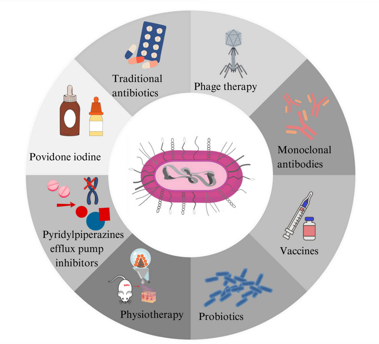
Fig. 4.
Diagram showing therapeutic strategies for treating infections caused by multidrug-resistant (MDR) K. pneumoniae. Own graphic design based on (Wantuch and Rosen, 2023; Lei et al., 2024 and Li et al., 2024).
4. Epidemiology of K. pneumoniae resistant to specific classes of antibiotics in Europe and Poland
Antibiotic resistance in K. pneumoniae bacilli is a major medical and epidemiological problem both across Europe and in Poland (Asokan et al. 2025, Shah et al. 2025). Data from national and international antibiotic resistance surveillance networks provide insights into the scale of this problem. In Europe, for the countries of the European Union (EU) and the European Economic Area (EEA) comprising Iceland and Norway, the monitoring of antibiotic resistance in bacterial strains cultured from blood and cerebro-spinal fluid samples is conducted by the European Centre for Disease Prevention and Control (ECDC) in Stockholm as part of the European Antimicrobial Resistance Surveillance Network (EARS-Net, https://www.ecdc.europa.eu) (EARS-Net 2024; ECDC 2026). EARS-Net uses standardized methodologies for data collection and analysis. However, given that each national surveillance system is shaped by country-specific protocols and practices, particular caution should be exercised when comparing antimicrobial resistance patterns across countries. In 2024, the EARS-Net network published data submitted by 30 European countries, from which 62 774 K. pneumoniae isolates derived from invasive infections (including blood) were reported. Data collected by EARS-Net laboratories, after being verified and prepared for transmission, are submitted to the EpiPulse database at the ECDC. The EARS-Net network in Europe is complemented by the CAESAR network (The Central Asian and European Surveillance of Antimicrobial Resistance Network) coordinated by the World Health Organization (WHO). Both networks: EARS-Net and CAESAR submit data to the WHO GLASS (Global Antimicrobial Resistance and Use Surveillance System) database to the extent that their systems overlap (https://www.who.int/initiatives/glass, https://www.who.int/) (EARS-Net 2024; WHO GLASS 2025; ECDC 2026).
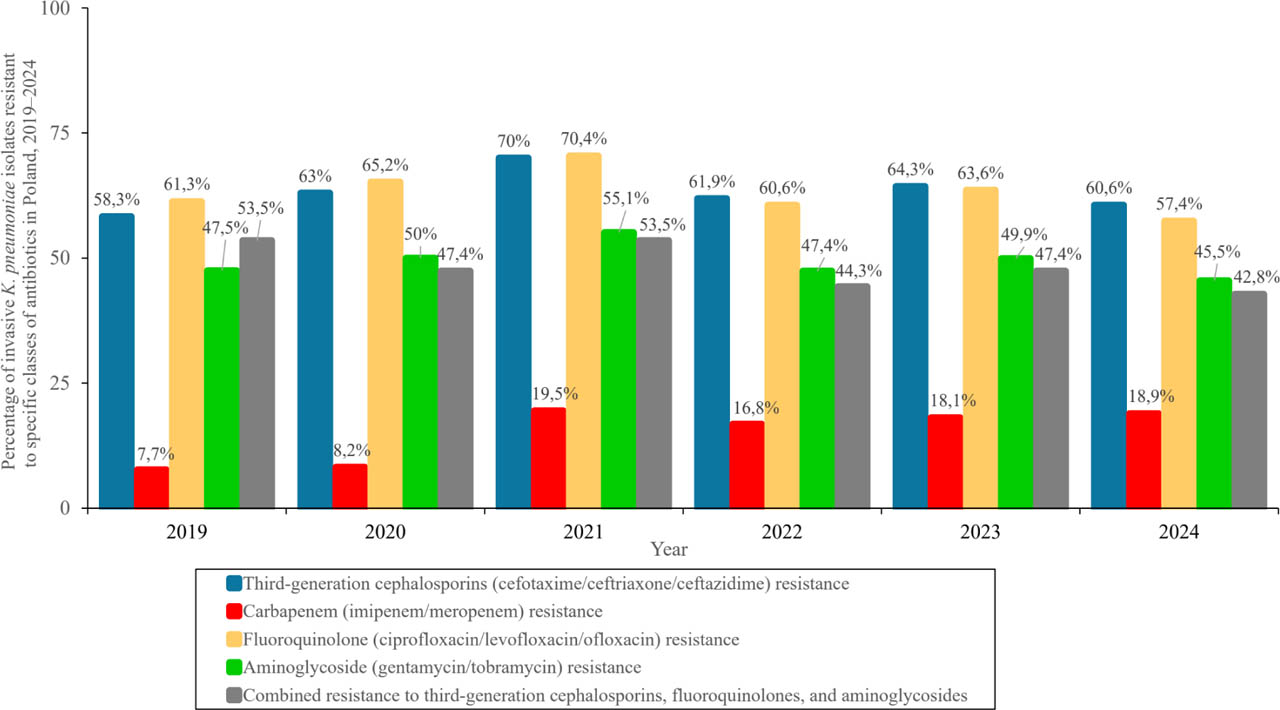
Fig. 5.
Graph showing the percentage of invasive K. pneumoniae isolates resistant to specific antibiotic classes in Poland, 2019–2024. Own graphic design based on data from EARS-Net (2024) and ECDC (2026).
In Poland, monitoring within the EARS-Net network includes laboratories from across the country that perform microbiological testing for selected university, provincial, and county hospitals. In 2024, data provided by 54 EARS-Net laboratories from across Poland, performing microbiological testing for 61 hospitals, were analyzed (EARS-Net 2024). In Poland, the dataset is funded from resources at the disposal of the Minister of Health as part of the implementation of the task titled “Preventing the Development of Antibiotic Resistance in Microorganisms” under the National Health Program for 2021–2025, and is coordinated by the Polish National Reference Centre for Antimicrobial Susceptibility Testing (KORLD) operating at the National Medicine Institute in Warsaw (Journal of Laws of the Republic of Poland, Warsaw, April 8, 2021, Item 642: Regulation of the Council of Ministers of March 30, 2021, on the National Health Program for 2021–2025, KORLD).
In EU/EEA countries, the estimated incidence of invasive K. pneumoniae isolates (per 100 000 population) was 19.2 cases in 2020, 19.3 in 2021, in 2022 – 20.5, in 2023 – 22.6, and in 2024 – 25.3, indicating an increase of +31.8% (EARS-Net 2024; ECDC 2026). According to the latest EARS-Net report, between 2019 and 2024, a statistically significant increase in the incidence rate of bloodstream infections caused by isolates belonging to this species was observed across the EU among invasive infections caused by K. pneumoniae. In 2024, when broken down by resistance phenotypes, the highest estimated prevalence of bloodstream infections caused by third-generation cephalosporin-resistant K. pneumoniae was reported in EU/EEA countries (9.03 per 100 000 population), followed by isolates resistant to fluoroquinolones (8.53 per 100 000 population) and, subsequently, isolates resistant to aminoglycosides (5.58 per 100 000 population). Worryingly, a statistically significant upward trend in the proportion of carbapenem-resistant isolates was observed among invasive K. pneumoniae isolates, with the estimated total incidence of carbapenem-resistant K. pneumoniae bloodstream infections in the EU in 2024 standing at 3.51 per 100 000 population (range across EU countries: 0.02–20.31) and was 61.0% higher compared to the baseline year of 2019 (EARS-Net 2024; ECDC 2026). This increase highlights the need to rapidly strengthen prevention and control measures in the EU, as emphasized in the Council Recommendation on stepping up EU action to combat antibiotic resistance through a One Health approach (2023/C 220/01) (Council of the European Union. Council Recommendation on stepping up EU actions to combat antimicrobial resistance in a One Health approach (2023/C 220/01). Brussels: Council of the European Union; 2023. Available at: https://eur-lex.europa.eu/legal-content/EN/TXT/PDF/?uri=OJ:JOC_2023_220_R_0001).
At the EU/EEA level for the period 2020–2024, as in previous years, significant differences were observed in the rates of AMR to specific antibiotic classes among invasive K. pneumoniae isolates, with higher resistance rates in this bacterial species reported in countries in Southern, Central, and Eastern Europe, such as Bulgaria, Croatia, Cyprus, Greece, Poland, Romania, and Italy (EARS-Net 2024; ECDC 2026). In this region, more than one-third (39.1%) of invasive K. pneumoniae isolates reported to the EARS-Net network in 2024 were resistant to at least one of the antimicrobial classes under surveillance, namely: fluoroquinolones, third-generation cephalosporins, aminoglycosides, and carbapenems. In 2024, the highest EU/EEA population-weighted mean AMR percentage was reported for third-generation cephalosporins (32.9%), followed by fluoroquinolones (31.4%), aminoglycosides (21.5%) and carbapenems (11.3%). Between 2020 and 2021, the emergence of antimicrobial-resistant (AMR) pathogenic K. pneumoniae coincided with the SARS-CoV-2 pandemic, which placed an enormous strain on the healthcare system and significantly impacted infection prevention and control programs. During this period, a significant increase in reported cases of invasive infections caused by all monitored bacterial pathogens was observed compared to 2020 (EARS-Net 2024; ECDC 2026).
In Poland in 2024, EARS-Net laboratories recorded a significant 63% increase in the number of invasive K. pneumoniae isolates (n=2066) under surveillance compared to 2019 (n=1172). In the same year, 2024, the incidence of invasive infections caused by K. pneumoniae in the country was 26.53 per 100 000 population, compared to 2019, when the rate was 17.96 per 100 000 population (EARS-Net 2024; ECDC 2026).
Compared to the rest of Europe, Poland ranks among the countries with a high percentage of invasive K. pneumoniae isolates resistant to third-generation cephalosporins, which stood at 60.6% in 2024. Between 2019 and 2024, the incidence rate per 100 000 population for infections caused by third-generation cephalosporin-resistant K. pneumoniae in Poland increased from 10.53 to 16.90 cases. This is confirmed by an analysis of epidemiological data from 2019–2021, which showed a statistically significant increase in the percentage of isolated invasive K. pneumoniae resistant to third-generation cephalosporins, from 58.3% in 2019, 63% in 2020, to 70% in 2021, representing 13.11 per 100 000 population. In the subsequent years 2022–2023, this percentage was 61.9% and 64.3%, respectively. In other European countries, the highest proportions of invasive K. pneumoniae isolates resistant to third-generation cephalosporins in 2024 were reported in Bulgaria (84.3%), Greece (70.1%), and Cyprus (68.1%). The lowest prevalence of third–generation cephalosporin resistance among invasive K. pneumoniae isolates in Europe in 2024 was recorded in: Norway (7.0%), Denmark (5.9%), and Iceland (4.9%). A statistically significant decrease in the population–weighted average for EU/EEA countries in the percentage of invasive K. pneumoniae isolates resistant to third–generation cephalosporins was observed, from 34.8% to 32.9% in 2019 and 2024, respectively (EARS–Net 2024; ECDC 2026).
As in Europe, a steady increase in the proportion of carbapenem–resistant invasive K. pneumoniae isolates was also observed in Poland between 2019 and 2024. In Poland in 2019, the incidence of carbapenem–resistant invasive K. pneumoniae isolates was 1.38 per 100 000 population; in 2020–1.45 per 100 000 population, in 2021 – 3.69 per 100 000 population, in 2022–3.30 per 100 000 population, in 2023–3.69 per 100 000 population, and in 2024–5.15 per 100 000 population (representing a 273.2% increase compared to 2019). In Poland, an analysis of data from 2019–2021 showed a statistically significant increase in the percentage of carbapenem resistance among isolated K. pneumoniae, from 7.7% in 2019, 8.2% in 2020 to 19.5% in 2021, which is more than double the rates observed in the preceding years of 2019–2020. In the subsequent years 2022–2024, the percentage of isolated invasive K. pneumoniae resistant to carbapenems was 16.8%, 18.1%, and 18.9%, respectively. In Europe during the 2022–2024 period, carbapenem resistance among isolated invasive K. pneumoniae varied widely, ranging from 0% (Finland and Iceland) to 72% in Greece, depending on the country. In 2024, the highest rates of carbapenem resistance among invasive K. pneumoniae isolates in Europe were recorded in countries such as Bulgaria (67.6%), Greece (60.2%), and Romania (50.3%), while the lowest rates of these isolates were recorded in Ireland (0.2%), Finland (0.2%), and Iceland (0%) (EARS–Net 2024; ECDC 2026). The results indicate the uncontrolled spread of invasive K. pneumoniae strains producing various carbapenemases (CPE) across Europe; consequently, immediate action is needed in the areas of infection control and prevention to halt this trend (EARS-Net 2024; WHO GLASS 2025; ECDC 2026). The emergence of invasive K. pneumoniae (CPE) strains has radically altered the map of Poland showing the distribution of strains of this species with dangerous resistance mechanisms. Current data from recent years, both in Europe and in Poland, indicate that the epidemiological situation regarding K. pneumoniae strains producing NDM-type carbapenemases persists, as well as strains of this species producing KPC-type carbapenemases, OXA-48-type carbapenemases, or ESBL-type beta-lactamases. This situation in Poland is confirmed by successive new isolations of K. pneumoniae KPC (+), NDM-1 (+), and OXA-48 (+) strains reported in various regions of the country (Ochońska et al. 2021; Wysocka et al. 2021; Biedrzycka et al. 2022; Brauncajs et al. 2022; Sarowska et al. 2022; Sękowska et al. 2024).
Poland also ranks among the leading European countries in terms of K. pneumoniae resistance to fluoroquinolones, where in 2024 the percentage of invasive isolates of this species resistant to this class of antibiotics was 57.4% (EARS–Net 2024; ECDC 2026). In the preceding years (2019–2023), the percentage of invasive K. pneumoniae isolates resistant to fluoroquinolones was 61.3% in 2019, 65.2% in 2020, 70.4% in 2021, 60.6% in 2022, and 63.6% in 2023. Since 2019, a statistically significant increase in the frequency of isolation of these fluoroquinolone–resistant bacteria has been observed. Between 2019 and 2024, the incidence rate per 100 000 population of infections caused by fluoroquinolone–resistant K. pneumoniae isolates in Poland increased from 11.00 to 15.82 cases. Furthermore, between 2019 and 2024, a statistically significant decrease was observed in the population–weighted average percentage of fluoroquinolone–resistant K. pneumoniae isolates across EU/EEA countries, from 34.6% to 31.4%, respectively. In 2024, a higher percentage of invasive K. pneumoniae isolates resistant to fluoroquinolones than in Poland was recorded only in Bulgaria (80.3%), Greece (69.9%), Cyprus (69.9%), and Romania (64.5%). Conversely, the lowest percentages of invasive K. pneumoniae isolates resistant to fluoroquinolones in Europe in 2024 were recorded in Ireland (9.7%), Denmark (8.2%), and Finland (7.1%) (EARS–Net 2024; ECDC 2026).
Poland is also among the European countries with a high-though, since 2019, stable-rate of aminoglyco-side resistance among invasive K. pneumoniae isolates, which stood at 47.5% in 2019, 50% in 2020, 55.1% in 2021, 47.4% in 2022, 49.9% in 2023, and 45.5% in 2024 (EARS–Net 2024; ECDC 2026). At the same time, between 2019 and 2024, a statistically significant decrease was observed in the population-weighted average for EU/EEA countries in the percentage of aminoglycoside resistance in the studied bacterial species, from 24.7% to 21.5%. During the 2019–2024 period, the incidence rate per 100 000 population for infections caused by aminoglycoside-resistant K. pneumoniae in Poland increased from 8.30 to 11.84 cases. In Europe, the highest rates of K. pneumoniae resistance to aminoglycosides in 2024 were recorded in countries such as Bulgaria (73.8%), Greece (59.6%), and Romania (53.8%). The lowest rates of aminoglycoside resistance among invasive K. pneumoniae isolates in Europe in the same year 2024 were recorded in Germany (3.5%), Finland (3.1%), and Denmark (3.0%) (EARS–Net 2024; ECDC 2026).
In Poland in 2024, the percentage of invasive K. pneumoniae isolates with MDR strains phenotype (resistant to third-generation cephalosporins, fluoroquinolones, and aminoglycosides, simultaneously) was 42.8%, whereas, in the preceding years 2019–2023, the percentage of invasive K. pneumoniae isolates with this MDR phenotype reached the following values: 45% in 2019, 47.4% in 2020, 53.5% in 2021, 44.3% in 2022, and 47.4% in 2023 (EARS-Net 2024; WHO GLASS 2025; ECDC 2026). A statistically significant decrease in the population-weighted average for EU/EEA countries in the proportion of MDR phenotype in K. pneumoniae was observed, from 21.8% to 18.8% in 2019 and 2024, respectively. During the 2019–2024 period, the incidence rate per 100 000 inhabitants of infections caused by MDR K. pneumoniae in Poland increased from 7.75 to 10.81 cases (EARS–Net 2024; ECDC 2026). In the EU/EEA region in 2024, the highest proportion of K. pneumoniae with MDR phenotype was reported in Bulgaria (71.5%), in Greece (57.5%), and in Romania (50.4%), while the lowest proportion of invasive K. pneumoniae isolates with this MDR phenotype was recorded in Finland (2.1%), Denmark (1.4%), and Iceland (0%) (EARS–Net 2024; ECDC 2026).
In Poland, information on the antibiotic resistance of invasive K. pneumoniae isolates is also provided in the annual reports on the country's sanitary conditions published by the Chief Sanitary Inspectorate (Report on the Activities of the State Sanitary Inspection in the Field of Public Health for 2024, Poland, https://www.gov.pl/web/gis/raport–––stan–sanitarny–kraju). The data contained in the reports indicate the occurrence of epidemic outbreaks in healthcare facilities in Poland. The latest report on the activities of the State Sanitary Inspection in the field of public health for 2024, containing data as of December 31, 2024, indicates that in 2024, as in previous years, a high incidence of outbreaks caused by K. pneumoniae was reported in Poland. According to this report, K. pneumoniae was the second most common bacterial pathogen causing outbreaks in Polish hospitals in 2024. In total, 249 outbreaks of K. pneumoniae were reported to state district health inspectors in 2024, accounting for 23.5% of all recorded infection outbreaks among the 1 060 outbreaks in which a biological agent was identified. In these outbreaks, 1 157 patients were infected, of whom 1 378 were found to have gastrointestinal colonization. In 209 outbreaks, K. pneumoniae bacteria producing carbapenemases: KPC, NDM, OXA-48, and OXA-181 were identified, while in 25 outbreaks, the presence of K. pneumoniae producing ESBL-type beta-lactamases was confirmed. Since 2015, there has been a noticeable upward trend in the reporting of outbreaks caused by K. pneumoniae (CPE) in Poland, with 59 outbreaks in 2019, 75 in 2020, 158 in 2021, 143 in 2022, and 167 in 2023. In 2024, K. pneumoniae outbreaks were most frequently reported from adult and pediatric anesthesiology and intensive care units, as well as from general medical wards, i.e., internal medicine, general medicine, and geriatrics and infectious disease wards in both level I, II, and III, as well as in oncology and rehabilitation hospitals (Report on the Activities of the State Sanitary Inspection in the Field of Public Health for 2024, Poland, https://www.gov.pl/web/gis/raport–––stan–sanitarny–kraju).
5. Summary
The constantly growing number of K. pneumoniae isolates resistant to antibiotics and/or other antimicrobial agents is currently one of the greatest challenges for modern medicine. Overuse of antibiotics and/or chemotherapeutics creates selective pressure for microorganisms, which accelerates the emergence and spread of K. pneumoniae strains with resistance mechanisms in the hospital environment and among outpatients. Due to the above, knowledge about the antibacterial spectrum of antibiotics and/or chemotherapeutics, mechanisms of acquiring resistance to antibiotics and/or other antimicrobial agents among K. pneumoniae and constant deepening of knowledge about the epidemiological occurrence of these mechanisms in Poland is crucial. It should be emphasized that only rational antibiotic therapy adapted to the resistance profile of a given strain creates real chances for the effective use of antibiotics and/or other antimicrobial agents in future generations. To sum up, studies on the molecular epidemiology of K. pneumoniae are important due to the need to organize epidemiological surveillance in the hospital environment and to monitor the transmission routes of the pathogen in the situation of the increase in isolation and spread of multidrug–resistant isolates observed in our country.