
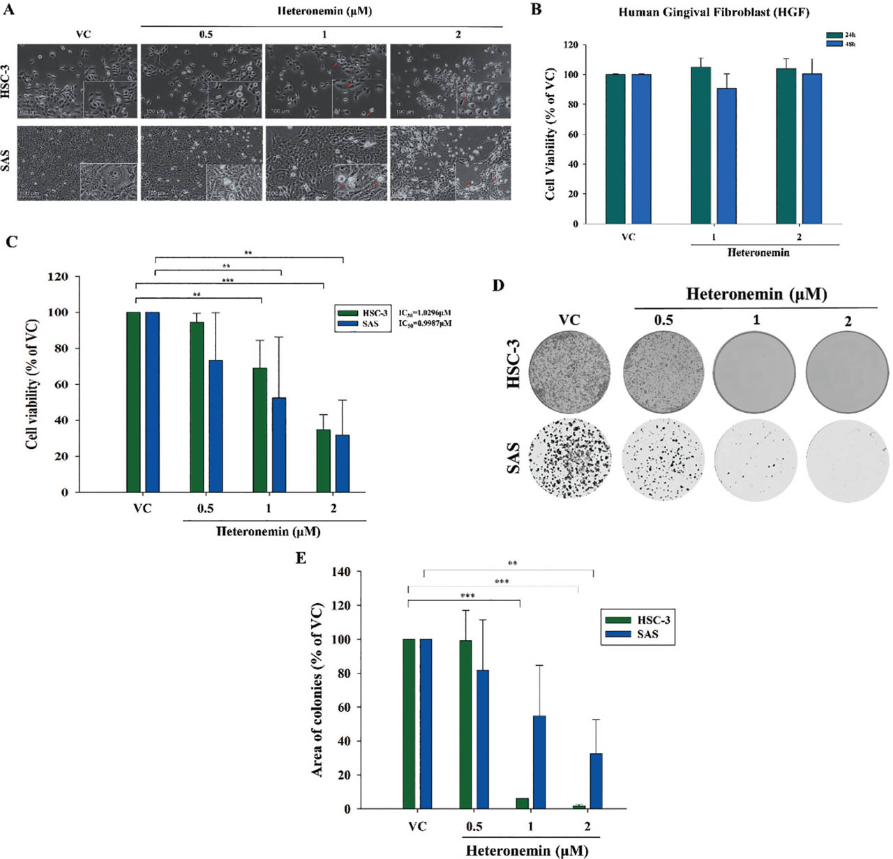
Fig 1.
Heteronemin reduces the viability and proliferation of OSCC cells. (A) Morphological change in OSCC cell lines treated with different concentrations of heteronemin. Red arrows: apoptotic cells. Blue arrows: vacuole formations in the cytoplasm. (B) Human gingival fibroblast cells treated with heteronemin did not exhibit a significant reduction in cell viability. (C) Concentration-dependent inhibition of OSCC cell viability. The IC50 of heteronemin in HSC-3 is 1.0296 μM, while the IC50 of heteronemin in SAS is 0.9987 μM. (D, E) Representative images of the colony formation and quantitative analyses, respectively, showing the concentration-dependent antiproliferative effect of prolonged treatment of heteronemin in OSCC cells. The data were obtained from three independent experiments and are expressed as means ± SDs. Asterisks indicate statistically significant differences between the treated group and the VC; *p < 0.05, **p < 0.01, and ***p < 0.001. OSCC, oral squamous cell carcinoma; VC: vehicle control.
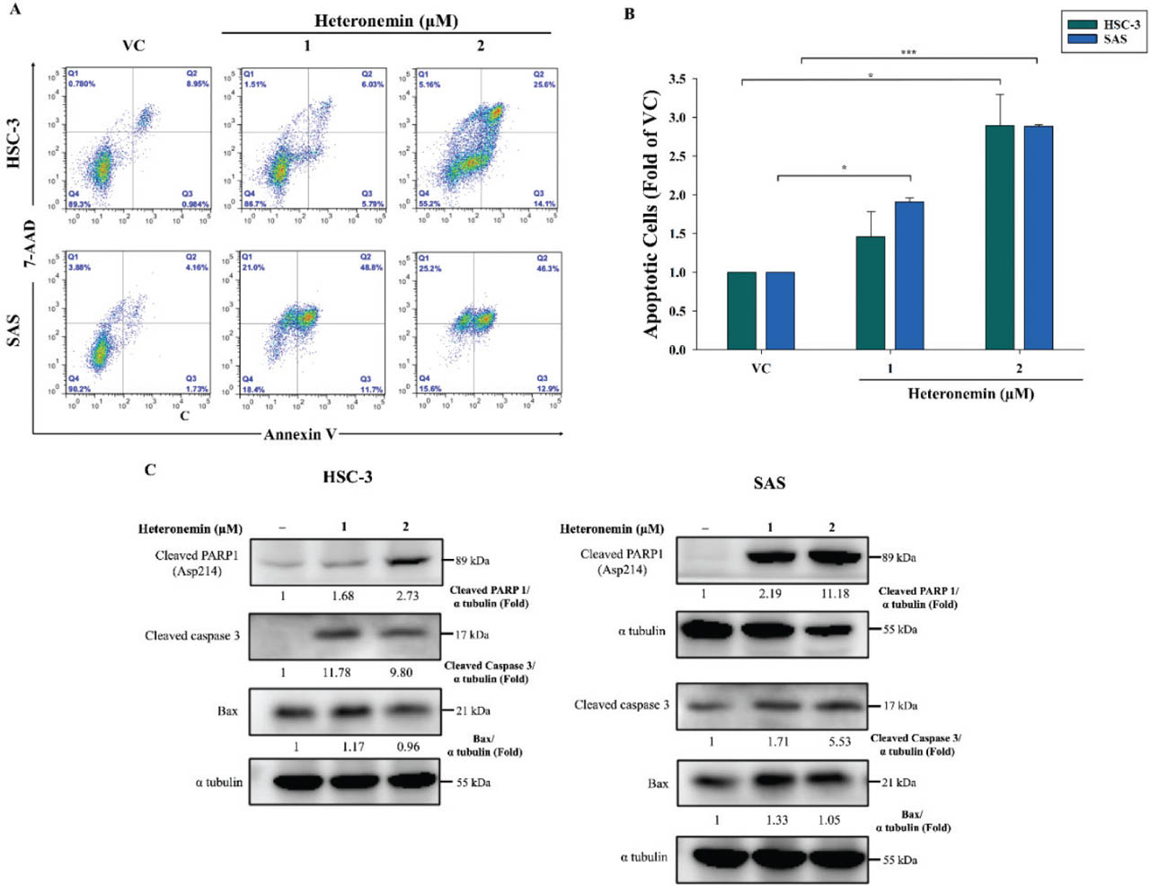
Fig 2.
Heteronemin induces apoptosis in OSCC cells. (A) OSCC cells treated with a control or 1 μM or 2 μM heteronemin were double-stained with Annexin V/7-AAD to detect cells undergoing early- and/or late-stage apoptosis as well as non-apoptotic cell populations. (B) Quantitative analysis of the apoptotic cell population. OSCC cells were treated with the corresponding concentrations of heteronemin, and cell lysates were collected after 24 h, stained with Annexin V/7AAD, and then analyzed via flow cytometry. (C) Cropped Western blotting data showing the protein expression of cleaved PARP1, cleaved caspase 3, and Bax in OSCC cells upon treatment with different concentrations of heteronemin. Alpha tubulin served as the loading control. 7-AAD, Annexin V/7-amino actinomycin D; OSCC, oral squamous cell carcinoma; VC, vehicle control.
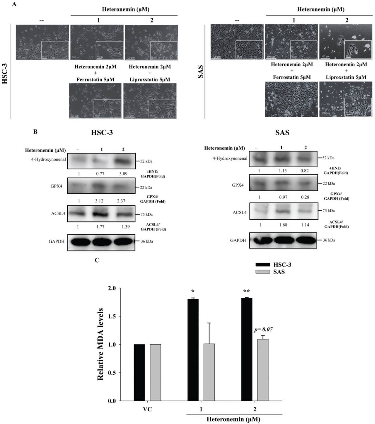
Fig 3.
Heteronemin potentially triggers the induction of ferroptosis in OSCC cells. (A) Treatment of the compound caused significant morphological changes and a reduction in viable cells, whereas pretreatment with the ferroptosis inhibitors, ferrostatin-1 and liproxstatin-1, relatively increased viable cells in both OSCC cell lines. (B) Cropped representative of the Western blot data showing an increase in the expression of the protein 4-HNE and ACSL4, and a decrease in GPX4 levels, which are significant markers of ferroptosis, in OSCC cells. (C) TBARS Assay was performed to measure MDA, which is an indicator of lipid peroxidation and oxidative stress in cells. Treatment of heteronemin increases MDA levels most significantly in HSC-3 cells. Asterisks indicate statistically significant differences between the treated group and the VC; *p < 0.05 and **p < 0.01. 4-HNE, 4-hydroxynonenal; ACSL4, Acyl-CoA Synthetase Long-Chain Family Member 4; GPX4, glutathione peroxidase 4; MDA, malondialdehyde; OSCC, oral squamous cell carcinoma; TBARS, thiobarbituric acid reactive substances; VC, vehicle control.
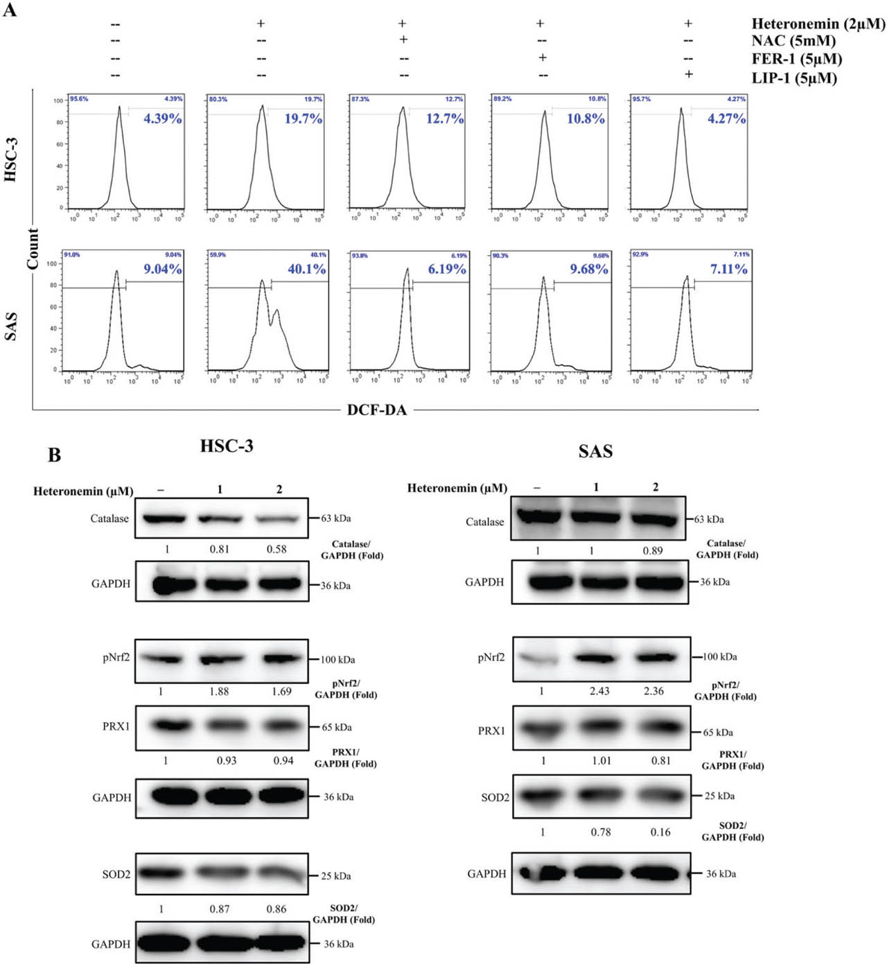
Fig 4.
Treatment with heteronemin results in the generation of ROS in OSCC cells. (A) DCF-DA staining analyzed via flow cytometry was performed to determine the ROS levels in OSCC cells treated with heteronemin for 24 h with or without pretreatment with NAC, ferrostatin-1, or liproxstatin-1. (B) Cropped representative of the Western blot data showing the expression levels of oxidative stress-related protein markers in OSCC cells after 24 h of treatment with indicated concentrations of heteronemin. OSCC, oral squamous cell carcinoma; ROS, reactive oxygen species.
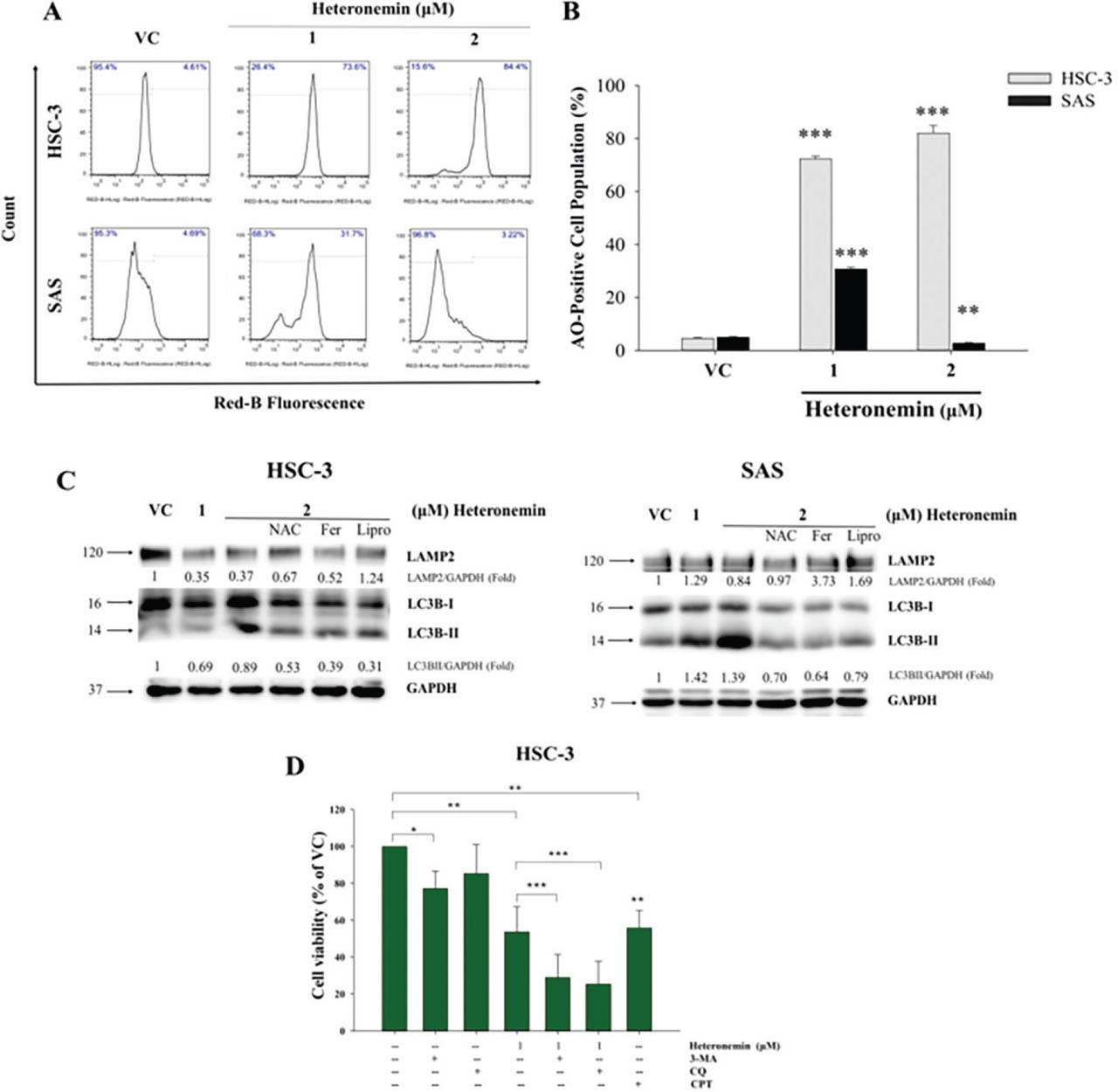
Fig 5.
Heteronemin induces autophagy in OSCC cells. (A) Representative results of Western blotting show the expression of autophagy-related proteins (LAMP2 and LC3B) in HSC-3 and SAS cells. OSCC cells were pretreated with NAC, Ferrostatin-1 (Fer), or Liproxstatin-1 (Lipro) before heteronemin administration. GAPDH as an internal control. (B) Cytometer-based AO staining revealed that heteronemin triggered the production of AVOs in OSCC cells. (C) Quantitative analysis of the AVOs. (D) Cell viability of HSC-3 cells treated with heteronemin in the presence or absence of autophagy inhibitors (3-MA, CQ) or a positive control (CPT). Data are presented as mean ± SD. Statistical difference among the treatment groups are denoted as *p < 0.05, **p < 0.01, ***p < 0.001. 3-MA, 3-methyladenine; AO, acridine orange; AVOs, acidic vesicular organelles; CQ, chloroquine; OSCC, oral squamous cell carcinoma; VC, vehicle control.
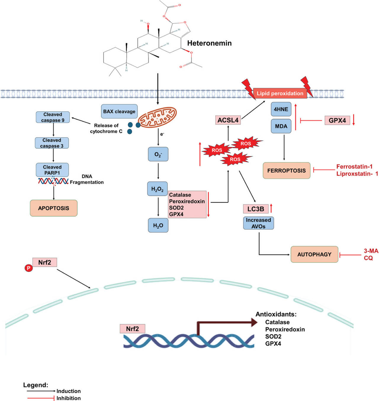
Fig 6.
The proposed model of heteronemin-induced apoptosis and non-apoptotic ferroptosis in OSCC cells. Heteronemin restricts OSCC cell growth by inducing caspase-mediated apoptosis and autophagy, as confirmed by the conversion of LC3. Increased ROS production causes oxidative stress, increasing autophagic stress and initiating ferroptosis in OSCC cells, with decreased GPX4 expression and increased lipid peroxidation. Finally, heteronemin-induced apoptosis and ferroptosis enhance OSCC cell death, suggesting promising therapeutic potential, particularly in apoptosis-resistant OSCC cells. Dot line: possible pathways. ACSL4, Acyl-CoA Synthetase Long-Chain Family Member 4; AVOs, acidic vesicular organelles; CQ, chloroquine; GPX4, glutathione peroxidase 4; MDA, malondialdehyde; OSCC, oral squamous cell carcinoma; ROS, reactive oxygen species.