
1. Introduction
Oral squamous cell carcinoma (OSCC) ranks as the most common form of oral cancer and is a primary contributor to morbidity and mortality in patients afflicted with head and neck squamous cell carcinoma (Gharat et al. 2016). The approximate survival rate among patients at the 5-year mark is approximately 50% (Almangush et al. 2021; Li et al. 2022). Present-day treatment options include surgical removal of the tumor followed by a combination of chemotherapy and radiation therapy; however, adverse effects such as trauma in the oral and maxillofacial region from surgery, nausea, vomiting, and hair loss caused by chemotherapy and the extent of permanent damage to healthy tissues dramatically affect patients' quality of life (Wu and Zhou 2015; Zhang et al. 2020a; Li et al. 2022). Moreover, despite advances in therapeutics, a major challenge in treating oral cancer is that resistance to chemotherapy and targeted therapies can lead patients to become less responsive or unresponsive to treatments. For example, resistance to platinum-based chemotherapy and endothelial growth factor receptor-targeted therapies is common in OSCC (Cao et al. 2022). Moreover, the rate of recurrence is high, and patient prognosis decreases, with a mortality rate of up to 92% in cases of relapse (Weckx et al. 2019). These limitations have fueled the search for novel approaches to therapeutics with improved efficacy and reduced side effects. In the modern era of drug discovery, natural products have demonstrated exceptional value as sources of potential compounds with anticancer properties that are less toxic or non-toxic to healthy normal cells (Hashem et al. 2022).
Marine organisms have also been explored in the search for lead compounds with anticancer properties, as their secondary metabolites have diverse biological functions. Heteronemin, a marine sesterterpenoid-type product, is the most abundant secondary metabolite isolated from sponges and has anticancer effects on multiple cancer cell lines, mostly by inducing apoptosis (Wu et al. 2015; Lee et al. 2018). Programmed cell death or apoptosis is an essential mechanism for maintaining homeostasis. Cells undergoing apoptosis undergo a series of defined molecular and morphological alterations, such as cellular contraction, chromatin condensation, DNA fragmentation, membrane blebbing, and the formation of apoptotic bodies (Bedoui et al. 2020). In cancer therapy, the primary goal is to eliminate cancer cells by activating apoptosis (Carneiro and El-Deiry 2020). Intriguingly, apoptosis can also fail. Several studies have reported the presence of apoptosis-resistant cancer cells, making it difficult for available treatments to succeed (Neophytou et al. 2021). Therefore, employing other forms of non-apoptotic cell death has become an interesting field of research to combat drug resistance in cancer.
This study thus aims to investigate whether heteronemin can induce apoptosis and non-apoptotic cellular death mechanisms in OSCC cells. We intended to uncover the molecular underpinnings of its action to better help OSCC patients overcome chemoresistance while potentially sparing healthy normal cells.
2. Materials and Methods
2.1. Cell culture
The human OSCC HSC-3 (Cell line no. JCRB0623) and SAS (cell line no. JCRB0260) cancer cell lines were purchased from the Japanese Collection of Research Bioresources Cell Bank (Osaka, Japan) and maintained in Dulbecco's modified Eagle's medium and Ham's F-12 nutrient mixture (DMEM/F12, 3:2; Gibco, Waltham, MA, USA) supplemented with 8% heat-inactivated fetal bovine serum (FBS; Gibco, Waltham, MA, USA), 1% penicillin/streptomycin and 2 mM glutamine at 37 °C under a humidified atmosphere with 5% CO2.
2.2. Heteronemin
Heteronemin (PubChem CID: 21589810) was isolated from Hippospongia sp. collected via scuba diving at a depth of 20 m from the coral reefs of Taitung, Taiwan. The extraction and characterization followed the same protocol as previously described (Chang et al. 2012; Lee et al. 2018). A stock of 20.3 mg of heteronemin with >98% purity was diluted in dimethyl sulfoxide (DMSO), aliquoted, and stored at −20°C until further use. The cell lines were treated with various concentrations of heteronemin, as indicated. The concentrations were selected based on the calculated IC50 against the OSCC cell lines.
2.3. Cell viability
The cell counting kit-8 (CCK-8) assay was conducted in triplicate to determine cell viability. The initial step involved cell counting, and then, approximately 3000 oral cancer cells were seeded per well in a 96-well cell culture plate and incubated at 37 °C in a humidified atmosphere with 5% CO2 for 24 h. After incubation, the culture medium was replaced with the corresponding culture medium with or without the respective inhibitors, such as ferrostatin-1 (Cat. No. T6500, TargetMol Chemicals Inc.), liproxstatin-1 (Cat. No. T2376, TargetMol Chemicals Inc.), N-acetylcysteine (NAC; Cas No. 616 – 91-1, Sigma-Aldrich, Co.), 3-methyladenine (3-MA; Cat. No. T1879, TargetMol Chemicals Inc.), and chloroquine (CQ; Cat. No. C6628 – 25 g, Sigma-Aldrich, Co.) for 6 h of pretreatment. Six replicates were performed for each concentration. Finally, 10 μL of CCK-8 reagent (IMT Formosa New Materials Co., Ltd., Kaohsiung, Taiwan) was added to each well, and the optical density (OD) at 450 nm was assessed with a multifunction microplate reader (BioTek Instruments Ltd., USA) after incubation for 1 h at 37 °C. The readings were subsequently standardized via blank medium, and the cell viability was computed as the ratio of the OD value of the treatment group to that of the control group. Data were analyzed using SigmaPlot 15.0 (Grafiti LLC, USA).
2.4. Colony formation
Colony formation or clonogenic assays were utilized to investigate the proliferative capacity of oral cancer cells upon treatment with heteronemin compared with control cells. The assay was performed by seeding HSC-3 and SAS cells at 500 cells/well. After 24 h, the cells were exposed to varying concentrations of heteronemin, with or without the addition of inhibitors. The cells were allowed to grow for 2 weeks and were given fresh medium every 2–3 days. To determine clonogenic survival, colonies consisting of HSC-3 and SAS cells were fixed, stained with crystal violet, and then incubated for at least 30 min or until distinct staining of the colonies became evident. Images were processed and analyzed via ImageJ 1.38e software (National Institutes of Health (NIH), Bethesda, Maryland).
2.5. Detection of apoptosis
Annexin V/7-amino actinomycin D (7-AAD) double staining was used to investigate the degree of apoptosis in heteronemin-treated cells. Membrane integrity and phosphatidylserine externalization are key factors in detecting apoptosis. Heteronemin-treated cells were harvested and stained with an Annexin V/7-AAD kit (Strong Biotech Corporation, Taipei, Taiwan). The cells were then examined via flow cytometry (Guava® easyCyte, Merck, Darmstadt, Germany) and FlowJo v7.5.5 software (FlowJo, LLC, Becton Dickinson and Company (BD), Ashland, Oregon, USA).
2.6. Reactive oxygen species (ROS) evaluation
To detect ROS generation in cells treated with heteronemin, 2′,7-dichlorofluorescein diacetate (DCF-DA; Cat# 302, IMT Formosa New Materials Co., Ltd., Kaohsiung, Taiwan) was used. After 24 h of treatment with heteronemin, the cells were exposed to 10 μM DCF-DA for 30 min. After incubation, the cells were washed with phosphate-buffered saline (PBS; Cat# BR110, Biomate, Taiwan), and the collected data were subjected to analysis via FlowJo v7.5.5 software (FlowJo, LLC, Becton Dickinson and Company (BD), Ashland, Oregon, USA).
2.7. Western blotting
The proteins were extracted from the cell lysates via a bicinchoninic acid protein assay kit (Cat. No. 23225, Thermo Scientific, Pierce, Rockford, IL, USA), size-fractionated via sodium dodecyl sulfate–polyacrylamide gel electrophoresis (SDS-PAGE), and electrophoretically transferred to polyvinylidene difluoride (PVDF) membranes (Ann Arbor, MI, USA). The membranes were then blocked with 5% non-fat milk in Tris-buffered saline containing Tween-20 (TBST) buffer (TBS containing 0.1% Tween 20), sequentially hybridized with relevant primary antibodies against 4-hydroxynonenal (4-HNE; 1:3000, Abcam Cat# ab46545), alpha tubulin (1:1000, Abclonal Cat# AC012), ACSL4 (1:20000, ABclonal Cat# A20414), Bax (1:8000, Proteintech Cat# 50599-2-Ig), catalase (1:5000, Merck Cat# 219010), cleaved caspase 3 (1:1000, ABclonal Cat# A11953), cleaved PARP1 (1:1000, Cell Signaling Technology Cat# 5625), GAPDH (1:20000, Affinity Biosciences Cat# AF0911), glutathione peroxidase 4 (GPX4; 1:5000, ABclonal Cat# A1933), LAMP2 (1:5000, ABclonal Cat# A1961), LC3B (1:1000, ABclonal Cat# A19665), pNrf2 (1:2000, ABclonal Cat# AP1133), peroxiredoxin (1:2000, ABclonal Cat# A4956), and superoxide dismutase 2 (SOD2; 1:2000, ABclonal Cat# A1340) overnight at 4 °C, washed and treated with horseradish peroxidase-conjugated secondary antibodies. Finally, the signals were detected and visualized via an enhanced chemiluminescence detection kit (Amersham Piscataway, NJ, USA). The Western blot data shown are representative of three individual experiments. The fold change reflects a representative blot. Image processing and quantification were done using ImageJ software.
2.8. Acridine orange (AO) staining for the detection of autophagy
Acridine orange (AO; Biotium, #40039) was utilized to investigate the induction of autophagy further. AO is a cell-permeable fluorescent dye protonated in acidic vesicular organelles (AVOs), such as lysosomes. AO dye emits red fluorescence (515 nm) upon blue light excitation (488 nm). This is a fast and reliable method for detecting autophagy induction (Thomé et al. 2016).
2.9. Thiobarbituric acid reactive substances (TBARS) assay
TBARS assay was conducted to measure malondialdehyde (MDA), an indicator of oxidative stress, by reacting with thiobarbituric acid, thereby detecting the occurrence of lipid peroxidation in heteronemin-treated OSCC cells. The assay was conducted following the manufacturer's standard protocol (Cat#309, IMT Formosa New Material Co., Ltd, Taiwan). Briefly, 2 × 10⁷ HSC-3 and SAS cells were collected and centrifuged at 300 × g at 4 °C to obtain the cell lysates. PBS was used to resuspend the lysates, and sonication was then performed to lyse the cells. After, the sonicated cells were centrifuged at 2000 × g at 4 °C, and the supernatant was collected for the succeeding analysis. Relevant reagents were added to the samples, and the results were obtained via detection of OD using a microplate reader at an absorbance of 530 nm.
2.10. Quantification of the results and statistical analysis
All the experimental procedures were performed in triplicate. The data are expressed as the means ± SDs. A two-tailed Student's t-test using SigmaPlot 15.0 (Grafiti LLC, USA) was used for comparison between two groups. One-way ANOVA and Holm-Sidak post hoc test were used for comparisons involving more than two groups, and a p-value of less than 0.05 indicated statistical significance. The significant differences are expressed as *p < 0.05, **p < 0.01, and ***p < 0.001. The IC50 of heteronemin in both cell lines was calculated using the AAT Bioquest IC50 calculator (https://www.aatbio.com/tools/ic50-calculator).
3. Results
3.1. Heteronemin selectively inhibits oral cancer cell growth and viability
Previous studies have shown the anticancer activity of heteronemin against various cancers, including lung cancer (Cheng et al. 2019; Chang et al. 2021), oral cancer (Huang et al. 2020), and prostate cancer (Wu et al. 2016). In this study, we investigated the effects of various concentrations of heteronemin (0.5 μM, 1 μM, and 2 μM) in HSC-3 and SAS oral cancer cells. Morphological changes were observed in the OSCC cells treated with the compound. Figure 1a shows the presence of apoptotic cells (red arrows) upon treatment in a concentration-dependent manner. Moreover, vacuole formations (blue arrows) were present in HSC-3 cells administered with 2 μM of heteronemin, which can possibly be indicative of the induction of autophagy (Maciel et al. 2018). To determine whether heteronemin is toxic in non-cancerous cells, we treated human gingival fibroblast cells with the compound for 24 h and 48 h and found that it is not significantly toxic in these cells (Figure 1b). We then tested the effect of heteronemin in HSC-3 and SAS cell lines. Our data reveal the concentration-dependent cytotoxicity of the compound with an IC50 of 1.0296 μM and 0.9987 μM in HSC-3 and SAS OSCC cell lines, respectively (Figure 1c).

Fig 1.
Heteronemin reduces the viability and proliferation of OSCC cells. (A) Morphological change in OSCC cell lines treated with different concentrations of heteronemin. Red arrows: apoptotic cells. Blue arrows: vacuole formations in the cytoplasm. (B) Human gingival fibroblast cells treated with heteronemin did not exhibit a significant reduction in cell viability. (C) Concentration-dependent inhibition of OSCC cell viability. The IC50 of heteronemin in HSC-3 is 1.0296 μM, while the IC50 of heteronemin in SAS is 0.9987 μM. (D, E) Representative images of the colony formation and quantitative analyses, respectively, showing the concentration-dependent antiproliferative effect of prolonged treatment of heteronemin in OSCC cells. The data were obtained from three independent experiments and are expressed as means ± SDs. Asterisks indicate statistically significant differences between the treated group and the VC; *p < 0.05, **p < 0.01, and ***p < 0.001. OSCC, oral squamous cell carcinoma; VC: vehicle control.
Moreover, a clonogenic assay was performed to test whether the drug can also modulate cell proliferation, and we found that heteronemin significantly inhibited the proliferation of both cell lines (Figures 1d,e), noting that HSC-3 was more sensitive compared to SAS, a contrasting observation in relation to the cell viability assay (Figure 1c). Altogether, these data demonstrate the selective toxicity of heteronemin against OSCC cells.
3.2. Heteronemin induces apoptotic cell death in OSCC cells
To determine the type of cell death induced by heteronemin, we conducted Annexin V/7-AAD double staining and analyzed the cells via flow cytometry. Figures 2a and 2b show that increasing concentrations of heteronemin caused the cells to undergo apoptosis. In addition, Western blot data (Figure 2c) revealed the increased expression of cleaved protein forms of PARP-1 and caspase 3. And while our data on Bax (p21) seem to show a decreased protein expression level with the administration of the higher concentration of heteronemin, this does not entirely mean that it is downregulated, as this can be considered that the p21 form of Bax is cleaved into its p18 fragment, which may not have been detected by the antibody used (Gao and Dou 2000).

Fig 2.
Heteronemin induces apoptosis in OSCC cells. (A) OSCC cells treated with a control or 1 μM or 2 μM heteronemin were double-stained with Annexin V/7-AAD to detect cells undergoing early- and/or late-stage apoptosis as well as non-apoptotic cell populations. (B) Quantitative analysis of the apoptotic cell population. OSCC cells were treated with the corresponding concentrations of heteronemin, and cell lysates were collected after 24 h, stained with Annexin V/7AAD, and then analyzed via flow cytometry. (C) Cropped Western blotting data showing the protein expression of cleaved PARP1, cleaved caspase 3, and Bax in OSCC cells upon treatment with different concentrations of heteronemin. Alpha tubulin served as the loading control. 7-AAD, Annexin V/7-amino actinomycin D; OSCC, oral squamous cell carcinoma; VC, vehicle control.
Nevertheless, this change in the expression of the mentioned apoptosis markers significantly suggests that heteronemin potentially induces the intrinsic apoptotic pathway in OSCC cells.
3.3. Heteronemin treatment causes ferroptotic cell death in OSCC
Several antineoplastic drugs induce apoptosis to kill cancer cells; however, many types of cancer have developed resistance (Mollaei et al. 2021). In this case, inducing non-apoptotic cell death mechanisms can provide an efficient alternative treatment strategy (Ye et al. 2018). Our Annexin V/7AAD double staining result (Figure 1a) reveals that heteronemin treatment not only triggers the apoptosis machinery but can also initiate other non-apoptotic cell death pathways, as evidenced by a significant increase of 7AAD-positive cell populations (upper left and right quadrants). To further investigate this phenomenon, we treated both OSCC cell lines with 2 μM of heteronemin with or without pretreatment of ferroptosis inhibitors ferrostatin-1 and liproxstatin-1. As shown in our data (Figure 3a), the pretreatment of the potent inhibitors of ferroptosis rescued the cells from the cytotoxic effect of the compound, implying the potential role of ferroptosis in inhibiting the OSCC cells' growth and proliferation. Ferroptosis is an iron-dependent cellular death mechanism that is highly driven by the peroxidation of membrane polyunsaturated phospholipids (Ding et al. 2023). Interestingly, our Western blot analysis (Figure 3b) revealed that heteronemin treatment increased the levels of 4-hydroxynonenal (4-HNE), a byproduct of lipid peroxidation, and Acyl-CoA Synthetase Long-Chain Family Member 4 (ACSL4), a crucial enzyme in fatty acid metabolism that plays a role in ferroptotic cell death by promoting the accumulation of oxidized phospholipids (Ding et al. 2023). Besides, GPX4 levels were downregulated (Figure 3b), As GPX4 plays a key role in inhibiting ferroptosis, its downregulation can mean that the administration of heteronemin triggers the induction of ferroptosis.

Fig 3.
Heteronemin potentially triggers the induction of ferroptosis in OSCC cells. (A) Treatment of the compound caused significant morphological changes and a reduction in viable cells, whereas pretreatment with the ferroptosis inhibitors, ferrostatin-1 and liproxstatin-1, relatively increased viable cells in both OSCC cell lines. (B) Cropped representative of the Western blot data showing an increase in the expression of the protein 4-HNE and ACSL4, and a decrease in GPX4 levels, which are significant markers of ferroptosis, in OSCC cells. (C) TBARS Assay was performed to measure MDA, which is an indicator of lipid peroxidation and oxidative stress in cells. Treatment of heteronemin increases MDA levels most significantly in HSC-3 cells. Asterisks indicate statistically significant differences between the treated group and the VC; *p < 0.05 and **p < 0.01. 4-HNE, 4-hydroxynonenal; ACSL4, Acyl-CoA Synthetase Long-Chain Family Member 4; GPX4, glutathione peroxidase 4; MDA, malondialdehyde; OSCC, oral squamous cell carcinoma; TBARS, thiobarbituric acid reactive substances; VC, vehicle control.
To better support our hypothesis so far, we performed a TBARS assay to biochemically evaluate ferroptotic lipid damage. TBARS detect and measure MDA, a byproduct of the degradation of lipid peroxides that is widely used as a lipid peroxidation biomarker in ferroptosis models (Chen et al. 2024b). Our data show that treatment of heteronemin can modulate the levels of MDA in OSCC cells (Figure 3c), further supporting the mechanistic involvement of ferroptotic cell death.
3.4. Heteronemin induces increased production of ROS
To determine the potential ability of heteronemin to induce oxidative stress, we treated oral cancer cells with different concentrations of heteronemin and conducted DCF-DA staining and Western blot analysis. Flow cytometry data revealed that heteronemin-treated cells presented higher levels of ROS compared to the vehicle control (VC), and pretreatment of oral cancer cells with 5 mM NAC, 5 μM ferrostatin 1 (Fer-1), or 5 μM liproxstatin-1 (Lip-1) significantly reduced ROS levels (Figure 4a). Our subsequent Western blot analysis demonstrated that heteronemin reduced the expression of the antioxidants peroxiredoxin (PRX1) and catalase while upregulating the protein expression of phosphorylated Nrf2, which is a key regulator of the body's antioxidant response (Figure 4b). Interestingly, despite upregulating Nrf2, the overwhelming oxidative stress renders the antioxidant response ineffective, ultimately causing cellular death.

Fig 4.
Treatment with heteronemin results in the generation of ROS in OSCC cells. (A) DCF-DA staining analyzed via flow cytometry was performed to determine the ROS levels in OSCC cells treated with heteronemin for 24 h with or without pretreatment with NAC, ferrostatin-1, or liproxstatin-1. (B) Cropped representative of the Western blot data showing the expression levels of oxidative stress-related protein markers in OSCC cells after 24 h of treatment with indicated concentrations of heteronemin. OSCC, oral squamous cell carcinoma; ROS, reactive oxygen species.
3.5. Heteronemin triggers autophagy in oral cancer cells
Autophagy plays an important role in maintaining cellular homeostasis under normal conditions. As a self-digesting process, it cleans damaged organelles or toxic cell components. In cancer, autophagy can play a protective role. It is triggered when cancer cells undergo oxidative stress, promoting survival and progression (Zada et al. 2021). Therefore, investigating the role of autophagy in treating oral cancer cells with heteronemin is important. To evaluate this phenomenon further, we performed AO staining to detect the presence of AVOs. AO stains and fluoresce red in AVOs such as autophagosomes, autolysosomes, and lysosomes, indicating the induction of autophagy. Our findings indicate that heteronemin treatment increased the production of AVOs (Figure 5a,b), suggesting that autophagy was activated.
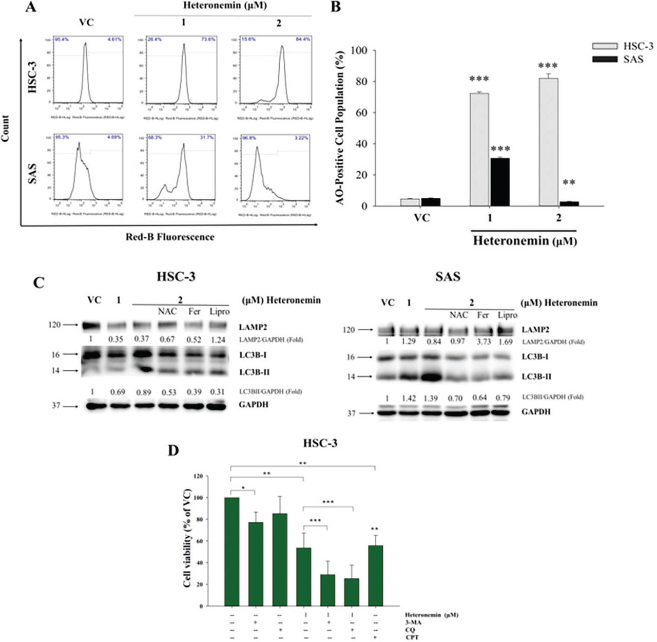
Fig 5.
Heteronemin induces autophagy in OSCC cells. (A) Representative results of Western blotting show the expression of autophagy-related proteins (LAMP2 and LC3B) in HSC-3 and SAS cells. OSCC cells were pretreated with NAC, Ferrostatin-1 (Fer), or Liproxstatin-1 (Lipro) before heteronemin administration. GAPDH as an internal control. (B) Cytometer-based AO staining revealed that heteronemin triggered the production of AVOs in OSCC cells. (C) Quantitative analysis of the AVOs. (D) Cell viability of HSC-3 cells treated with heteronemin in the presence or absence of autophagy inhibitors (3-MA, CQ) or a positive control (CPT). Data are presented as mean ± SD. Statistical difference among the treatment groups are denoted as *p < 0.05, **p < 0.01, ***p < 0.001. 3-MA, 3-methyladenine; AO, acridine orange; AVOs, acidic vesicular organelles; CQ, chloroquine; OSCC, oral squamous cell carcinoma; VC, vehicle control.
Since ROS has been reported to play a part in triggering multiple cell death pathways and mediating the interaction between these pathways. We hypothesize that ROS trigger autophagy in the context of heteronemin treatment. Autophagy induction can be achieved by converting LC3B-I to lipid-bound LC3B-II through proteolytic cleavage and lipidation (Peña-Martinez et al. 2022). Hence, we pretreated cells with NAC, an ROS scavenger, and detected a decrease in LC3B II levels, indicating that heteronemin-induced autophagy can be strongly attributed to the regulation of ROS. Hence, NAC reduces ROS levels and inhibits autophagy induction, decreasing LC3BII levels (Figure 5c). Similarly, as shown in Figure 5c, pretreatment with the ferroptosis inhibitors ferrostatin-1 and liproxstatin-1 reduced the expression levels of LC3BII. Ferroptosis is an iron-dependent form of cell death characterized by lipid peroxidation (Su et al. 2019; Endale et al. 2023). Thus, the decrease in LC3BII levels upon pretreatment with ferroptosis inhibitors suggests that the effect of heteronemin on the induction of autophagy may also be related to mechanisms involving ferroptosis, possibly through the ROS pathway. We also performed a cell viability assay in which HSC-3 cells were pretreated with the autophagy inhibitors 3-methyladenine (3-MA) and chloroquine (CQ), and we found that this pretreatment further reduced the viability of the oral cancer cell lines, suggesting a protective role of autophagy in cancer cells treated with heteronemin (Figure 5d).
4. Discussion
In this study, we investigated the role of heteronemin, a scalarane sesterterpenoid isolated from marine sponges, in highly aggressive and metastatic HSC-3 (Mahjour et al. 2019), and the poorly differentiated but stem-like properties of SAS oral cancer cell lines (Ohnishi et al. 2018; Lau et al. 2021). Research has shown that heteronemin not only promotes programmed cell death or apoptosis in several types of cancer cells (Cao et al. 2003; Lee et al. 2018; Cheng et al. 2019; Chang et al. 2021; Wang et al. 2022) but also induces ferroptosis in HA22T and HA59T hepatocellular carcinoma cells (Chang et al. 2021) and autophagy in human renal carcinoma cells (HRCCs) (Wu et al. 2015). Huang et al. also demonstrated the effectiveness of heteronemin in OEC-M1 and SCC-25 oral cancer cell lines, highlighting that combined treatment with heteronemin and tetrac (3,3′,5,5′-tetraiodothyroacetic acid) inhibited ERK1/2 activation, increased p53 phosphorylation, inhibited Homo sapiens Thrombospondin 1, and suppressed transforming growth factor β expression, thereby negatively affecting the proliferation and metastasis of oral cancer cells (Huang et al. 2020). Our study, on the other hand, revealed that heteronemin treatment alone could accelerate the apoptotic process by cleaving p21 Bax to p18 and could activate autophagy. Moreover, heteronemin also triggered the induction of ferroptosis, a non-apoptotic cell death process. The data reported here are the first to suggest that heteronemin induces apoptosis, autophagy, and ferroptosis in oral cancer cell lines, further supporting its potent activity. More importantly, we provided mechanistic explanations as to how the compound elicits the mentioned processes, augmenting the current scientific knowledge regarding the therapeutic activity of heteronemin.
Two known pathways modulate apoptosis: the intrinsic (mitochondrial) and extrinsic (death receptor) pathways. While the extrinsic pathway is triggered by external signals, such as the binding of ligands to death receptors on the surface of the cell and the subsequent activation of caspases, the intrinsic pathway involves the mitochondria (Wang et al. 2022). Bcl-2 family proteins govern the permeability of the external part of the mitochondrial membrane and can be categorized as either pro- or anti-apoptotic proteins. Bax and Bak, which are proapoptotic proteins, engage in homo-oligomerization and contribute to the creation of pores in the outer mitochondrial membrane. These pores serve as pathways for the escape of proapoptotic molecules, including cytochrome c (Brunelle and Letai 2009; Wu et al. 2015). Cytochrome c and apoptotic protease-activating factor 1 are released, activating caspase-9 as it interacts with it, leading to the caspase cascade (Saelens et al. 2004). Activated caspase 9, in turn, activates caspase 3, which cleaves PARP1. Cleavage of PARP1 impairs DNA repair, causing cells to undergo apoptosis (Wood and Newcomb 2000). In fact, the cleavage of PARP1 is considered a hallmark of apoptosis (Wongchitrat et al. 2019). Our study sought to determine how heteronemin triggers apoptosis in oral cancer cells, and our findings revealed the upregulation of cleaved caspase 3, cleaved PARP, as well as the potential cleavage of p21 Bax, which implies that heteronemin treatment results in the apoptosis of oral cancer cells by activating the intrinsic apoptosis pathway.
The proteolytic cleavage of p21 Bax to its p18 form is a late event that is dependent upon the caspase-mediated activation of calpain following cytotoxic stress. Compelling evidence has shown that the cleaved p18 fragment has a potent ability to initiate apoptosis (Wood and Newcomb 2000), and while this cleavage is not a prerequisite for Bax to instigate apoptosis, the p18 variant expedites apoptotic progression (Cao et al. 2003; Wongchitrat et al. 2019). When tested against sarcomas in a phase 1 clinical trial, ZSTK474, a pan-phosphatidylinositol 3-kinase (PI3K) inhibitor, was found to cleave p21Bax to p18, progressing to apoptosis (Isoyama et al. 2023). Furthermore, previous studies in tumor cell lines confirmed that blocking the cleavage of Bax significantly impedes drug-induced programmed cell death, whereas enhancing the inhibition of p18 Bax degradation does not increase the ability of drugs to induce apoptosis (Cao et al. 2003). In primary chronic lymphocytic leukemia cells, combined treatment with ibrutinib and nutlin-3 led to the cleavage of p21 Bax to p18 and increased apoptosis, overcoming the protective effect of the tumor microenvironment (Rimondi et al. 2021). These similar findings support the current knowledge that heteronemin can be a potent regulator of Bax expression, increasing its ability to kill oral cancer cells via apoptosis. Hence, novel strategies to enhance cancer chemotherapy may include a focus on either increasing the cleavage of p21 Bax to its p18 fragment or inhibiting its degradation (Soond et al. 2021).
Standard cytotoxic used to treat OSCC primarily cause cell cycle arrest and engage apoptosis in rapidly dividing cells (e.g., cisplatin). However, chemoresistance has commonly been reported, with cisplatin-resistant cells becoming apoptosis-refractory as well (Cheng et al. 2021; Chen et al. 2024a). Hence, looking for new strategies to trigger non-apoptotic cell death pathways addresses a clinically unmet need. Previously, we demonstrated that heteronemin induces ferroptosis in hepatocellular carcinoma by reducing GPX4 expression (Chang et al. 2021). GPX4 is an antioxidant defense enzyme that plays a crucial role in repairing damage caused by lipid peroxidation and acts as a key suppressor of ferroptosis, an iron-dependent form of non-programmed cell death (Chen et al. 2015). Hence, GPX4 acts as a “cleaner,” eliminating ROS in cells and thereby preventing lipid peroxidation (Zhang et al. 2020b). Ferroptosis is characterized by the depletion of GSH, impairment of GPX4 redox defense function, inflammation, and the formation of harmful lipid-derived ROS (Jelinek et al. 2018; Li et al. 2020). In ferroptosis, a reduction in GPX4 may lead to uninhibited lipid peroxidation of the cell membrane, further killing the cancer cells, consequently making ferroptosis a valuable strategy to overcome drug resistance in cancer treatment. In fact, Chen and colleagues highlighted in their review the undeniable potential therapeutic effect of triggering ferroptosis and that combined therapy with ferroptosis inducers and chemotherapy could significantly increase the 5-year survival rate of OSCC patients (Chen et al. 2024a). Moreover, Zhang et al. (2022) described several anticancer drugs that contain compounds that induce ferroptosis by suppressing GPX4 or blocking the synthesis of GSH. For example, temozolomide has curcumin analogs that ubiquitinate androgen receptors and suppress GPX4 to induce ferroptosis as well as overcome temozolomide resistance in glioblastoma (Chen et al. 2020). In our study, we presented that treatment of heteronemin markedly downregulated GPX4 expression – evidence of ferroptosis induction.
Interestingly, we demonstrate here also that not only does heteronemin regulate the expression of GPX4, it also potentially increases 4-HNE levels, implying the presence of lipid peroxidation. ROS have been found to induce cell damage via the peroxidation of polyunsaturated fatty acids (PUFAs) in cell membranes, which can be triggered directly or via ROS decomposition into highly reactive and cell-damaging radicals (Mustafa et al. 2018). PUFAs, integral components of cell membranes, play a vital role in modulating various biological processes. Owing to weak C–H bonds at the bis-allylic positions, PUFAs are susceptible to oxidation. Additionally, membrane PUFAs serve as the primary target for ROS assault and are the primary substrate of lipid peroxidation during ferroptosis (Yin et al. 2011; Yan et al. 2021). The oxidation of PUFAs results in a diverse range of primary lipid oxidation products, and of all the products resulting from lipid peroxidation, 4-HNE is one of the most biologically active (Zhong and Yin 2015).
Furthermore, we present here the upregulated expression of ACSL4, which catalyzes the biosynthesis of arachidonoyl-coenzyme A (CoA), mediating the induction of ferroptosis via lipid peroxidation (Ding et al. 2023). Thus, heteronemin putatively causes lipid peroxidation, eventually leading to ferroptosis. It is noteworthy to say that ferroptosis is also considered a form of immunogenic cell death. Ferroptosis signaling has been linked to increasing the immunogenicity of cancer cells, which promotes an anti-tumor response involving the infiltration of immune cells, therefore augmenting the efficacy of immunotherapy (Zhai et al. 2024). Further determining the exact mechanism of how ferroptosis affects the tumor microenvironment landscape in OSCC via heteronemin is a future study worthy of pursuit as we delve deeper and progress to developing treatment strategies to combat resistant OSCC cells, benefiting OSCC patients through precision oncology.
The interplay between ferroptosis and apoptosis in this study can be seen as mediated by ROS, in which increased production of ROS causes oxidative stress targeting the mitochondria via Bax activation, triggering the caspase cascade and, eventually, apoptosis. Similarly, increased production of ROS leads to lipid peroxidation of the cell membrane, a hallmark of ferroptosis. ROS play a dynamic role in maintaining homeostasis. At moderate levels, ROS are essential for normal cellular functions such as signal transduction and immune responses. However, ROS can cause oxidative stress at excessive levels, eventually leading to cell death (Redza-Dutordoir and Averill-Bates 2016). Various cell death mechanisms, such as apoptosis, ferroptosis, necroptosis, and autophagy, share one predominant feature—their dependence on ROS signaling and control. ROS act as rheostats that determine cell death and the type of cell death the cells will undergo (Villalpando-Rodriguez and Gibson 2021). To investigate whether heteronemin can increase ROS generation, DCF-DA staining was analyzed via flow cytometry. The data presented here revealed that heteronemin administration led to an increase in ROS generation, while pretreatment of cancer cells with N-acetylcysteine (NAC), an ROS scavenger, and the ferroptosis inhibitors Fer-1 and Lip-1 remarkably decreased the ROS levels (Figure 4a).
Our study uncovers an intriguing result on how HSC-3 and SAS react differently to heteronemin treatment and therefore offers mechanistic insights into their sensitivity to ferroptosis, via oxidative stress, but ultimately pointing to a preferable result, that is, cell death. To elaborate, treatment of heteronemin decreases the expression of catalase, a key antioxidant that eliminates ROS, albeit more pronounced in HSC-3 compared to SAS (Figure 4b). PRX1, which is a hydrogen peroxide scavenger that protects cells from oxidative damage, can be seen as modestly downregulated as well. Unexpectedly, phosphorylated nuclear factor erythroid 2-related factor 2 (pNrf2) was found to have been upregulated. Nrf2 is a master regulator of the body's antioxidant response, where it generally helps in managing oxidative stress. It has been reported that its upregulation is linked to poor response of patients to chemotherapy (Yu et al. 2025). Our results show a paradoxical Nrf2 response upon heteronemin treatment. Nrf2 targets catalase; however, heteronemin treatment reduces catalase despite an increase in Nrf2. Aside from the fact that catalase is not a strong Nrf2 target (catalase is mainly regulated by FOXO, PGC-1α, and PPARγ), catalase is highly sensitive to oxidative inactivation. For example, 4-HNE can directly inactivate catalase (Pigeolet et al. 1990; Bauer and Zarkovic 2015). What's more, we show here that heteronemin can downregulate SOD2 (Figure 4b). SOD2 plays a biphasic role in cancer, where it can be oncogenic or a tumor suppressor, depending on the disease context. For instance, overexpression of SOD2 suppressed the malignant phenotype in human pancreatic cancer cells (Weydert et al. 2003). In contrast, SOD2 was found to reduce temozolomide-induced ROS production, resulting in apoptosis inhibition in glioblastoma cells (Chien et al. 2019). Besides, SOD2 overexpression reportedly drives γ-irradiation-induced cancer cell invasion (Jung et al. 2019). Thus, downregulating SOD2 may prove to be beneficial in treating OSCC. The mechanism implied here suggests that upon treatment of heteronemin, OSCC cells try to compensate by activating their antioxidant machinery. Unfortunately, this compensatory mechanism proves to be insufficient and hence ineffective. This shows that the compound can potently trigger an apoptosis-ferroptosis cell death switch leading to preferential ferroptotic cell death, especially when the cells' antioxidant defenses are compromised.
An interesting study by Lee et al. (2020) described the possible interplay of apoptosis and ferroptosis, in which combined treatment with the ferroptotic agent erastin and the apoptotic agent TRAIL led to increased apoptosis. These findings suggest that this combined treatment disrupted the mitochondrial membrane potential, which can be attributed to the increased oligomerization of BAX and its accumulation in the mitochondria, further activating the caspase cascade and inducing apoptosis (Lee et al. 2020). Therefore, investigating the effect of heteronemin on the mitochondrial membrane potential is an area worth investigating in the future.
As a dynamic physiologic and pathologic process, autophagy, when it occurs at low levels, enables cells to adjust to ongoing stress and elude cell death. Nevertheless, under certain cellular conditions and when elevated, autophagy presents an alternative route for eliminating abnormal cells (Wu et al. 2015; Feng et al. 2022). This process is also activated in cancer therapeutics, promoting cell death in cancer cells. Interestingly, autophagy in cancer cells does not always indicate cell death; it can also serve as a protective mechanism in the response of cancer cells to treatment (Masui et al. 2016; Xiao et al. 2021). Since current evidence shows the ability of heteronemin to induce autophagy, as evidenced by the conversion of soluble LC3-I to lipid-bound LC3-II (Wu et al. 2015), we also suspected that the drug could actively induce autophagy in oral cancer cells and investigated the possible process by which autophagy is triggered. Our results show that treatment with heteronemin induces autophagy in oral cancer cells, as indicated by the upregulated expression of the LC3-II protein. The conversion of LC3-I to LC3-II through proteolytic cleavage can serve as an indicator of the induction of autophagy (Hu et al. 2014). Moreover, our AO staining results further confirmed that autophagy was induced, as evidenced by the increased production of AVOs. The increased levels of ROS could have activated the autophagic process, which plays a protective role in this interaction, as pretreatment with 3-MA and CQ did not increase oral cancer cell viability.
Our findings (Figure 6) on the effect of heteronemin in OSCC cells suggest several promising therapeutic strategies. Given the dual-inducing potential of heteronemin on both apoptosis and ferroptosis, it might be possible to get around resistance to single-pathway therapies. For example, combining heteronemin with pro-oxidant therapies may enhance its cytotoxicity, and coadministering with autophagy inhibitors could improve the efficacy of anti-OSCC drugs. Further structural modifications of heteronemin and the development of targeted delivery systems could increase its potency and selectivity for treating OSCC.

Fig 6.
The proposed model of heteronemin-induced apoptosis and non-apoptotic ferroptosis in OSCC cells. Heteronemin restricts OSCC cell growth by inducing caspase-mediated apoptosis and autophagy, as confirmed by the conversion of LC3. Increased ROS production causes oxidative stress, increasing autophagic stress and initiating ferroptosis in OSCC cells, with decreased GPX4 expression and increased lipid peroxidation. Finally, heteronemin-induced apoptosis and ferroptosis enhance OSCC cell death, suggesting promising therapeutic potential, particularly in apoptosis-resistant OSCC cells. Dot line: possible pathways. ACSL4, Acyl-CoA Synthetase Long-Chain Family Member 4; AVOs, acidic vesicular organelles; CQ, chloroquine; GPX4, glutathione peroxidase 4; MDA, malondialdehyde; OSCC, oral squamous cell carcinoma; ROS, reactive oxygen species.
The lipophilic nature of heteronemin, while advantageous for passive diffusion across the lipid bilayer of OSCC cells, necessitates specialized delivery platforms for clinical translation. Hence, translational perspectives consider the utilization of lipid nanoparticles (LNPs) to improve the systemic stability and tumor-targeted delivery of the compound via the enhanced permeability and retention effect (Seo et al. 2023). LNPs, including solid LNPs and nanostructured lipid carriers, provide a biocompatible lipid matrix that can effectively encapsulate hydrophobic molecules, thereby enhancing their stability, solubility, and bioavailability. Furthermore, given the accessibility of the oral cavity, mucoadhesive nanoemulsions or lipid-based hydrogels could offer a localized delivery approach. Such systems would maintain prolonged contact with the oral mucosa, increasing the local concentration of heteronemin at the tumor site while minimizing systemic exposure and associated toxicities (Seo et al. 2023; Chaudhary et al. 2024; Wilar et al. 2024).
Abbreviations
- ANOVA
Analysis of Variance
- BAX
BCL2 Associated X, Apoptosis Regulator
- CPT
Camptothecin
- DCF-DA
2′,7′-Dichlorodihydrofluorescein diacetate
- Fer-1
Ferrostatin-1
- FOXO
Forkhead box O
- GAPDH
Glyceraldehyde 3-phosphate dehydrogenase
- GPX4
Glutathione Peroxidase
- 4 HGF
Human Gingival Fibroblasts
- HSC-3
Human Squamous Carcinoma-3
- LAMP2
Lysosome-Associated Membrane Protein 2
- LIP-1
Liproxstatin-1
- NAC
N-acetylcysteine
- OECM-1
Oral Epidermoid Carcinoma-1
- PARP-1
Poly [ADP-ribose] polymerase 1
- PGC-1α
Peroxisome proliferator-activated receptor gamma coactivator 1-alpha
- PPARγ
Peroxisome Proliferator-Activated Receptor
- SCC-25
Squamous Cell Carcinoma-25
- SDs
Standard Deviations
- TRAIL
Tumor necrosis factor-related apoptosis-inducing ligand
Acknowledgment
The authors express their profound gratitude for the technical assistance provided by Ms. Yen-Chun Chen, Ms. Chen-Xi He, and Ms. Yun-Ning Hung. The authors would like to acknowledge the use of artificial intelligence (AI) tools for language editing and grammatical refinement during the preparation of this manuscript. All experimental design, data collection, analysis, and interpretation were conducted solely by the human authors, who maintain full responsibility for the scientific integrity and accuracy of the results presented.
Notes
[1] Financial disclosure Funding Statement
The authors express their genuine appreciation to the following institutions for providing financial support: the National Science and Technology Council, Taiwan (Grant numbers: NSTC 114-2622-B-037-001 and NSTC 114-2314-B-037-017-MY3); NSYSU-KMU joint grant (NSYSUKMU115P-32); Kaohsiung Medical University Research Center, Taiwan (KMU-TC113A04); Kaohsiung Medical University, Taiwan (KMU-TB114009-2); and the ChiMei-KMU Joint Research Project (grant number 111-CM-KMU-03).
[2] Contributed by Author Contributions
Rovelyn Gallego, Chien-Chih Chiu, and Sheng-Kai Hsu conceptualized the study. Methodology and software development were carried out by Rovelyn Gallego and Chun-Tzu Hung, who also contributed to formal analysis, data curation, and visualization. Validation was performed by Rovelyn Gallego, Chun-Tzu Hung, and Chien-Chih Chiu. The investigation was conducted by Rovelyn Gallego, Chun-Tzu Hung, Yi-Hua Chang, Ciao-Ping Chen, and En-De Shu, with resources provided by Mei-Chin Lu. Rovelyn Gallego prepared the original draft, while Rovelyn Gallego, Chien-Chih Chiu, Ching-Ming Chien, and Ching-Chung Ko undertook review and editing. Supervision was provided by Chien-Chih Chiu and Ching-Chung Ko, with project administration and funding acquisition managed by Chien-Chih Chiu. All authors reviewed and approved the final version of the manuscript.