Have a personal or library account? Click to login
Antinuclear Antibodies in Non-Rheumatic Diseases Cover

Figures & Tables

Fig 1.

ANAs in non-rheumatic diseases. ALBIA, addressable laser bead immunoassay; ANAs, antinuclear antibodies; ELISA, enzyme-linked immunosorbent assay; EPF, endemic pemphigus foliaceous; IIF, indirect immunofluorescence; PBC, primary biliary cholangitis.

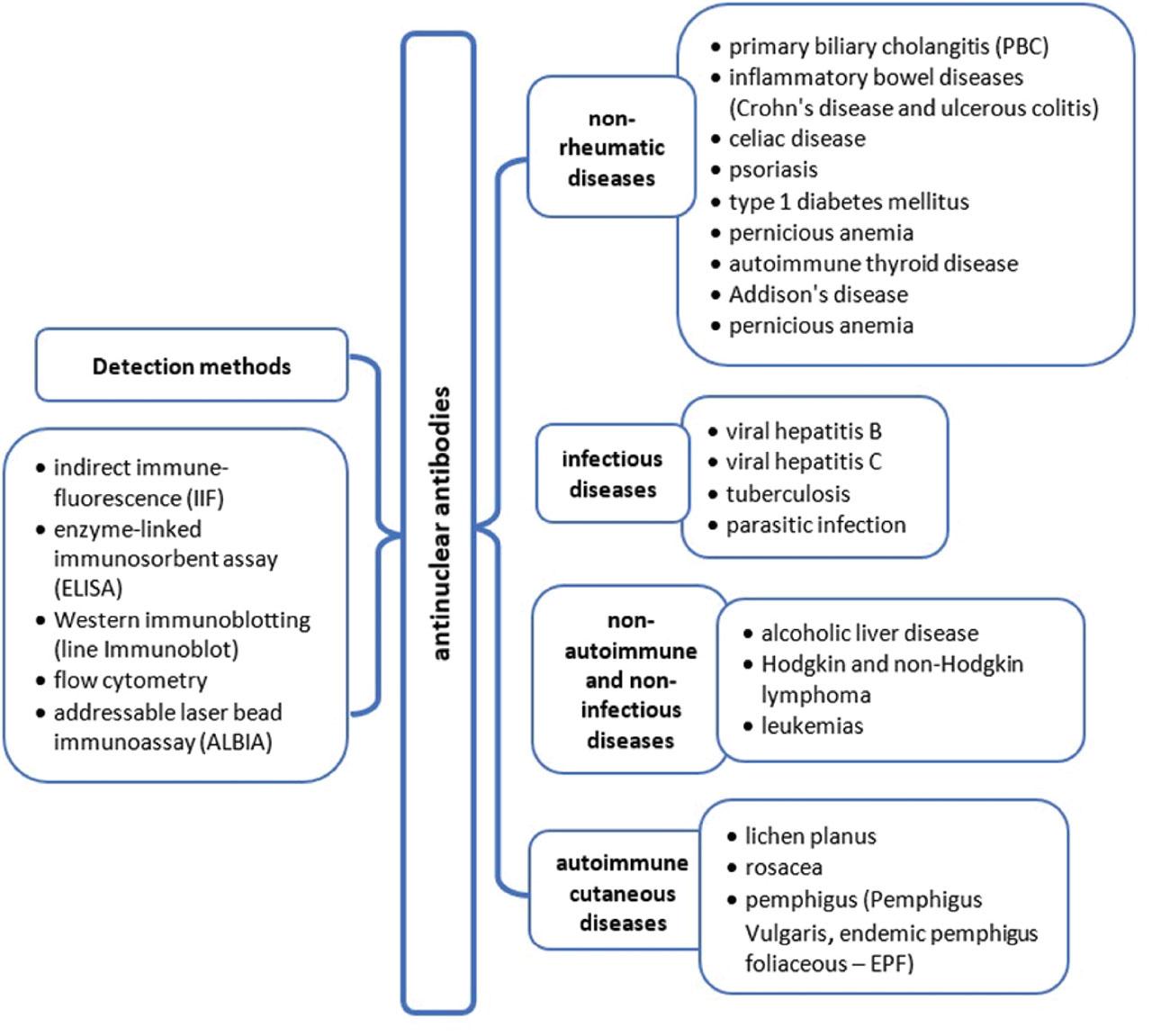
Methods used for ANA detection

Methods for ANA measurement
MethodMethod descriptionAdvantagesLimitations
IIF
  • Recommended by ACR and EULAR as the gold standard for ANA testing (Meroni and Schur 2010)

  • The most established method for ANA screening (Kumar et al. 2009; Pisetsky 2011)

  • Uses human epithelial (Hep-2) cells

  • Confirms nuclear antigens and fluorescence patterns using a fluorescence microscope (e.g., speckled, homogeneous, nuclear mixture, and cytoplasmic mixture patterns; Peng et al. 2014)

  • A negative IIF result is conclusive

  • All positive results must be confirmed by a second independent method (e.g., ELISA or ImmunoBlot) (Choi et al. 2020)

  • Determines staining patterns and titers concurrently (Agmon-Levin et al. 2014)

  • High sensitivity test

  • A time-consuming and labor-intensive test

  • A lack of adequate standardization (Damoiseaux et al. 2005; Agmon-Levin et al. 2014)

  • Poor specificity—false-positive results due to the non-specific binding of antibodies and the presence of closely related antigens sharing similar epitopes (Kątnik-Prastowska 2009)

  • Low positive predictive value

  • Operator-dependent test—high inter and intra-laboratory variabilities of pattern recognition resulted from the individual abilities of investigators (Infantino et al. 2017; Rigon et al. 2017)

ELISA
  • Test based on antigen–antibody reaction

  • Automated methods (Shah and Maghsoudlou 2016)

  • Monospecific tests identifying various specific antigens in CTD such as dsDNA, SSA/Ro (52 kDA and 60 kDA), SSB/La, U1-RNP, Sm, centromere, Jo-1, Scl-70, Rib-P, fibrillarin, RNA Pol III, PM-Scl, PCNA and Mi-2 (Robier et al. 2015)

  • More specific and sensitive than IIF (Murdjeva et al. 2011; Aydin 2015; Kohl and Ascoli 2017)

  • Simple procedure

  • High specificity and sensitivity caused by an antigen–antibody reaction

  • High efficiency caused by lack of necessity of complicated sample pre-treatment

  • Safe and eco-friendly (radioactive substances and large amounts of organic solvents are not required)

  • Cost-effective assay (low-cost reagents)

  • Tedious/laborious assay procedure

  • Expensive preparation of antibodies, which requires specific culture cell media

  • The risk of antibodies inactivation caused by their labeling

  • High possibility of false positive/negative antigens detection resulting from cross-reactivity of secondary antibody (false results caused by insufficient blocking of immobilized antigen)

  • Antibody instability if not refrigerated transport and inadequate storage is performed (Konstantinou 2017)

Western immunoblotting (line ImmunoBlot)
  • Enables the detection of protein expression levels based on its molecular weight and immunoreactivity with a specific antibody (Butler et al. 2019)

  • Identifies antigen specificity (reactivity) on a membrane in distinct lines (Kroon et al. 2022)

  • Consists of a series of interrelated steps (Murdjeva et al. 2011)

  • More sensitive than DID and CIE

  • Particularly useful in detecting ANAs in non-rheumatic diseases (Pillai-Kastoori et al. 2020)

  • High sensitivity, specificity, ease, and rapidity of execution

  • Potential for automation

  • Precisely identifies antibody reactions against nuclear antigens (Lallier et al. 2019)

  • Expensive

  • Time consuming

  • Detects linear epitopes only

  • Minor variations in a series of interrelated steps can alter the quality of the blot and introduce errors (Pillai-Kastoori et al. 2020)

  • Reproducibility and trustworthiness (caused by non-precise test performance) (Butler et al. 2019; Kroon et al. 2022)

Flow cytometry
  • Evaluates antibody reactivity using beads with fluorescent signals and antigens

  • (Vignali 2000; Naides et al. 2020)

  • Concurrent antigen recognition

  • High sensitivity (Vignali 2000; Avaniss-Aghajani et al. 2007; Bonilla et al. 2007)

  • Economical multi-antigen testing

  • Has the potential for automation (Bonilla et al. 2007)

  • Lack of uniformity in testing may be a cause of misdirecting results (Naides et al. 2020)

  • Provides a single result at a time (Vignali 2000; Avaniss-Aghajani et al. 2007; Bonilla et al. 2007)

ALBIA
  • Based on the application of autoantigen array and flow technologies (Giavedoni 2005; Swana et al. 2019)

  • Is used in various immunoassay, genomic, and proteomic analyses (Giavedoni 2005; Satoh et al. 2015; Swana et al. 2019).

  • One serum may react with many autoantigens in a multiplexed ALBIA (Fritzler et al. 2006)

  • The ability to assess several antibody specificities simultaneously in a small serum sample

  • Reliable, accurate, cost-saving

  • Efficient

  • Has a rapid turnaround time

  • High sensitivity

  • Less dependence on highly skilled operators (Fritzler et al. 2006; Satoh et al. 2015; Swana et al. 2019).

Characteristics of ANAs antibodies in infectious and non-infectious diseases

ANAs antibodies in infectious diseases
DiseaseNumber of patientsNumber of ANAs (+) patients/clinical characteristicsMethod of ANAs measurementReference
Hepatitis C
  • N = 89

  • control: n = 100

  • 20.2% (18 patients)

IIFde Castro et al. (2022)
N = 48
  • 11 patients (23%)

IIFPisetsky (2011)
TB
  • n = 83

  • control: n = 83

  • 33% with TB (mainly present anti-Scl-70)

  • 20% in control group

ELISAShen et al. (2013)
n = 1 (case report)
  • ANAs titer: 1:1280 in pleural fluid

  • ANA IgG and IgM levels positive at 1:160 and 1:40, respectively

IIFWin et al. (2003)
Parasitic infectionn = 613
  • ANA titer inversely linked to schistosome infection

  • ↑ ANA level after anti-helminthic treatment

ELISAMutapi et al. (2011)
n = 125
  • ↑ ANA prevalence in patients with the infection progression

  • 6.7% in acute stage

  • 23.3% in chronic stage

  • 70.0% in late-stage

IFATWang et al. (2018)
ANA in non-infectious diseases
DiseasesNumber of patientsANA-characteristicsMethodReference
ALDn = 9063.8% (44 out of 69)IIFLian et al. (2013)
n = 47
  • 22% with AID

  • 71% with CAH

  • Anti-ssDNA and anti-dsDNA in 60% of patients with ALD and CAH

Quantafluor fluorescent autoantibody test kit.Laskin et al. (1990)
NHL
  • NHL: n = 119

  • HL: n = 60

  • ANAs presence of patients with NHL without the risk of autoimmune disease development

IIFAltintas et al. (2008)
  • n = 347

  • control: n = 213

  • ANAs occurrence targeting mitotic proteins

  • ANAs in 19% of NHL patients with particular subgroups, such as ANAs directed against mitotic proteins or associated mitotic proteins

IIF—Hep2 cellsGuyomard et al. (2003)
Leukemian = 196
  • ANA positive in 34% with leukemia

  • ANAs with a nucleolar pattern were linked to a two-fold increase in the risk of death compared to negative ANA results in leukemia

IIFWang et al. (2021)
n = 216
  • ANA positivity in 30 (13.9%) patients

  • The correlation of positive ANA and TP53 disruption improves accuracy in predicting OS

IIFTSun et al. (2019)
ANAs in autoimmune cutaneous diseases
EPF
  • n = 120

  • control: n = 372

  • ANA detected in three EPF patients (2.5%)

IIFNisihara et al. (2003)
PV
  • n = 50

  • control n = 50

  • ANA positivity in 40% of patients

  • The most common patterns: homogenous or speckled

IIFSaleh et al. (2017)
LPn = 47
  • Rat esophagus is a preferred substrate for ANAs detection in LP patients, (ANA positive in 40.42% LP patients)

  • Monkey esophagus—ANA-positive in a 27.6% subjects, while Hep-2 cells and rat liver are unsuitable

IIF on Rat esophagus, Monkey esophagus, Hep-2 cells, and rat liverCarrizosa et al. (1997)
n = 100
  • ANA-positivity of 22%

IIFRambhia et al. (2018)
Rosacean = 101-ANAs with titre 1:160
  • ANA ≥1:160 in 53.5%

  • High ANAs titers are more prevalent in females (55.8%) than in males (44.15%)

IIF on Hep-2Woźniacka et al. (2013)

The prevalence of ANAs non-rheumatic autoimmune diseases

DiseaseAnalyzed groupsANAs presence/detection of specific antibodiesANAs detection methodReference
PBCn = 32
  • 10 patients

IIFWalker et al. (1965)
Psoriasis
  • Ps: n = 101

  • PsA: n = 100

  • control: n = 50

  • 25.7% Ps ANAs positive

  • 15% PsA ANAs positive

IIFPatrikiou et al. (2020)
  • n = 118

  • control: n = 50

  • ↑ prevalence of ANAs in males (M: F = 57.1% vs. 42.9%)

ELISASingh et al. (2010)
DMt1
  • n = 70

  • control: n = 20

  • 27% of patients

IIFHeras et al. (2010)
  • -

    Pediatric: n = 80

  • -

    Pediatric: control n = 473

  • -

    Adult: control n = 1125

  • Significantly higher prevalence (16.2%) of ANAs among pediatric diabetics

IIFNotsu et al. (1983)
CDn = 101
  • 24% of subjects with CD (P < 0.001)

  • ANA positivity is associated with the presence of the HLA DQ2/DQ8 haplotypes (P < 0.001).

IIFCarroccio et al. (2015)
n = 161
  • 8.4% ANA positive

  • Showed distinct nuclear staining patterns: homogeneous (6), fine-speckled (7), and coarse-speckled (1)

IIFAlmeida et al. (2019)
Autoimmune thyroid disease
  • -

    GD: n = 256

  • -

    Grave's orbitopathy n = 156

  • control: n = 78

  • ANA positive in 212 (80%) GD patients vs. ANA positive in no GO (91%)

IIFLanzolla et al. (2023)
  • n = 168

  • control: n = 75

  • ANAs in 35% of patients with ATD (anti-_ dsDNA and anti-Ro)

IIF using Hep-2 cells as substrateTektonidou (2004)
n = 104
  • 60% of patients have a speckled ANA pattern. (ANAs related to higher prevalence of ATG and ATPO in 30% of children with chronic lymphocytic thyroiditis without rheumatologic conditions

IIF using Hep-2 cells as substrateTorok and Arkachaisri (2010)
PA
  • n = 124

  • control: 41

  • 16.1% ANA

Indirect IIFMorawiec-Szymonik et al. (2019)
ADn = 1
  • Positive ANAs (392.23 IU/mL; N = < 23) present in the course of antiphospholipid syndrome

Serological screeningYazdi et al. (2021)
29-year-old with clinical features of acute Addisonian crisis and SLE
  • AD co-exist with SLE

Serology screeningGodswill and Odigie (2014)
Language: English
Submitted on: Jun 1, 2024
Accepted on: Nov 4, 2024
Published on: Jan 19, 2025
Published by: Hirszfeld Institute of Immunology and Experimental Therapy
In partnership with: Paradigm Publishing Services
Publication frequency: 1 issue per year

© 2025 Nikita Niranjan Kumar, Samir Ahmad Dit Al Hakim, Bogna Grygiel-Górniak, published by Hirszfeld Institute of Immunology and Experimental Therapy
This work is licensed under the Creative Commons Attribution-NonCommercial-NoDerivatives 4.0 License.