
INTRODUCTION
Cerebral palsy (CP) is a broad term that describes persistent disorder of movement resulting from an insult to the immature brain, leading to sensory and motor disorders, with/without intellectual disability, hearing impairment or speech problems1.
Spastic hemiplegic cerebral palsy (SHCP) is a type of spastic CP in which one side of the child’s body is affected. Approximately 20%–30% of children with CP have spastic hemiplegia2. It affects the movement and muscle tone on one side of the body causing stiffness, decreased muscle strength, impaired bone growth and weakness, which lead to difficulties with walking, coordination and daily tasks3. In children with SHCP, spasticity and weakness of muscles lead to a variety of problems affecting gait patterns4. One of these primary gait anomalies is genu recurvatum (GR)5.
GR is a position of the knee joint in which the range of motion (ROM) exceeds the neutral position or 0° of extension. It can also be quite disabling, causing deformity of the proximal tibia. GR has been described to occur in 8%–40% of children with CP. About 40%–68% of hemiplegic children have GR6. Uncontrolled locking of the knee joint can lead to abnormal gait, ligament injury, knee pain, arthritis and instability, affecting independency in activities of daily living (ADL)7.
GR is a result of several causes. One of these causes is weakness in the quadriceps muscle, which is essential for extending and stabilising the knee during the stance phase. During walking, weak quadriceps leads to buckling of the knee joint under body weight and diminished knee movement (excursion)8. Another cause is weakness of the gluteus maximus muscle. This weakness leads to anterior pelvic tilting, which is compensated for by an extension moment at the knee, resulting in GR9.
Universal exercise unit (UEU) is a novel technique for strengthening weak muscles by isolating the target muscle10. It consists of the spider cage, a pulley system, straps and weights for resistance. The numerous therapeutic benefits of the UEU include enhancing the ROM, passive or active, improving the flexibility of muscles, and increasing muscle strength without inducing any compensatory or undesired movement, thereby enhancing functional skills11.
Rebound therapy is the therapeutic use of rebound and rebound-like devices. It is an effective and enjoyable modality that can be integrated into rehabilitation programmes for children with CP12. It can increase proprioception, which is essential for joint sense of position and movement. In addition, it is effective in increasing muscle strength, endurance, balance and functional capacity13. Multiple studies have suggested using the UEU as a rehabilitation modality for children with CP to increase strength, balance and functional mobility10,11. Conversely, other studies have suggested the usage of rebound therapy12,13.
To the best of our knowledge, this is the first study that has aimed to compare the effects of UEU versus rebound therapy on GR in children with hemiplegic CP. It was hypothesised that there would be no difference between the effects of UEU and rebound therapy on GR in children with hemiplegic CP.
MATERIALS AND METHODS
Study design and participants
This study is a comparative randomised controlled trial that was carried out from March 2025 to January 2026. Fifty-one children with SHCP from both genders participated in the current study. They were recruited from Banha Educational Hospital, Banha University Hospital and Outpatient Clinics for Pediatric Physical Therapy, Cairo University. The study was carried out by the Ethical Committee of the Faculty of Physical Therapy, Cairo University (PT/REC/012/005355); informed consent from all children’s parents were obtained before starting the study. The study was retrospectively registered on ClinicalTrials.gov under ID: NCT07008521 on May 2025.
Children were aged 5–7 years, and their degree of spasticity was mild according to the Modified Ashworth Scale14. They were on Levels I and II on the Gross Motor Function Classification Scale (GMFCS)15. They had mild to moderate GR (15°–30° as identified by plain X-Ray)16, and were able to follow instructions. Children were excluded if they had visual or hearing impairment, lower limb surgical interference, severe deformities of the lower limb, or had been given spastic muscles botulinum injection during the past 6 months.
In all, 60 children with SHCP were screened for eligibility; 9 children were excluded as they did not meet the inclusion criteria (Fig 1), while the remaining 51 were randomly allocated into three groups: the control, and study groups A and B. Each child in the three groups received a designed physical therapy programme. The children in the control group received traditional progressive strengthening exercises, while group A received UEU (pulley system part) and group B received rebound therapy. Randomisation was done by closed, sealed, opaque envelopes numbered as 1, 2 and 3, and allocated as follows: 1 = control group, 2 = group A and 3 = group B. The selection of envelopes was done by the child’s parents. This process was performed by an evaluator who was uninformed about the selection and allocation of subjects.
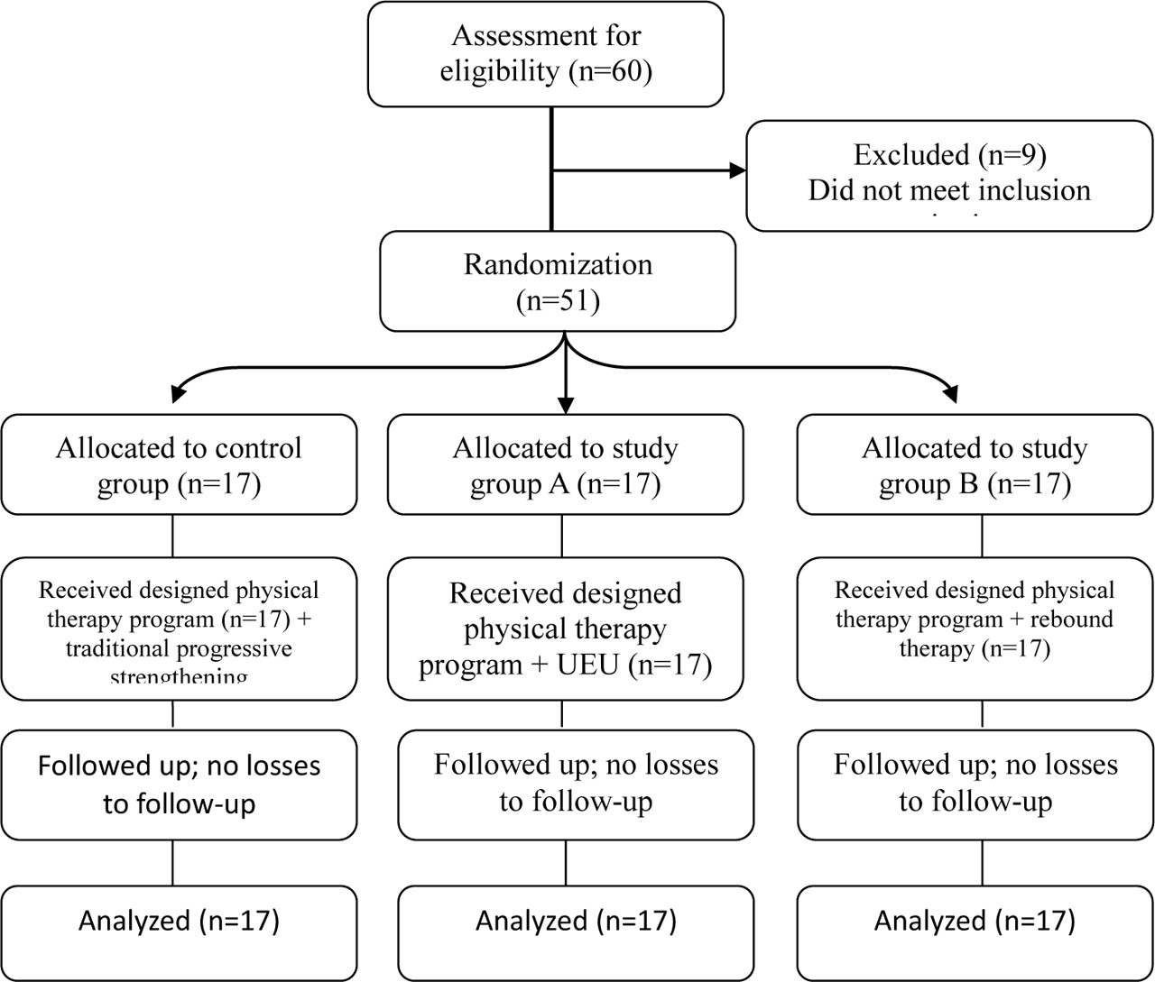
Figure 1.
CONSORT flow chart for participants in this study.
Sample size
The calculation of sample size was done using G*POWER statistical software (version 3.1.9.2; Franz Faul, Universitat Kiel, Germany). The requested size of the current study was N = 51 (17 in each group). Calculation was done as α = 0.05, power = 90% and effect size = 0.4. Sample size was calculated based on the primary outcome measure and on previous studies done on a similar population17.
Materials and equipment
In the current study, this part is consistent with the abstract that mentioned that degree of GR is the primary outcome, while muscle strength, balance skills, functional mobility skills and quality of life were the secondary outcome measures.
Procedures
Evaluation and outcome measures
The evaluation of all measured outcomes was done by blinded assessors.
Evaluation of muscle strength
The Lafayette MMT (model: 01163, Parkway North, USA) was selected to assess the muscle strength (quadriceps and gluteus maximus) in this study. It is a new ergonomic handheld strength measurement system that is developed for measuring the muscle strength with automatic storage of data. It gives more objective, reliable and accurate results18.
In this study, muscle strength assessment of the quadriceps and gluteus maximus was carried out as follows.
For quadriceps muscle: Each child was made to sit in an erect position on a plinth, with the hips and knees flexed at 90°, and the hands resting on their lap. The examiner placed the handheld dynamometer against the child’s tested leg anteriorly, just above the lateral malleolus, resisting the knee extension movement as the child extended their knee19.
For gluteus maximus muscle: Each child lay down in the prone position with the examined knee flexed at 90°, while stabilising the pelvis and trunk with belts, while the untested limb was in a relaxed and extended position. Then, the child was asked to perform hip extension against a dynamometer, which was positioned over the posterior aspect of the thigh (10 cm above knee joint) to resist hip extension. Three trials were performed for both the muscles, and the average of three measurements was calculated, based on Azab et al.19.
Evaluation of degree of GR
DONG FANG Plain-Radiographic Equipment (Model F92-1JB, X051-20-40/125, 32 mm, JAPAN) was used to determine the degree of GR of each child in this study. It is used for assessing ROM in children with CP and represents the gold standard for evaluating GR in the paediatric population, as it allows for weight-bearing and full-length alignment views that capture the deformity20.
Each child was positioned in unilateral standing (weight-bearing) with a chair in front of them for support during the evaluation procedures. The X-ray radiographs were obtained from a lateral view, at a distance of 90 cm from the device. Then, the angle was measured by intersecting the distal lateral axes of the femur and the shaft of tibia, based on Newton20. The assessment was performed by a blinded technician who was unaware of the participants’ allocation or selection.
Evaluation of balance skills
The Pediatric Balance scale (PBS) was used for evaluating balance skills in this study. It is used for assessment of balance in children aged from 5 years to 15 years, with mild to moderate motor impairment21. It includes 14 items which are scored from 0 to 4; higher total scores suggest that the child has good balance ability, based on Cesar et al.22.
Evaluation of functional mobility
Functional mobility was assessed by using the Mobility Questionnaire (MobQuest-28). It is a parent-reported questionnaire which consists of 28 items with a 5-point rating scale (0 = without any difficulty to 4 = impossible without help) with a maximum raw score of 112. It is used for evaluating functional mobility in children with CP23.
Evaluation of quality of life
The Pediatric quality of life inventory (PedsQL) (Arabic version parent-report) was used to evaluate the quality of life. It is used to assess the quality of life in children aged from 2 years to 18 years24. It is a multidimensional child self-report and parent-report scales, which is composed of seven domains. In this study, four domains of quality of life (ADL, movement, pain and fatigue) were assessed. Increased scores indicate an improvement in the quality of life skills, based on Varni et al.25.
Interventions
The protocol of treatment for all participating children was conducted at Banha Educational hospital, Banha University Hospital and Outpatient Clinics for Pediatric Physical Therapy, Cairo University.
For control group: Each child in the control group received a designed physical therapy programme according to their functional mobility, which was conducted for 1 hour, three sessions per week and for 3 consecutive months. The designed programme consisted of the following exercises, based on Madbouly et al.26: step standing, single limb stand, standing on balance board, dynamic standing (supine and prone to stand), abdominal and core stability exercises on wedge, and gait training forward, backwards and sideways. In addition, traditional progressive strengthening exercises for the quadriceps and gluteus maximus were done for 30 minutes, at three sessions per week and for 3 consecutive months, based on Madbouly et al.26, as follows: free weights in the form of adaptable weight cuffs were joined to the child’s lower leg by straps resisting knee extension or hip extension. Each child exercised at a load of approximately 60%–80% of their one repetition maximum (1-RM), performing 1–2 sets of 10–15 repetitions with rest intervals of 1 minute between the sets, based on Verschuren et al.27.
For group (A): Each child in this group was given the same designed exercises programme as described in the control group as well as UEU (pulley system part) to strength the quadriceps and gluteus maximus muscles, three times per week for 3 consecutive months. The pulley system part was performed for 30 minutes (15 minutes for each muscle) instead of the traditional strengthening programme.
The weight used in the training programme is detected by the 1-RM technique. 1-RM is considered as the maximum weight that can be lifted through full ROM. To evaluate 1-RM, the starting weight is estimated to be lower than the child’s maximum lifting capacity, which is gradually increased until reaching 1-RM. The included weight was from 1 kg to 3 kg regarding the assessed muscle group, with (1–5 minutes) periods of rest, based on Faigenbaum et al.28. Children carried out a mild to moderate load (about 60% 1-RM), for 1–2 sets with 10–15 repetitions). The workload was progressively graduated by an increment of 5%–10% of the weight, maintaining a target range of two sets of 10–15 repetitions, based on David et al.29.
The exercise programme was conducted as follows.
For quadriceps muscle: Each child was in a supine position on the table within a spider cage. The child was secured by straps at the chest, pelvis and non-involved limb, while the other limb was free to be moved, starting in a flexed position. A strap around the foot was connected to the upper part of the cage by a rope, and a weight to push at the end of this rope. The child was told to push their leg against the weight performing extension movement of limb. The exercises were conducted for 1–2 sets for 10–15 repetitions for each set, based on David et al.29.
For gluteus maximus: Each child was in a supine position on the table within a spider cage. The child was secured by straps on the chest, pelvis and non-involved limb, while the other limb was free to be moved, starting with an extended knee, which was maintained by a knee brace. A strap around the foot was connected to the upper part of the cage by a rope and a weight to push at the end of this rope. The child was told to push their leg against the weight, performing extension movement (lowering of leg from flexed hip to extended hip). The exercises were carried out for 1–2 sets for 10–15 repetitions for each set29.
For group (B): Each child in this group was given the same designed exercise programme as the control group, as well as rebound therapy in place of the traditional strengthening in the control group, for three sessions per week for 3 consecutive months. Every session was divided into warming-up for 5 minutes, 15–20 minutes of rebounding exercises and 5 minutes of cooling down. In the warming-up session, the child was asked to perform sitting transitions (side sitting, high kneeling and long sitting), bouncing at a slower rate or bouncing without leaving the surface of the trampoline. The rebounding programme comprised three sets of exercises, with each set lasting for 3 minutes of rebounding exercises and two sets of rest between the rebounding sets (3 minutes each). Each set of rebounding exercises started slowly and gradually progressed. The therapist stood behind the child in the trampoline, bouncing with him, guiding the movement and providing support. The child bounced up and down, holding on to the hand bar for maintaining steady balance when alone. During cool-down (5 minutes), the child was instructed to perform slow bouncing and/or controlled breathing exercises, based on Mohamed et al.16.
Statistical analysis
One-way ANOVA was conducted to compare continuous baseline characteristics, and the chi-square test was used to compare sex distribution between the groups. Data normality was assessed using the Shapiro–Wilk test, and homogeneity of variances was examined using Levene’s test. A mixed MANOVA was performed to evaluate the effects of treatment group, time and the treatment × time interaction on quadriceps strength, gluteus maximus strength, GR angle, PBS, Mob Quest 28 and the four PedsQL domains (ADL, movement, pain, and fatigue). A multivariate approach was used because these prespecified outcomes were clinically related, allowing treatment effects to be evaluated simultaneously while limiting inflation of Type I error associated with multiple separate analyses.
The assumptions relevant to the multivariate analysis, including multivariate normality and multicollinearity among dependent variables, were assessed before interpreting the MANOVA results. Given the sample size, the multivariate findings were interpreted cautiously, with consideration of the assumption testing and effect sizes. Post hoc pairwise comparisons following significant omnibus effects were performed with Bonferroni adjustment. Specifically, the paired t-test was used to analyze the within-group pre- and post-treatment changes presented in tables 2–4. Statistical significance was set at p < 0.05, and effect sizes were expressed as partial eta squared (ηp2). All analyses were conducted using SPSS version 27 (IBM SPSS, Chicago, IL, USA).
RESULTS
Subject characteristics
Table 1 shows the subject characteristics of the control group, and groups A and B. There was no significant difference between groups in age and sex distribution (p > 0.05).
Effect of treatment on quadriceps and gluteus maximus strength, GR angle, PBS, MobQuest-28 and PedsQL
Mixed MANOVA revealed that there was a significant interaction of treatment and time (Wilks’ Lambda = 0.003, F(18, 80) = 74.69, p = 0.001, ηp2 = 0.94). There was a significant main effect of time (Wilks’ Lambda = 0.001, F(9, 40) = 2979.48, p = 0.001, ηp2 = 0.99). There was a significant main effect of treatment (Wilks’ Lambda = 0.28, F(18, 80) = 3.89, p = 0.001, ηp2 = 0.47).
Within group comparison
All groups demonstrated significant post-treatment increases in quadriceps strength (p < 0.001), with percentage increases of 17.51% in the control group, 38.43% in group A and 45.65% in group B. Gluteus maximus strength also improved significantly in all the groups (p < 0.001), with gains of 16.97%, 41.97% and 49.45% for the control, A and B groups, respectively. GR angle decreased significantly across all groups (p < 0.001), with reductions of 10.42%, 26.58% and 26.42% for the control, A and B groups, respectively (Table 2).
Table 2.
Mean quadriceps and gluteus maximus strength and GR angle pre- and post-treatment of the control group, and groups A and B
| Control group | Group A | Group B | |
|---|---|---|---|
| Mean ± SD | Mean ± SD | Mean ± SD | |
| Quadriceps strength (kg) | |||
| Pre treatment | 15.08 ± 1.90 | 14.91 ± 2.16 | 14.94 ± 2.31 |
| Post treatment | 17.72 ± 2.13 | 20.64 ± 2.84 | 21.76 ± 3.13 |
| MD (% of change) | −2.64 (17.51%) | −5.73 (38.43%) | −6.82 (45.65%) |
| 95% CI (p ˂ 0.001) | −2.98: −2.31 | −6.07: −5.40 | −7.16: −6.49 |
| Gluteus maximus strength (kg) | |||
| Pre-treatment | 9.19 ± 1.25 | 9.22 ± 1.33 | 9.06 ± 1.28 |
| Post-treatment | 10.75 ± 1.33 | 13.09 ± 1.68 | 13.54 ± 1.64 |
| MD (% of change) | −1.56 (16.97%) | −3.87 (41.97%) | −4.48 (49.45%) |
| 95% CI (p ˂ 0.001) | −1.79: −1.33 | −4.10: −3.63 | −4.71: −4.24 |
| GR angle (degrees) | |||
| Pre-treatment | 20.82 ± 3.54 | 21.71 ± 3.98 | 21.35 ± 4.31 |
| Post-treatment | 18.65 ± 3.22 | 15.94 ± 2.63 | 15.71 ± 3.26 |
| MD (% of change) | 2.17 (10.42%) | 5.77 (26.58%) | 5.64 (26.42%) |
| 95% CI (p ˂ 0.001) | 1.64: 2.71 | 5.23: 6.30 | 5.11: 6.18 |
PBS significantly increased by 6.63% in the control group, 40.29% in group A and 66.71% in group B (p < 0.001). Similarly, MobQuest-28 scores improved significantly by 12.98%, 53.02% and 60.46% across the control, A and B groups, respectively (p < 0.001). (Table 3).
Table 3.
Mean PBS and MobQuest-28 pre-and post-treatment of the control group, and groups A and B
| Control group | Group A | Group B | |
|---|---|---|---|
| mean ± SD | mean ± SD | mean ± SD | |
| PBS | |||
| Pre-treatment | 26.53 ± 7.57 | 25.12 ± 7.28 | 25.65 ± 8.40 |
| Post-treatment | 28.29 ± 8 | 35.24 ± 6.91 | 42.76 ± 7.71 |
| MD (% of change) | −1.76 (6.63%) | −10.12 (40.29%) | −17.11 (66.71%) |
| 95% CI (p ˂ 0.001) | −2.74: −0.79 | −11.09: −9.14 | −18.09: −16.14 |
| MobQuest-28 | |||
| Pre-treatment | 48.94 ± 14.99 | 47.59 ± 16.34 | 46.41 ± 15.49 |
| Post-treatment | 55.29 ± 17.07 | 72.82 ± 20.33 | 74.47 ± 19.85 |
| MD (% of change) | −6.35 (12.98%) | −25.23 (53.02%) | −28.06 (60.46%) |
| 95% CI (p ˂ 0.001) | −8.12: −4.59 | −27.00: −23.47 | −29.83: −26.29 |
All PedsQL domains showed significant post-treatment gains in all groups (p < 0.001). ADL improved by 12.48%, 29.25% and 27.68%; movement by 17.76%, 37.32% and 45.87%; pain by 10.18%, 24.57% and 20.78%; and fatigue by 13.75%, 27.03% and 31.29% in the control, A and B groups, respectively (Table 4).
Table 4.
Mean ADL, movement, pain and fatigue pre- and post-treatment of control group, and groups A and B
| Control group | Group A | Group B | |
|---|---|---|---|
| Mean ± SD | Mean ± SD | Mean ± SD | |
| ADL | |||
| Pre-treatment | 59.94 ± 15.97 | 59.59 ± 15.47 | 61.53 ± 12.47 |
| Post-treatment | 67.42 ± 12.35 | 77.02 ± 9.37 | 78.56 ± 9.41 |
| MD (% of change) | −7.48 (12.48%) | −17.43 (29.25%) | −17.03 (27.68%) |
| 95% CI (p ˂ 0.001) | −13.29: −1.66 | −23.25: −11.62 | −22.84: −11.21 |
| Movement | |||
| Pre-treatment | 62.94 ± 9.02 | 61.47 ± 11.29 | 59.28 ± 17.37 |
| Post-treatment | 74.12 ± 12.02 | 84.41 ± 11.30 | 86.47 ± 11.01 |
| MD (% of change) | −11.18 (17.76%) | −22.94 (37.32%) | −27.19 (45.87%) |
| 95% CI (p ˂ 0.001) | −16.50: −5.85 | −28.26: −17.62 | −32.51: −21.87 |
| Pain | |||
| Pre-treatment | 71.54 ± 6.18 | 71.84 ± 6.93 | 72.57 ± 7.57 |
| Post-treatment | 78.82 ± 7.36 | 89.49 ± 9.66 | 87.65 ± 9. 75 |
| MD (% of change) | −7.28 (10.18%) | −17.65 (24.57%) | −15.08 (20.78%) |
| 95% CI (p ˂ 0.001) | −11.20: −3.36 | −21.57: −13.72 | −19.00: −11.15 |
| Fatigue | |||
| Pre-treatment | 58.82 ± 4.97 | 58.46 ± 3.08 | 59.93 ± 3.17 |
| Post-treatment | 66.91 ± 5.75 | 74.26 ± 8.81 | 78.68 ± 8.57 |
| 0MD (% of change) | −8.09 (13.75%) | −15.80 (27.03%) | −18.75 (31.29%) |
| 95% CI (p ˂ 0.001) | −11.81: −4.36 | −19.54: −12.08 | −22.48: −15.02 |
Between group comparison
Pre-treatment comparisons reported no significant differences between groups A and B and the control group in quadriceps strength, gluteus maximus strength, GR angle, PBS, MobQuest-28 and PedsQL (p = 0.97, p = 0.92, p = 0.81, p = 0.87 and p = 0.89, respectively).
Post-treatment comparisons revealed no significant differences between groups A and B in quadriceps strength, gluteus maximus strength, GR angle, MobQuest-28, or any PedsQL domains (p > 0.05). However, group B showed a significantly greater increase in PBS score compared with group A (p = 0.01).
Both groups A and B demonstrated significantly greater increases than the control group in quadriceps strength (p = 0.008 and p < 0.001, respectively), gluteus maximus strength (both p < 0.001) and reductions in GR angle (group A: p = 0.03; group B: p = 0.01). PBS scores in both the groups were also significantly increased than those of the control group (p = 0.02 and p < 0.001, respectively).
Similarly, for MobQuest-28, groups A and B showed significantly greater improvements than the control group (p = 0.02 and p = 0.01, respectively). For PedsQL outcomes, both groups A and B exhibited significantly greater gains than the control group in ADL (p = 0.02 and p = 0.009, respectively), movement (p = 0.03 and p = 0.008, respectively), pain score (p = 0.003 and p = 0.01, respectively) and fatigue score (p = 0.02 and p = 0.001, respectively). The effect sizes for the outcomes were large (ηp2 = 0.17–0.39), indicating substantial statistical effects. (Table 5).
Table 5.
Comparison of quadriceps and gluteus maximus strength, GR angle, PBS, MobQuest-28 and PedsQL between groups A, B and Control post-treatment
| Outcome | Group A versus B | Group A versus C | Group B versus C | ||||
|---|---|---|---|---|---|---|---|
| MD (95% CI) | p value | MD (95% CI) | p value | MD (95% CI) | p value | ||
| Quadriceps strength (kg) | −1.12 (−3.45: 1.20) | 0.46 | 2.92 (0.60: 5.25) | 0.008 | 4.04 (1.72: 6.37) | 0.001 | 0.39 |
| Gluteus maximus strength (kg) | −0.45 (−1.77: 0.88) | 0.68 | 2.34 (1.01: 3.66) | 0.001 | 2.79 (1.46: 4.11) | 0.001 | 0.17 |
| GR angle (degrees) | 0.23 (−2.36: 2.83) | 0.97 | −2.71 (−5.30: −0.11) | 0.03 | −2.94 (−5.54: −0.35) | 0.01 | 0.39 |
| PBS | −7.52 (−14.00: −1.06) | 0.01 | 6.95 (0.47: 13.41) | 0.02 | 14.47 (8.00: 20.94) | 0.001 | 0.18 |
| MobQuest28 | −1.65 (−17.93: 14.63) | 0.97 | 17.53 (1.25: 33.81) | 0.02 | 19.18 (2.89: 35.46) | 0.01 | 0.19 |
| ADL | −1.54 (−10.44: 7.37) | 0.90 | 9.60 (0.70: 18.51) | 0.02 | 11.14 (2.23: 20.05) | 0.009 | 0.19 |
| Movement | −2.06 (−11.80: 7.68) | 0.86 | 10.29 (0.55: 20.04) | 0.03 | 12.35 (2.61: 22.10) | 0.008 | 0.22 |
| Pain | 1.84 (−5.81: 9.49) | 0.82 | 10.67 (3.01: 18.31) | 0.003 | 8.83 (1.17: 16.48) | 0.01 | 0.29 |
| Fatigue | −4.42 (−11.08: 2.25) | 0.24 | 7.35 (0.69: 14.02) | 0.02 | 11.77 (5.10: 18.43) | 0.001 | 0.39 |
DISCUSSION
The aim of this study was to compare the effects of UEU and rebound therapy on GR, muscle strength, balance skills, functional mobility and quality of life in children with spastic hemiplegic CP. The treatment protocols were successfully completed by all groups, resulting in significant improvements across all measured outcomes post-treatment compared with that of pre-treatment. Post-treatment comparison between groups A and B revealed no significant difference in most measured variables, except for the PBS scores, which significantly improved in favour of group B.
The significant improvement in all outcome measures within the control group may be attributed to the effects of progressive strengthening exercises targeting the quadriceps and gluteus maximus muscles, which help improve GR, balance, functional mobility and quality of life. This is endorsed by Kushwaha and Suresh30, who have reported that progressive resistance training improves lower limb strength, balance and functional independence, thereby enhancing mobility, independence and overall quality of life. Also, this is analogous to the work of Merino-Andres et al.31, who revealed that strength training in children with CP has positive effects on the lower limb muscles, gross motor function and standing balance.
The significant decline in the degree of GR among children of group A may be due to the effect of UEU. This is established by Hamouda et al.32, who found that UEU could enhance muscle strength, knee alignment and decreased GR angle in children with spastic CP, which is achieved by increasing the muscle architecture and the cross-sectional area. This is also reported by Elsharkawy et al.33, who mentioned that UEU is effective for treating GR in children with CP by increasing the muscle strength of the lower limbs. Afzal et al.11 claim that strength training by UEU may stimulate the joint receptors, helping to stabilise and control joint movement.
The improvement in balance among children in group A may be due to the impact of UEU, as documented by Elnahas et al.34, who reported that UEU may be applied for children with spastic CP to increase the muscle strength and balance through neural function improvement, increasing the muscle cross-sectional area and architecture and therefore, improving the ability of standing and balance in children with spastic CP.
The significant improvement of functional mobility and quality of life was also stated by Mohamed et al.35, whose work show that UEU is a beneficial tool for rehabilitation of children with spastic CP to enhance muscle strength, gait and functional mobility skills. This is similar to the work of Elshahat et al.36, who said that UEU improves balance in children with spastic CP, and this may help such children to walk more efficiently without falling, and improve their performance in their ADLs.
Elhady et al.37 stated that UEU improves muscle strength and ROM by eliminating the effect of gravity and enhancing functional mobility among children with spastic CP; this supports the current findings regarding functional mobility among children with spastic CP.
The current results regarding the improvement of GR among children in group B is supported by Mohamed et al.16, whose work proved that rebound therapy is effective in correcting GR through increasing muscle strength and stimulation of visual, proprioceptive and vestibular systems, and thereby improving joint awareness and decreasing GR angle.
The improvement in balance skills, functional mobility and quality of life in group B is also shown in the work of El-Nemr and Kora38, who reported that rebound therapy has a great impact on improving balance in children with CP.
Abd Elmonem and Abd Elhady39 report that rebounding exercises are an effective modality for enhancing balance in children with CP. Kora et al.40 state that rebound therapy can be included in rehabilitation of children with spastic CP to enhance gross motor functions, gross motor coordination and balance. The current findings are supported by Romero-Franco et al.41, who have documented that rebound therapy stimulates the skin, joint and muscle receptors as well as the vestibular system, leading to joint stability improvement, muscle tone modulation, core stability enhancement and improved equilibrium reactions.
Regarding the clinical significance of the GR angle, clinical gait analysis guidelines widely accept a Minimal Detectable Change for 5° as the threshold required to indicate a true meaningful clinical change42. As shown in our results (Table 2), the mean difference (MD) in knee hyperextension angle achieved a reduction of 5.77° in group A and 5.64° in group B. Both outcomes successfully surpassed the MCD threshold. This concludes that UEU and rebound may be effective modalities for controlling knee joint during walking in children with CP.
While the minimal clinically important difference (MCID) for PBS in children with CP ranges from 3.66 to 5.83 points43, as demonstrated in our results, group A achieved a mean improvement of 10.12 points and group B achieved 17.11 points. Both intervention groups exceeded the upper threshold of MCID, suggesting that the balance gains are robust and clinically meaningful, with the rebound therapy being superior over UEU. Additionally, the MCID for MobQuest-28 is generally recognised as approximately 11 points, or a percentage of change exceeding 15%–20%, as a true clinical transformation beyond measurement error44. Our findings show exceptional clinical responsiveness, with group A improving by 25.23 points and group B by 28.06 points. These substantial gains across both scales directly translate into reduced mobility restrictions in ADL and improved postural safety.
For the PedsQL, the MCID required is 4.50 points for the parent proxy-report45. As demonstrated in our results (Table 4), the MD for ADL and health-related skills exceed the MCID threshold. Therefore, we suggest that the implemented UEU and rebound therapy may achieve a clinically meaningful enhancement in the children’s daily independence and overall quality of life.
Finally, this study demonstrated that children with spastic hemiplegic CP who participated in this trial showed a significant decrease in GR angle, along with increased muscle strength, balance skills, functional mobility and quality of life. These multidimensional improvements can be attributed to the cumulative effects of the standard physical therapy programme, the UEU, and rebound therapy. Both interventions produced beneficial changes compared with the control group, while rebound therapy demonstrated an additional statistically significant advantage over UEU, specifically for balance.
Limitations
There were several limitations to this study. First, it was conducted on a single clinical type of CP within a specific age group (from 5 years to 7 years). Second, there was a lack of long-term follow-up for participants. Finally, the study was constrained by a relatively small sample size, single-country recruitment and absence of therapist blinding. Hence, further studies are recommended to be performed on other types of CP with different age groups. Further researches are needed to compare both modalities on other outcome measures and to determine and illustrate the maintenance of the gained improvement.
CONCLUSIONS
From the obtained results in this study, both UEU and rebound therapy may be included within the treatment programme of children with spastic hemiplegic CP to manage GR, enhance muscle strength, balance, functional mobility and quality of life, while rebound therapy demonstrated an additional statistically significant advantage over UEU, specifically for balance in such children
ACKNOWLEDGEMENTS
The autors express their gratitude to all the children and their parents or legal guardians for their participation and for providing informed consent to participate in this study.