
Introduction
The supine bridge exercise (SBE) entails elevating the lumbopelvic region from a supine position1,2. Owing to its ease of execution and proven ability to enhance trunk stability, it has become a staple line intervention in clinical practice3,4. SBE is a closed-chain exercise that improves the stability of the trunk and strengthens the lumbar and pelvic muscles by maintaining a bridge posture against gravity5,6. It helps prevent injuries to the spinal soft tissues, including the paraspinal muscles and ligaments, and enhances the functional performance of the spinal joints7. Furthermore, it is useful for strengthening the lower-extremity muscles, such as the glutes and hamstrings, and can improve the performance capacity of both the lumbar and hip extensors8,9.
During SBE, lumbopelvic instability diminishes gluteus maximus (GM) activation and elicits compensatory increases in the activity of the erector spinae (ES) and hamstrings10,11. In patients with low back pain, the onset of GM contraction during hip extension is delayed relative to that of the multifidus and hamstring muscles, resulting in compensatory anterior pelvic tilt and an increased lumbar lordotic angle12–14. Abdominal muscle weakness and reduced activation should be considered as important contributors to these compensatory patterns15,16. Reduced activation of the transversus abdominis (TrA) and internal oblique (IO) muscles, in particular, aggravates lumbopelvic instability, imposes mechanical stress on the spinal joints and results in faulty lumbopelvic motion17. Hence, during SBE it is crucial to establish a biomechanical environment that allows the TrA and IO muscles to activate normally3,18.
Recent studies have emphasised that intra-abdominal pressure and breathing techniques play essential roles in maintaining trunk stability during various resistance and functional movements. Blazek et al.19 report that breathing strategies such as the Valsalva maneuver markedly increase intra-abdominal and intrathoracic pressures, thereby supporting trunk stabilisation during high-intensity resistance exercises. In addition, specific breathing patterns have been shown to influence force production and movement efficiency, with pronounced effects during movement phases that require greater stability, such as the sticking region20. Furthermore, abdominal wall tension has been found to vary across different resistance exercises, highlighting its importance for postural control and efficient force transmission21. Given the central role of abdominal pressure and abdominal wall tension in stabilising the lumbopelvic region, exercise strategies that effectively engage the deep abdominal musculature are of particular interest.
The abdominal draw-in maneuver (ADIM) is widely employed as an exercise method to strengthen the TrA and IO muscles22,23. The ADIM, which entails pulling the navel towards the spine, selectively strengthens the TrA and IO muscles, thereby enhancing lumbopelvic stability24,25. Furthermore, ADIM enhances coordination between the local and global muscles, prevents anterior pelvic tilt and reduces lumbar lordosis11,22,26.
Manual resistance (MR) can be employed as a motor-control technique to selectively activate specific muscles10. An adequate level of resistance is essential for muscle strengthening, for which MR is particularly useful to effectively reinforce weakened muscles during the early stages of rehabilitation7. MR provides precise vectors and allows appropriate modulation of load, and thereby demonstrates superior effectiveness in enhancing activation of the target muscles10,27. Furthermore, by precisely modulating the intensity of the resistance, the therapist can minimise compensatory movements, and by flexibly altering the point of resistance application, he can strengthen the muscles across a range of angles7.
Till date, several studies have investigated the selective activation of GM and the activity of the abdominal muscles during SBE. Youdas et al.2 report that performing SBE on one leg increases activation of the GM, compared with the two-legged SBE, relative to the hamstring muscles. Lee and Lee28 report that SBE combined with ADIM increased the thickness of the TrA muscle and improved balance ability. Dafkou et al.4 applied external loads of 10 and 20 kg using dumbbells during the performance of SBE combined with ADIM, and reported that the external load did not affect the recruitment of the abdominal muscles. However, no studies have yet investigated the combined application of MR and ADIM during SBE. Therefore, this study was conducted to examine the effects of applying MR and the ADIM technique together during SBE on the GM, ES, IO and hamstring muscles.
MATERIALS AND METHODS
Subjects
This study was conducted on 20 healthy adult males (Table 1). The required sample size was estimated based on previous studies with similar research designs22,29. Using G*Power software (Heinrich Heine University Düsseldorf, Düsseldorf, Germany) and assuming a medium effect size (f = 0.3) according to Cohen’s criteria, which was consistent with prior studies reporting sample sizes of 16–22 participants and comparable effect magnitudes, a statistical power of 0.80 and a significance level of 0.05, the minimum number of participants was calculated to be 18. To account for potential dropouts, a total of 20 participants were recruited.
Table 1.
General characteristics of subjects (n = 20)
| Characteristics | Subjects |
|---|---|
| Age (year) | 23.24 ± 1.43 |
| Height (cm) | 174.37 ± 3.24 |
| Weight (kg) | 67.72 ± 4.76 |
| BMI | 23.01 ± 1.21 |
The exclusion criteria were participants with low back, pelvic or leg pain; those with orthopaedic or neurological disorders; deformities or functional impairments; or any other conditions that would prevent them from performing SBE. Participants reported engaging only in light recreational physical activity, such as walking 1–2 times/week, and none participated in regular resistance training, core-stability exercises or glute-focused strengthening programmes.
This study was approved by the Institutional Review Board of Daegu University (No: 1040621-202505-HR-037), and written informed consent was obtained from all participants prior to participation.
Surface EMG recording and data processing
During the performance of SBE, muscle activation of the GM, ES, IO and hamstring muscles was measured using a wireless surface electromyography (EMG) device, TeleMyo DTS EMG (Noraxon Inc., Scottsdale, AZ, USA). To minimise skin resistance, hair was removed using a fine razor, followed by abrasion of the skin’s stratum corneum with sandpaper. The skin surface was then thoroughly cleaned of oils using an alcohol swab. Disposable Ag/AgCl surface electrodes were attached to each muscle. The electrode placements for each muscle were as follows: the GM electrodes were attached at the midpoint between the greater trochanter and the sacrum12; the ES electrodes were placed 2 cm lateral to the spinous process of the first lumbar vertebra12; the hamstring electrodes were positioned at the midpoint between the popliteal crease and the gluteal fold24; while the IO electrodes were attached diagonally 2 cm distal and inferior to the anterior superior iliac spine22.
EMG signal processing was performed using Myo-Research Master Edition 1.06 XP software (Noraxon Inc., Scottsdale, AZ, USA), with a sampling rate set at 1,500 Hz. The frequency bandwidth for signal analysis was set from 20 to 400 Hz, and a notch filter was applied to eliminate 60 Hz noise. The collected data were processed by converting the signals into root mean square values. EMG signals obtained from each muscle were normalised as a percentage of maximal voluntary isometric contraction (%MVIC). The MVIC measurements followed Kendall’s protocol27, with each measurement performed for 5 seconds. The mean value of the central 3 seconds, excluding the first and last 1 second, was calculated. Three trials were conducted for each muscle, and the average of these was used as the final value.
Experimental procedures
Before the experiment, all participants were instructed in the SBE and ADIM procedures, followed by a 10-minute training session to ensure that they could perform SBE while maintaining ADIM without difficulty. The participants then performed SBE under three randomised conditions: ADIM, MR and ADIM combined with MR. A 5-minute rest period was given between each condition to prevent muscle fatigue and learning effects22. In the ADIM condition, participants lay supine with knees bent at 90°, feet shoulder-width apart and flat on the floor with soles parallel to the ground, head and trunk aligned straight and the gaze directed towards the ceiling. Upon the examiner’s verbal cue, they performed ADIM by drawing their navel towards the spine, lifted their hips maximally and held the position for 5 seconds while maintaining ADIM (Figure 1A). Data were collected during the 5 seconds of maximal hip elevation, with the middle 3 seconds used for analysis, across three trials separated by 30 seconds of rest. In the MR condition, participants performed SBE without ADIM, while MR was applied opposing the hip elevation to induce isometric contraction (Figure 1B). MR was applied after the participants reached maximal hip elevation, and the intensity was adjusted to a level that allowed SBE to be maintained in an isometric position. Because the ability to sustain an isometric contraction varies considerably among individuals, quantifying MR as a fixed external load was deemed inappropriate, as it would not reflect each participantts actual performance capacity. Therefore, the magnitude of MR was individually adjusted so that each participant could maintain a stable maximal hip elevation. To ensure consistency in the application of MR, all resistance was provided by the first author, a licensed physical therapist with more than 15 years of clinical experience. All other procedures were identical to those in the SBE with ADIM condition. In the ADIM combined with MR condition, participants performed SBE with ADIM while receiving MR during maximal hip elevation (Figure 1C), following the same procedure as in the other conditions.
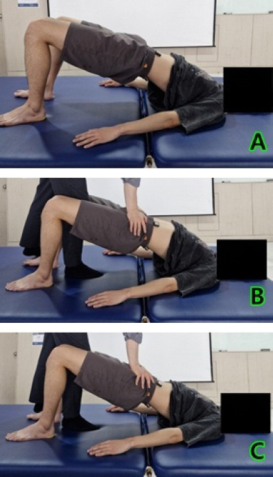
Figure 1.
SBE under three experimental conditions: (A) with ADIM; (B) with MR; (C) with ADIM combined with MR. ADIM - abdominal draw-in maneuver, MR - manual resistance, SBE - supine bridge exercise
Statistical analyses
Statistical analyses were performed using PASW Statistics 18 (SPSS Inc., Chicago, IL, USA). The Shapiroicslg test was used to assess the normality of data. To examine differences in the EMG amplitudes of the GM, ES, IO and hamstring muscles across the three conditions, a one-way repeated-measures ANOVA was conducted. Because this study employed a pure within-subjects design, Levene, s test for homogeneity of variance was not applicable, as all participants completed every experimental condition. The assumption of sphericity was evaluated using Mauchlyes test, and when this assumption was violated, the GreenhouseaGeisser correction was applied. Post hoc multiple comparisons were performed using the Bonferroni correction, with the level of significance set at p < 0.05.
RESULTS
In SBE performed under the three conditions, significant differences were observed in activation of the GM, ES, IO and hamstring muscles, and in the GM/ES ratio (p < 0.05; Table 2). Post hoc analyses revealed that GM activity was significantly greater in the ADIM combined with MR condition compared with both ADIM and MR (p < 0.001), and significantly higher in the MR condition than in the ADIM condition (p < 0.001). ES activity was significantly elevated in the MR and ADIM combined with MR conditions compared with the ADIM condition (p < 0.001). IO activity was significantly increased in the ADIM combined with MR condition compared with both other conditions (p < 0.001), and greater in the MR condition than in the ADIM condition (p < 0.001). Hamstring activity was significantly elevated in the MR and ADIM combined with MR conditions compared with the ADIM condition (p < 0.001). Finally, the GM/ES ratio was significantly higher in the ADIM combined with MR condition compared with both the ADIM (p =0.01) and MR (p < 0.001) conditions.
Table 2.
EMG activity of each muscle under three conditions during SBE
| Muscle activity (%MVIC) | ADIM | MR | ADIM + MR | F | p | η²p |
|---|---|---|---|---|---|---|
| GM | 31.51 ± 7.15a, c (28.16~34.86) | 67.30 ± 6.87b (64.08~70.52) | 71.85 ± 7.18 (68.49~75.21) | 282.01 | < 0.001* | 0.937 |
| ES | 37.41 ± 7.68a, c (33.82~41.00) | 74.21 ± 9.05 (69.97~78.45) | 72.50 ± 8.47 (68.54~76.46) | 458.44 | < 0.001* | 0.960 |
| IO | 17.36 ± 4.52a, c (15.24~19.48) | 38.52 ± 6.30b (35.57~41.47) | 48.48 ± 5.86 (45.74~51.22) | 359.64 | < 0.001* | 0.950 |
| Hamstring | 30.54 ± 6.22a, c (27.63~33.45) | 63.24 ± 8.69 (59.17~67.31) | 61.86 ± 7.31 (58.44~65.28) | 229.57 | < 0.001* | 0.924 |
| GM/ES ratio | 0.85 ± 0.14c | 0.92 ± 0.19b | 1.01 ± 0.19 | 8.22 | 0.006* | 0.302 |
DISCUSSION
The application of ADIM during SBE enhances activation of the TrA and IO muscles, thereby increasing the stability of the lumbopelvic region and aiding in selective activation of the GM3,4,28. MR is an exercise control technique that selectively activates muscles by providing precise vectors and appropriately adjusting loads, and thus demonstrating excellent efficacy in increasing activation of target muscles10,27. In this study, we aimed to compare the activity of the GM, ES, IO and hamstring muscles during SBE, by providing ADIM and MR, both simultaneously and separately.
In the study, both conditions in which MR was provided showed a pronounced increase in muscle activity for all muscles, compared with the condition where only ADIM was applied (p < 0.05), with notable differences in the means. Therefore, it can be suggested that the application of MR during SBE significantly increases the muscle activity of the GM, ES, IO and hamstrings. Resistance exercises induce neuromuscular adaptations by enhancing motor unit recruitment and firing rate, as well as improving coordination between the agonist and antagonist muscles, and thereby increasing muscle activity and coordination30,31. SBE, a closed-chain movement, inherently requires coordination of the muscles in the lower extremities and the torso for effective execution and stability32. Performing SBE isometrically against MR likely required the recruitment and substantial activation and coordination of various muscles. This may explain the marked increase in muscle activity observed in all muscles under the conditions where MR was applied in this study.
It was seen that the ADIM combined with MR condition elicited the greatest GM activation, compared with either ADIM alone or MR alone (p < 0.05). It also significantly enhanced the GM/ES muscle activation ratio compared with other conditions (p < 0.05). Therefore, it can be suggested that ADIM combined with MR helps to selectively strengthen the GM. The reasons for these results can be considered from two perspectives. First, the application of ADIM is believed to increase the activity of the TrA and IO muscles, thereby enhancing lumbopelvic stability15,17,22,24. Supporting this, studies by Grenier and McGill15 and Kim and Kim22 report that ADIM increases lumbopelvic stability, reducing compensatory activity of the lumbar joints, and enhancing the muscle activity of the gluteal muscles. In addition, Oh et al.24 have indicated that the application of ADIM increases lumbopelvic stability, and reported an increase in the muscle activity of the GM during prone hip extension. However, in this study, the ADIM combined with MR condition resulted in a significantly greater ES activity compared with the ADIM condition alone. This might be because, despite applying ADIM, the action of lifting the hips against MR had to be maintained in an isometric exercise form, leading to higher absolute ES activation demands.
The second reason is that ADIM likely reduced the anterior pelvic tilt, creating a favourable environment for GM activation. ADIM contracts the TrA and IO muscles, causing a posterior pelvic tilt1,28. Although the pelvic angle was not measured in this study, ADIM combined with MR showed a significantly greater GM and IO activity compared with the other conditions (p < 0.05), and the GM/ES ratio was also elevated (p < 0.05). This suggests that the increased IO activity from ADIM helped prevent anterior pelvic tilt, allowing the GM and IO muscles to work together to perform the bridge exercise as a force couple. Consequently, this likely required greater activity of the GM relative to the ES muscle.
Dafkou et al.4 provided external resistances of 10 and 20 kg while performing SBE with ADIM, and found that external resistance did not aid in recruitment of the abdominal muscles. In this study, ADIM combined with MR condition resulted in a significantly higher IO muscle activity than the conditions with only ADIM or only MR (p < 0.05). The reason for the differing results compared with Dafkou et al.4 could be that external resistance from objects merely provides a uniform load, whereas MR allows for adjusting the amount of resistance so that the participant can perform isometric contractions. This makes it easier to engage the targeted muscles and potentially enhances muscle activation by allowing directional vector adjustments.
Moreover, increases in abdominal pressure and abdominal wall tension are recognized as fundamental mechanisms that contribute to trunk stability across a broad range of resistance exercises19,21. Movements such as squats, deadlifts and pressing tasks require coordinated activation of the deep abdominal musculature to maintain spinal stiffness and optimise force transfer throughout the kinetic chain20,21. Although the present study examined these mechanisms only within the context of SBE, the enhanced IO and GM muscle activation observed with ADIM combined with MR may reflect stabilisation strategies that are similarly relevant during other load-bearing or multi-joint exercises. This broader perspective suggests that integrating ADIM with appropriately applied resistance may have practical implications not only for SBE but also for improving trunk stability during diverse rehabilitative and strength-training tasks.
The limitations of this study are as follows. First, the study was conducted exclusively on healthy adult males, making it difficult to generalise the findings to patients of other age groups. Second, the amount of MR could not be quantified. Third, since the pelvic angle and the TrA muscle were not measured, our ability to explain lumbopelvic stability solely on the basis of IO muscle activity was limited. Fourth, as a cross-sectional study, only the immediate effects were observed. Future research should investigate age and gender differences and include short- and long-term assessments of patients with lumbar and hip joint dysfunction and pain. Additionally, exploring the impact of ADIM combined with MR in other exercises is necessary.
CONCLUSIONS
The application of MR during SBE can effectively enhance muscle activation. Moreover, the simultaneous application of ADIM with MR increases the activation of the IO and GM muscles, suggesting that this combined approach improves lumbopelvic stability and selectively enhances GM activity. These findings highlight the clinical potential of incorporating ADIM with MR during SBE as an effective strategy for rehabilitation programmes aimed at improving trunk and pelvic stability.
Notes
[8] Conflicts of interest CONFLICT OF INTEREST
The authors declare no conflicts of interest.