
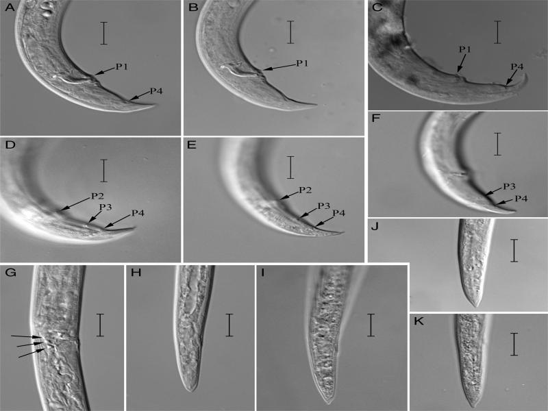
Figure 1:
Bursaphelenchus yongensis paratypes (C, F) and a population from Beijing (A, B, D, E, G-K): A-F: Male tail by different focus show caudal papillae; G: Lateral view of female vulval regions show a three-celled structure; H-K: Female tail. (Scale bars = 10 μm)